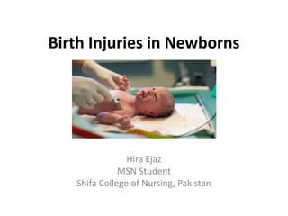
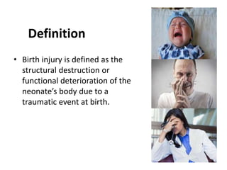
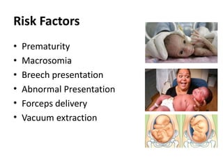
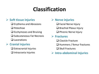
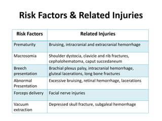
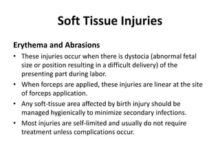
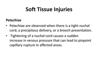
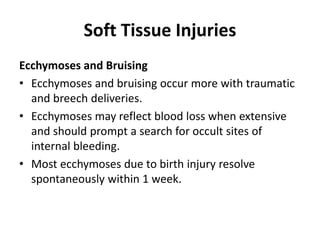
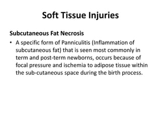
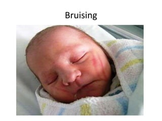
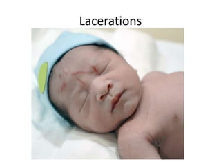
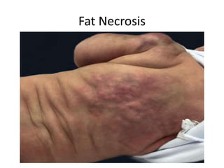
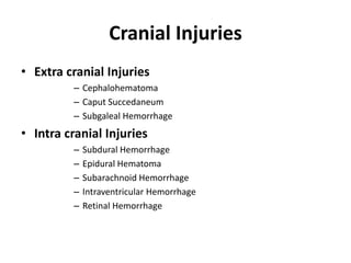
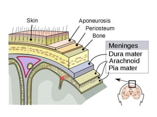
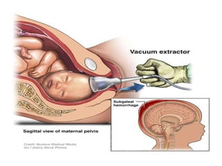
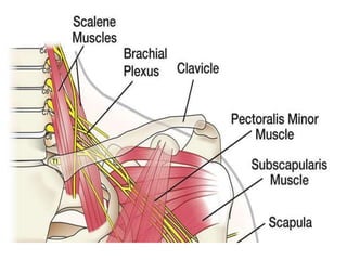
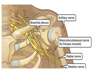
Birth Injuries in Newborns
Hira Ejaz
MSN Student
Shifa College of Nursing, Pakistan
Objective
At the end of this session students would be
able to:
• Identify different types of birth injuries in
newborns including:
– Soft tissue injuries
– Cranial injuries
– Nerve injuries
– Fractures
– Intra-abdominal injuries
Definition
• Birth injury is defined as the
structural destruction or
functional deterioration of the
neonate’s body due to a
traumatic event at birth.
Risk Factors
• Prematurity
• Macrosomia
• Breech presentation
• Abnormal Presentation
• Forceps delivery
• Vacuum extraction
Classification
 Soft tissue injuries
 Erythema and Abrasions
 Petechiae
 Ecchymoses and Bruising
 Subcutaneous Fat Necrosis
 Lacerations
 Cranial injuries
 Extracranial Injuries
 Intracrania Injuries
 Nerve injuries
 Facial Nerve Injury
 Brachial Plexus Injury
 Phrenic Nerve Injury
 Fractures
 Clavicle Fracture
 Hummers / femur fractures
 Skull Fractures
 Intra-abdominal Injuries
Risk Factors & Related Injuries
Risk Factors Related Injuries
Prematurity Bruising, intracranial and extracranial hemorrhage
Macrosomia Shoulder dystocia, clavicle and rib fractures,
cephalohematoma, caput succedaneum
Breech
presentation
Brachial plexus palsy, intracranial hemorrhage,
gluteal lacerations, long bone fractures
Abnormal
Presentation
Excessive bruising, retinal hemorrhage, lacerations
Forceps delivery Facial nerve injuries
Vacuum
extraction
Depressed skull fracture, subgaleal hemorrhage
Soft Tissue Injuries
Erythema and Abrasions
• These injuries occur when there is dystocia (abnormal fetal
size or position resulting in a difficult delivery) of the
presenting part during labor.
• When forceps are applied, these injuries are linear at the site
of forceps application.
• Any soft-tissue area affected by birth injury should be
managed hygienically to minimize secondary infections.
• Most injuries are self-limited and usually do not require
treatment unless complications occur.
Soft Tissue Injuries
Petechiae
• Petechiae are observed when there is a tight nuchal
cord, a precipitous delivery, or a breech presentation.
• Tightening of a nuchal cord causes a sudden
increase in venous pressure that can lead to pinpoint
capillary rupture in affected areas.
Soft Tissue Injuries
Ecchymoses and Bruising
• Ecchymoses and bruising occur more with traumatic
and breech deliveries.
• Ecchymoses may reflect blood loss when extensive
and should prompt a search for occult sites of
internal bleeding.
• Most ecchymoses due to birth injury resolve
spontaneously within 1 week.
Soft Tissue Injuries
Subcutaneous Fat Necrosis
• A specific form of Panniculitis (Inflammation of
subcutaneous fat) that is seen most commonly in
term and post-term newborns, occurs because of
focal pressure and ischemia to adipose tissue within
the sub-cutaneous space during the birth process.
Scalp Abrasion
Petechiae
Bruising
Lacerations
Fat Necrosis
Cranial Injuries
• Extra cranial Injuries
– Cephalohematoma
– Caput Succedaneum
– Subgaleal Hemorrhage
• Intra cranial Injuries
– Subdural Hemorrhage
– Epidural Hematoma
– Subarachnoid Hemorrhage
– Intraventricular Hemorrhage
– Retinal Hemorrhage
Cranial Injuries
Caput Succedaneum
• During labor, increased pressure on the fetal head
results in accumulation of blood and serum above
the periosteum and below the skin, leading to caput
succedaneum
• Causes edematous swelling of the scalp, Usually
resolves in 4 to 6 days
Subgaleal Hemorrhage
• It is collection of blood between the aponeurosis and
periosteum of the skull
Cranial Injuries
Cephalohematoma
• It is caused by a subperiosteal collection of blood
due to rupture of vessels beneath the periosteum.
• On physical examination, the affected area is
typically largest on postnatal day 3.
• Cephalohematomas resolve over the course of 3 to 4
weeks.
Intracranial injuries
Subdural Hemorrhage
• It is bleeding between the dura mater and the arachnoid layer
of brain. It is caused by rupture of bridging veins and is the
most common intracranial hemorrhage in term newborns.
• Affected infants may become symptomatic in the first 24 to 48
hours after birth. Presenting findings generally include
respiratory depression, apnea, and/or seizures.
Intracranial injuries
• Epidural Hemorrhage
• Epidural hematoma is very rare in neonates and is caused by
injury to the middle meningeal artery.
• Most cases also involve a corresponding linear skull fracture.
Presenting signs include hypotonia, seizures, bulging
fontanelles, and a change in the neonate’s level of
consciousness.
• Diagnosis is usually made by head CT scan.
Intracranial injuries
Subarachnoid Hemorrhage
• This is the second most common intracranial hemorrhage in
neonates caused by rupture of bridging veins in the
subarachnoid space.
• Symptoms may appear at 24 to 48 hours after birth and may
include apnea or seizures.
Intracranial Hemorrhages
Nerve Injuries
Facial Paralysis
• Results from pressure on the facial nerve by forceps.
• The mandibular branch of the facial nerve is most
commonly affected, resulting in less muscle
contraction on the affected side.
• Loss of the nasolabial fold, partial closing of the eye,
and the decreased ability to contract the lower facial
muscles on the affected side with the appearance of a
“drooping” mouth are common features. The mouth is
drawn over to the unaffected side with crying.
• Traumatic facial paralysis resolve in 2 to 3 weeks.
Nerve Injuries
Brachial plexus injury
• Causes brachial palsy also called Erb’s Palsy
• It involves paralysis of upper arm muscles
• Diagnosed by the presence of unilateral arm weakness
• Testing the Moro reflex in an affected infant produces
arm movement asymmetry because the affected arm
does not rise as high as the unaffected arm
• Most brachial plexus injuries resolve spontaneously.
• Improvement in movement can often be noted within
the first 24 hours of birth.
Nerve Injuries
Phrenic nerve injury
• Is associated with brachial plexus injury.
• It occurs most often with breech delivery and lateral
extension of the neck, with avulsion of C3 through C5 nerve
roots.
• Clinical signs include recurrent episodes of cyanosis
followed by respiratory distress.
• Phrenic nerve injury impairs diaphragmatic excursion on
the involved side, resulting in ineffective respiration.
• Bulging of the abdomen does not occur with inspiration.
Diagnosis is made by a chest radiograph showing elevation
of the diaphragm in an infant who has an associated
brachial plexus injury
Fractures
Clavicle Fracture
• This is the most common fracture in newborns.
• The risk factors for clavicle fracture are use of vacuum and forceps,
shoulder dystocia, higher birth weight, and increased maternal age
• The diagnosis is based on a displaced fracture in the newborn
period; it is often associated with tactile crepitus or petechiae over
the affected side and asymmetric Moro reflex
• No specific treatment is necessary. Infants may experience
increased pain on the affected side for 5 to 7 days. This pain is
usually amenable to oral or rectal acetaminophen. Once the callus
forms (usually by 7 to 10 days), the pain subsides. Prognosis is
excellent without any long-term sequelae.
Fractures
Skull Fracture
• There are 2 types of skull fractures: linear and depressed.
• A depressed fracture increases the possibility of an
intracranial process, especially when the fracture is greater
than 1 cm.
• Subdural hematoma is the most common intracranial injury
with depressed skull fracture.
• Forceps delivery is one of the major risk factors.
• If the fracture is less than 1 cm, depressed, and with no
neurological deficit, it can be managed with close
monitoring.
• CT scan is required to determine the presence or absence
of intracranial lesions.
Fractures
Long bone fractures
• Other than the clavicle, humerus or femur
fracture are the most common bony injuries
during the birth process.
• Clinical features include decreased movement
of the effected part, localized crepitus, pain
with palpation and on movement.
• Intra-abdominal Injuries
Intra-abdominal Injuries
• Liver and spleen injuries have been reported as birth-
related intra-abdominal injuries.
• Maternal trauma 1 to 2 weeks before delivery and
trauma during the delivery process is major causes of
intra-abdominal injuries.
• The most common presentation is shock, pallor,
anemia, and abdominal distension.
• Ultrasonography and CT scan of the abdomen are the
preferred diagnostic modalities
• Treatment involves fluid resuscitation, blood
component therapy that includes packed red blood cell
transfusion, and clotting factor rep
Nursing Management
• Physical assessment of the newborn
• Incubator care
• Monitoring of vital signs
• Record keeping
• Observation
• Early recognition of danger signs
• Pediatrician/ Neonatologist consultation
• Carry important investigations
• Help mother in feeding the infant
• Prevent drying of the eye by instilling artificial tears
Birth Injuries

Birth Injuries

  • 1.
    Birth Injuries inNewborns Hira Ejaz MSN Student Shifa College of Nursing, Pakistan
  • 2.
    Objective At the endof this session students would be able to: • Identify different types of birth injuries in newborns including: – Soft tissue injuries – Cranial injuries – Nerve injuries – Fractures – Intra-abdominal injuries
  • 3.
    Definition • Birth injuryis defined as the structural destruction or functional deterioration of the neonate’s body due to a traumatic event at birth.
  • 4.
    Risk Factors • Prematurity •Macrosomia • Breech presentation • Abnormal Presentation • Forceps delivery • Vacuum extraction
  • 5.
    Classification  Soft tissueinjuries  Erythema and Abrasions  Petechiae  Ecchymoses and Bruising  Subcutaneous Fat Necrosis  Lacerations  Cranial injuries  Extracranial Injuries  Intracrania Injuries  Nerve injuries  Facial Nerve Injury  Brachial Plexus Injury  Phrenic Nerve Injury  Fractures  Clavicle Fracture  Hummers / femur fractures  Skull Fractures  Intra-abdominal Injuries
  • 6.
    Risk Factors &Related Injuries Risk Factors Related Injuries Prematurity Bruising, intracranial and extracranial hemorrhage Macrosomia Shoulder dystocia, clavicle and rib fractures, cephalohematoma, caput succedaneum Breech presentation Brachial plexus palsy, intracranial hemorrhage, gluteal lacerations, long bone fractures Abnormal Presentation Excessive bruising, retinal hemorrhage, lacerations Forceps delivery Facial nerve injuries Vacuum extraction Depressed skull fracture, subgaleal hemorrhage
  • 7.
    Soft Tissue Injuries Erythemaand Abrasions • These injuries occur when there is dystocia (abnormal fetal size or position resulting in a difficult delivery) of the presenting part during labor. • When forceps are applied, these injuries are linear at the site of forceps application. • Any soft-tissue area affected by birth injury should be managed hygienically to minimize secondary infections. • Most injuries are self-limited and usually do not require treatment unless complications occur.
  • 8.
    Soft Tissue Injuries Petechiae •Petechiae are observed when there is a tight nuchal cord, a precipitous delivery, or a breech presentation. • Tightening of a nuchal cord causes a sudden increase in venous pressure that can lead to pinpoint capillary rupture in affected areas.
  • 9.
    Soft Tissue Injuries Ecchymosesand Bruising • Ecchymoses and bruising occur more with traumatic and breech deliveries. • Ecchymoses may reflect blood loss when extensive and should prompt a search for occult sites of internal bleeding. • Most ecchymoses due to birth injury resolve spontaneously within 1 week.
  • 10.
    Soft Tissue Injuries SubcutaneousFat Necrosis • A specific form of Panniculitis (Inflammation of subcutaneous fat) that is seen most commonly in term and post-term newborns, occurs because of focal pressure and ischemia to adipose tissue within the sub-cutaneous space during the birth process.
  • 11.
  • 12.
  • 13.
  • 14.
  • 15.
  • 16.
    Cranial Injuries • Extracranial Injuries – Cephalohematoma – Caput Succedaneum – Subgaleal Hemorrhage • Intra cranial Injuries – Subdural Hemorrhage – Epidural Hematoma – Subarachnoid Hemorrhage – Intraventricular Hemorrhage – Retinal Hemorrhage
  • 19.
    Cranial Injuries Caput Succedaneum •During labor, increased pressure on the fetal head results in accumulation of blood and serum above the periosteum and below the skin, leading to caput succedaneum • Causes edematous swelling of the scalp, Usually resolves in 4 to 6 days Subgaleal Hemorrhage • It is collection of blood between the aponeurosis and periosteum of the skull
  • 20.
    Cranial Injuries Cephalohematoma • Itis caused by a subperiosteal collection of blood due to rupture of vessels beneath the periosteum. • On physical examination, the affected area is typically largest on postnatal day 3. • Cephalohematomas resolve over the course of 3 to 4 weeks.
  • 25.
    Intracranial injuries Subdural Hemorrhage •It is bleeding between the dura mater and the arachnoid layer of brain. It is caused by rupture of bridging veins and is the most common intracranial hemorrhage in term newborns. • Affected infants may become symptomatic in the first 24 to 48 hours after birth. Presenting findings generally include respiratory depression, apnea, and/or seizures.
  • 26.
    Intracranial injuries • EpiduralHemorrhage • Epidural hematoma is very rare in neonates and is caused by injury to the middle meningeal artery. • Most cases also involve a corresponding linear skull fracture. Presenting signs include hypotonia, seizures, bulging fontanelles, and a change in the neonate’s level of consciousness. • Diagnosis is usually made by head CT scan.
  • 27.
    Intracranial injuries Subarachnoid Hemorrhage •This is the second most common intracranial hemorrhage in neonates caused by rupture of bridging veins in the subarachnoid space. • Symptoms may appear at 24 to 48 hours after birth and may include apnea or seizures.
  • 28.
  • 29.
    Nerve Injuries Facial Paralysis •Results from pressure on the facial nerve by forceps. • The mandibular branch of the facial nerve is most commonly affected, resulting in less muscle contraction on the affected side. • Loss of the nasolabial fold, partial closing of the eye, and the decreased ability to contract the lower facial muscles on the affected side with the appearance of a “drooping” mouth are common features. The mouth is drawn over to the unaffected side with crying. • Traumatic facial paralysis resolve in 2 to 3 weeks.
  • 31.
    Nerve Injuries Brachial plexusinjury • Causes brachial palsy also called Erb’s Palsy • It involves paralysis of upper arm muscles • Diagnosed by the presence of unilateral arm weakness • Testing the Moro reflex in an affected infant produces arm movement asymmetry because the affected arm does not rise as high as the unaffected arm • Most brachial plexus injuries resolve spontaneously. • Improvement in movement can often be noted within the first 24 hours of birth.
  • 36.
    Nerve Injuries Phrenic nerveinjury • Is associated with brachial plexus injury. • It occurs most often with breech delivery and lateral extension of the neck, with avulsion of C3 through C5 nerve roots. • Clinical signs include recurrent episodes of cyanosis followed by respiratory distress. • Phrenic nerve injury impairs diaphragmatic excursion on the involved side, resulting in ineffective respiration. • Bulging of the abdomen does not occur with inspiration. Diagnosis is made by a chest radiograph showing elevation of the diaphragm in an infant who has an associated brachial plexus injury
  • 37.
    Fractures Clavicle Fracture • Thisis the most common fracture in newborns. • The risk factors for clavicle fracture are use of vacuum and forceps, shoulder dystocia, higher birth weight, and increased maternal age • The diagnosis is based on a displaced fracture in the newborn period; it is often associated with tactile crepitus or petechiae over the affected side and asymmetric Moro reflex • No specific treatment is necessary. Infants may experience increased pain on the affected side for 5 to 7 days. This pain is usually amenable to oral or rectal acetaminophen. Once the callus forms (usually by 7 to 10 days), the pain subsides. Prognosis is excellent without any long-term sequelae.
  • 39.
    Fractures Skull Fracture • Thereare 2 types of skull fractures: linear and depressed. • A depressed fracture increases the possibility of an intracranial process, especially when the fracture is greater than 1 cm. • Subdural hematoma is the most common intracranial injury with depressed skull fracture. • Forceps delivery is one of the major risk factors. • If the fracture is less than 1 cm, depressed, and with no neurological deficit, it can be managed with close monitoring. • CT scan is required to determine the presence or absence of intracranial lesions.
  • 40.
    Fractures Long bone fractures •Other than the clavicle, humerus or femur fracture are the most common bony injuries during the birth process. • Clinical features include decreased movement of the effected part, localized crepitus, pain with palpation and on movement. • Intra-abdominal Injuries
  • 41.
    Intra-abdominal Injuries • Liverand spleen injuries have been reported as birth- related intra-abdominal injuries. • Maternal trauma 1 to 2 weeks before delivery and trauma during the delivery process is major causes of intra-abdominal injuries. • The most common presentation is shock, pallor, anemia, and abdominal distension. • Ultrasonography and CT scan of the abdomen are the preferred diagnostic modalities • Treatment involves fluid resuscitation, blood component therapy that includes packed red blood cell transfusion, and clotting factor rep
  • 42.
    Nursing Management • Physicalassessment of the newborn • Incubator care • Monitoring of vital signs • Record keeping • Observation • Early recognition of danger signs • Pediatrician/ Neonatologist consultation • Carry important investigations • Help mother in feeding the infant • Prevent drying of the eye by instilling artificial tears

Editor's Notes

  • #11 Subcutaneous fat necrosis is hard and well-circumscribed. Usually it is surrounded by erythema, but it can be flesh-colored or blue. Resolution occurs spontaneously by 6 to 8 weeks of age. Affected infants require long-term follow-up evaluation for the development of hypercalcemia, which can occur up to 6 months after the initial presentation of the skin lesions
  • #12 Scalp Abrasion
  • #13 Petechiae
  • #14 Bruising
  • #15 Lacerations
  • #16 Fat Necrosis
  • #18 Anatomy of skull (aponeurosis is a connective tissue sheet) Bone outer surface a sheet covering all the bones
  • #30 7th cranial nerve Ptosis
  • #31 Nerve Injuries
  • #32 If a newborn with brachial palsy has tachypnea and requires oxygen, the possibility of phrenic nerve involvement increases and ultrasonographic studies of hemidiaphragmatic movement may be indicated. Types Upper plexuses injury, lower and total plexuses injury
  • #33 Send signals from spinal cord to the shoulder arm and hand
  • #36 brachial plexus injury
  • #40 Linear fractures are non-depressed and usually need only close follow-up evaluation and monitoring. Neurosurgical consultation should be obtained within 12 to 24 hours of diagnosis for those with evidence of an intracranial process and if the depression is greater than 1 cm.