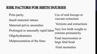
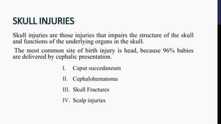
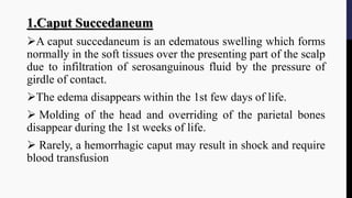
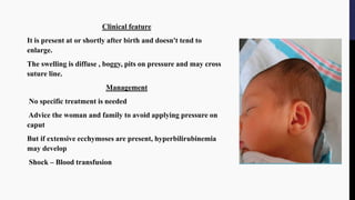
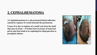
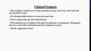
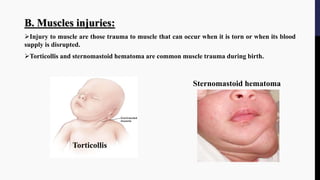
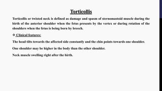
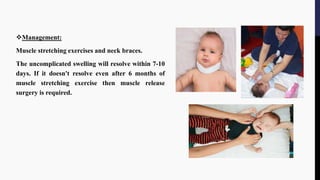
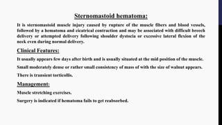
Newborn Birth Injuries: The Untold Story
Introduction:
As a result of the birth process some injuries occur that may be minor, whereas other may be more serious. Despite skilled midwifery and obstetric care in developed, birth trauma still occurs.
Definition:
An impairment of the infants body function or structure due to adverse influences that occur at birth.
Risk factors:
Primi parity,
Small maternal stature
Prolonged or usually rapid labor
Malpresentation of the fetus
Use of mid forceps or vaccum extraction
Fetal macrosomia or large fetal head
Classification:
Based on areas involved:
1. trauma to skin and superficial tissues
2. muscle trauma
3. nerve trauma
4. fractures
PREVENTION OF BIRTH INJURIES
- To prevent or to detect early intrauterine fetal asphyxia.
- To avoid premature delivery.
- To avoid traumatic vaginal delivery.
- To extend the use of caesarean section in abnormal & complicated presentation more liberally.
- Improve the level of doctor and nurses
THANK YOU.
BIRTH INJURIES IN NEWBORN: Definition of birth injuries , statistics, etiology, classification of birth injuries , head injuries: cephalhematoma and Caput succedaneum, skull fractures
, nerve injuries: erb's palsy and klumpke's palsy, bone injuries: clavicular and long bone fracture , intra-abdominal and soft tissue injuries, management and prevention of birth injuries
Birth Injuries are the common complications of Instrumental Delivery. So intrapartum management should be done very carefully in ordered to ensure healthy and good outcome of baby.
Birth injuries are inflicted during the time of delivery of the baby. it can occur in different parts of the body such as head, shoulder, eyes, nerves, etc. these injuries may be minor which resolve themselves with time while others are major and require prompt treatment. it is also very important to focu upon the prevention of occurence of such birth injuries. these injuries can be head injury, paralysis, fracture, soft tissue injury, visceral injury etc.
Prolonged labor is the inability of a woman to proceed with childbirth upon going into labor. Prolonged labor typically lasts over 20 hours for first time mothers, and over 14 hours for women that have already had children.
The female pelvis is ideal for childbearing. Complete knowledge on it helps a obstetrician or midwife to conduct normal labour as well as detect any abnormalities related to abnormal pelvis.
An impairment of the infants body function or structure due to adverse influences that occur at birth. Injuries to the infant may result from mechanical forces (i.e., compression, traction) during the birth process. 0.7% (Seven of every 1,000) births result in birth injuries, though most women give birth in modern hospitals surrounded by medical professionals. Birth injuries account for fewer than 2% of neonatal deaths. Infant mortality resulting from birth trauma fell from 64.2 to 7.5 deaths per 100,000 live births from 1970-1985. Birth injuries can be classified to Soft tissue injuries, Head and neck injuries, Facial injuries, Cranial nerve injuries, Spinal cord injuries, Peripheral Nerve injury, Fractures & Torticollis, Intra-abdominal injury. Proper management neccissates, early recognition of trauma, Careful physical and neurological evaluation, Establish whether additional injuries exist, Injury may result from resuscitation, Assess Symmetry of structure & function, Specific examination such as cranial nerve, joint range of motion, scalp/skull integrity.
Birth Injury during delivery or high risk infants.pptxformanite2
Nursing care for high risk newborn, birth injury, high risk infants, intrcranial hemorrhage, brachial palsy, cerebral palsy, head injuries during birth or during delivery
Flu Vaccine Alert in Bangalore Karnatakaaddon Scans
As flu season approaches, health officials in Bangalore, Karnataka, are urging residents to get their flu vaccinations. The seasonal flu, while common, can lead to severe health complications, particularly for vulnerable populations such as young children, the elderly, and those with underlying health conditions.
Dr. Vidisha Kumari, a leading epidemiologist in Bangalore, emphasizes the importance of getting vaccinated. "The flu vaccine is our best defense against the influenza virus. It not only protects individuals but also helps prevent the spread of the virus in our communities," he says.
This year, the flu season is expected to coincide with a potential increase in other respiratory illnesses. The Karnataka Health Department has launched an awareness campaign highlighting the significance of flu vaccinations. They have set up multiple vaccination centers across Bangalore, making it convenient for residents to receive their shots.
To encourage widespread vaccination, the government is also collaborating with local schools, workplaces, and community centers to facilitate vaccination drives. Special attention is being given to ensuring that the vaccine is accessible to all, including marginalized communities who may have limited access to healthcare.
Residents are reminded that the flu vaccine is safe and effective. Common side effects are mild and may include soreness at the injection site, mild fever, or muscle aches. These side effects are generally short-lived and far less severe than the flu itself.
Healthcare providers are also stressing the importance of continuing COVID-19 precautions. Wearing masks, practicing good hand hygiene, and maintaining social distancing are still crucial, especially in crowded places.
Protect yourself and your loved ones by getting vaccinated. Together, we can help keep Bangalore healthy and safe this flu season. For more information on vaccination centers and schedules, residents can visit the Karnataka Health Department’s official website or follow their social media pages.
Stay informed, stay safe, and get your flu shot today!
- Video recording of this lecture in English language: https://youtu.be/lK81BzxMqdo
- Video recording of this lecture in Arabic language: https://youtu.be/Ve4P0COk9OI
- Link to download the book free: https://nephrotube.blogspot.com/p/nephrotube-nephrology-books.html
- Link to NephroTube website: www.NephroTube.com
- Link to NephroTube social media accounts: https://nephrotube.blogspot.com/p/join-nephrotube-on-social-media.html
Tom Selleck Health: A Comprehensive Look at the Iconic Actor’s Wellness Journeygreendigital
Tom Selleck, an enduring figure in Hollywood. has captivated audiences for decades with his rugged charm, iconic moustache. and memorable roles in television and film. From his breakout role as Thomas Magnum in Magnum P.I. to his current portrayal of Frank Reagan in Blue Bloods. Selleck's career has spanned over 50 years. But beyond his professional achievements. fans have often been curious about Tom Selleck Health. especially as he has aged in the public eye.
Follow us on: Pinterest
Introduction
Many have been interested in Tom Selleck health. not only because of his enduring presence on screen but also because of the challenges. and lifestyle choices he has faced and made over the years. This article delves into the various aspects of Tom Selleck health. exploring his fitness regimen, diet, mental health. and the challenges he has encountered as he ages. We'll look at how he maintains his well-being. the health issues he has faced, and his approach to ageing .
Early Life and Career
Childhood and Athletic Beginnings
Tom Selleck was born on January 29, 1945, in Detroit, Michigan, and grew up in Sherman Oaks, California. From an early age, he was involved in sports, particularly basketball. which played a significant role in his physical development. His athletic pursuits continued into college. where he attended the University of Southern California (USC) on a basketball scholarship. This early involvement in sports laid a strong foundation for his physical health and disciplined lifestyle.
Transition to Acting
Selleck's transition from an athlete to an actor came with its physical demands. His first significant role in "Magnum P.I." required him to perform various stunts and maintain a fit appearance. This role, which he played from 1980 to 1988. necessitated a rigorous fitness routine to meet the show's demands. setting the stage for his long-term commitment to health and wellness.
Fitness Regimen
Workout Routine
Tom Selleck health and fitness regimen has evolved. adapting to his changing roles and age. During his "Magnum, P.I." days. Selleck's workouts were intense and focused on building and maintaining muscle mass. His routine included weightlifting, cardiovascular exercises. and specific training for the stunts he performed on the show.
Selleck adjusted his fitness routine as he aged to suit his body's needs. Today, his workouts focus on maintaining flexibility, strength, and cardiovascular health. He incorporates low-impact exercises such as swimming, walking, and light weightlifting. This balanced approach helps him stay fit without putting undue strain on his joints and muscles.
Importance of Flexibility and Mobility
In recent years, Selleck has emphasized the importance of flexibility and mobility in his fitness regimen. Understanding the natural decline in muscle mass and joint flexibility with age. he includes stretching and yoga in his routine. These practices help prevent injuries, improve posture, and maintain mobilit
Factory Supply Best Quality Pmk Oil CAS 28578–16–7 PMK Powder in Stockrebeccabio
Factory Supply Best Quality Pmk Oil CAS 28578–16–7 PMK Powder in Stock
Telegram: bmksupplier
signal: +85264872720
threema: TUD4A6YC
You can contact me on Telegram or Threema
Communicate promptly and reply
Free of customs clearance, Double Clearance 100% pass delivery to USA, Canada, Spain, Germany, Netherland, Poland, Italy, Sweden, UK, Czech Republic, Australia, Mexico, Russia, Ukraine, Kazakhstan.Door to door service
Hot Selling Organic intermediates
Pulmonary Thromboembolism - etilogy, types, medical- Surgical and nursing man...VarunMahajani
Disruption of blood supply to lung alveoli due to blockage of one or more pulmonary blood vessels is called as Pulmonary thromboembolism. In this presentation we will discuss its causes, types and its management in depth.
The prostate is an exocrine gland of the male mammalian reproductive system
It is a walnut-sized gland that forms part of the male reproductive system and is located in front of the rectum and just below the urinary bladder
Function is to store and secrete a clear, slightly alkaline fluid that constitutes 10-30% of the volume of the seminal fluid that along with the spermatozoa, constitutes semen
A healthy human prostate measures (4cm-vertical, by 3cm-horizontal, 2cm ant-post ).
It surrounds the urethra just below the urinary bladder. It has anterior, median, posterior and two lateral lobes
It’s work is regulated by androgens which are responsible for male sex characteristics
Generalised disease of the prostate due to hormonal derangement which leads to non malignant enlargement of the gland (increase in the number of epithelial cells and stromal tissue)to cause compression of the urethra leading to symptoms (LUTS
Explore natural remedies for syphilis treatment in Singapore. Discover alternative therapies, herbal remedies, and lifestyle changes that may complement conventional treatments. Learn about holistic approaches to managing syphilis symptoms and supporting overall health.
ARTIFICIAL INTELLIGENCE IN HEALTHCARE.pdfAnujkumaranit
Artificial intelligence (AI) refers to the simulation of human intelligence processes by machines, especially computer systems. It encompasses tasks such as learning, reasoning, problem-solving, perception, and language understanding. AI technologies are revolutionizing various fields, from healthcare to finance, by enabling machines to perform tasks that typically require human intelligence.
Title: Sense of Smell
Presenter: Dr. Faiza, Assistant Professor of Physiology
Qualifications:
MBBS (Best Graduate, AIMC Lahore)
FCPS Physiology
ICMT, CHPE, DHPE (STMU)
MPH (GC University, Faisalabad)
MBA (Virtual University of Pakistan)
Learning Objectives:
Describe the primary categories of smells and the concept of odor blindness.
Explain the structure and location of the olfactory membrane and mucosa, including the types and roles of cells involved in olfaction.
Describe the pathway and mechanisms of olfactory signal transmission from the olfactory receptors to the brain.
Illustrate the biochemical cascade triggered by odorant binding to olfactory receptors, including the role of G-proteins and second messengers in generating an action potential.
Identify different types of olfactory disorders such as anosmia, hyposmia, hyperosmia, and dysosmia, including their potential causes.
Key Topics:
Olfactory Genes:
3% of the human genome accounts for olfactory genes.
400 genes for odorant receptors.
Olfactory Membrane:
Located in the superior part of the nasal cavity.
Medially: Folds downward along the superior septum.
Laterally: Folds over the superior turbinate and upper surface of the middle turbinate.
Total surface area: 5-10 square centimeters.
Olfactory Mucosa:
Olfactory Cells: Bipolar nerve cells derived from the CNS (100 million), with 4-25 olfactory cilia per cell.
Sustentacular Cells: Produce mucus and maintain ionic and molecular environment.
Basal Cells: Replace worn-out olfactory cells with an average lifespan of 1-2 months.
Bowman’s Gland: Secretes mucus.
Stimulation of Olfactory Cells:
Odorant dissolves in mucus and attaches to receptors on olfactory cilia.
Involves a cascade effect through G-proteins and second messengers, leading to depolarization and action potential generation in the olfactory nerve.
Quality of a Good Odorant:
Small (3-20 Carbon atoms), volatile, water-soluble, and lipid-soluble.
Facilitated by odorant-binding proteins in mucus.
Membrane Potential and Action Potential:
Resting membrane potential: -55mV.
Action potential frequency in the olfactory nerve increases with odorant strength.
Adaptation Towards the Sense of Smell:
Rapid adaptation within the first second, with further slow adaptation.
Psychological adaptation greater than receptor adaptation, involving feedback inhibition from the central nervous system.
Primary Sensations of Smell:
Camphoraceous, Musky, Floral, Pepperminty, Ethereal, Pungent, Putrid.
Odor Detection Threshold:
Examples: Hydrogen sulfide (0.0005 ppm), Methyl-mercaptan (0.002 ppm).
Some toxic substances are odorless at lethal concentrations.
Characteristics of Smell:
Odor blindness for single substances due to lack of appropriate receptor protein.
Behavioral and emotional influences of smell.
Transmission of Olfactory Signals:
From olfactory cells to glomeruli in the olfactory bulb, involving lateral inhibition.
Primitive, less old, and new olfactory systems with different path
2. DEFINE BIRTH INJURIES
Birth injuries are an impairment of the infant’s body
function or structure due to adverse influences that
occurred at birth.
Birth injuries may be severe enough to cause neonatal
deaths, still births or number of morbidities.
3. RISK FACTORS FOR BIRTH INJURIES
Prim parity
Small maternal stature
Maternal pelvic anomalies
Prolonged or unusually rapid labor
Oligohydramnios
Malpresentation of the fetus
Use of mid forceps or
vaccum extraction
Versions and extractions
Very low birth weight or
extreme prematurity
Fetal macrosomia or
large fetal head
Fetal anomalies
4. Skull injuries are those injuries that impairs the structure of the skull
and functions of the underlying organs in the skull.
The most common site of birth injury is head, because 96% babies
are delivered by cephalic presentation.
I. Caput succedaneum
II. Cephalohematoma
III. Skull Fractures
IV. Scalp injuries
5. 1.Caput Succedaneum
A caput succedaneum is an edematous swelling which forms
normally in the soft tissues over the presenting part of the scalp
due to infiltration of serosanguinous fluid by the pressure of
girdle of contact.
The edema disappears within the 1st few days of life.
Molding of the head and overriding of the parietal bones
disappear during the 1st weeks of life.
Rarely, a hemorrhagic caput may result in shock and require
blood transfusion
6. Clinical feature
It is present at or shortly after birth and doesn't tend to
enlarge.
The swelling is diffuse , boggy, pits on pressure and may cross
suture line.
Management
No specific treatment is needed
Advice the woman and family to avoid applying pressure on
caput
But if extensive ecchymoses are present, hyperbilirubinemia
may develop
Shock – Blood transfusion
7. 2. CEPHALHEMATOMA
A Cephalohematoma is a sub periosteal blood collection
caused by rupture of vessels beneath the periosteum.
Causes It is due to rupture of a small vein from the skull
that may be from : • Friction between bones of maternal
pelvis and fetal skull as in cephalopelvic disproportion or
precipitate labour.
8. Clinical Features
The swelling is usually never at birth, gradually develops a few hours after birth and
may persist for weeks.
It is incompressible and never crosses the suture line.
The overlying scalp may show discoloration.
The condition may be confused with caput succedaneum or meningocele. Meningocele
lies over a suture line or fontanels and there is impulse on crying.
Rarely suppuration occurs.
9. Management
No active treatment is necessary unless it becomes infected or complicated.
A head CT should be obtained if neurological symptoms are present.
Vitamin K 1-2 mg IM should be given to correct any coagulation defect.
In case of infected hematoma, the condition is treated with incision and drainage, systematic
antibiotics and monitoring of hematocrit and bilirubin level.
Advice the woman and family to avoid hot compress by using oil.
10. 3. Scalp Injuries
Scalp injuries are those injuries that are characterized by impairment in integrity of the
scalp tissue.
Causes
• Forceps delivery (tip of the blades)
• Incised wound inflicted during cesarean section
• Scalp-electrode placement
• Episiotomy
Management
The wound should be dressed with an antiseptic solution like 2% mercurochrome.
On occasion, the incised wound may cause risk hemorrhage and requires stitches.
11. 4. Skull Fracture
Fracture of the vault of the skull (frontal bone or anterior part of the parietal bone) is defined
as distortion in the continuity of skull bone which may be of fissure/linear or depressed type.
Causes
Effect of difficult forceps delivery or due to wrong application of forceps.
Projected sacral promontory of the flat pelvis.
12. Clinical Features
Fissure fracture if uncomplicated is usually symptomless.
Depressed fracture may be associated with neurological manifestations.
Signs of associated complications such as intracranial hemorrhage, raised intracranial
pressure, leakage of CSF.
Diagnosis
History of type of delivery, other injuries to head during birth. •
Physical examination
X-ray can confirm diagnosis.
13. Management
Linear or fissure fracture requires no treatment.
Depressed fracture may require surgical elevation.
If there is leakage of cerebral fluid through nose, antibiotic
therapy is indicated.
14. Intracranial injuries are the
injuries to the structures inside the
cranium during the process of the
birth that is characterized by
abnormal neurological
manifestations within first 48
hours of life.
15. Traumatic Intracranial Hemorrhage
It is defined as hemorrhage inside cranium due to trauma and it can be extradural or
subdural hemorrhage.
Extradural hemorrhage: It is defined as hemorrhage in space between cranial bones and outer
layer of duramater. It is usually associated with fractured skull bone.
Subdural hemorrhage: It is defined as hemorrhage in the space between arachnoid mater and
inner layer of duramater.
16. Causes of Traumatic ICH
Excessive moulding in deflexed vertex.
Rapid compression of the head during delivery of the after-coming head of breech or in
precipitate labor.
Forcible forceps traction following wrong application of the blades.
Clinical Features of Traumatic ICH
The hemorrhage may be fatal and the baby is delivered stillborn or with severe respiratory
depression(APGAR score:0-3).
Gradually, the features of cerebral irritation appear.
Hydrocephalus and mental retardation may be a late sequelae.
17. Anoxic Intracranial Hemorrhage
It is defined as hemorrhage inside the cranium due to perinatal asphyxia, trauma and ischemia.
It can be intraventricular, subarachnoid and intracerebral.
Causes :
Perinatal asphyxia, trauma and ischemia
Clinical features:
Altered level of consciousness
Focal neurological defecits
Seizures
18. Diagnosis of ICH
Doppler ultrasonography can detect any change in cerebral circulation.
CT scan is useful to detect cortical neuronal injury.
Magnetic resonance imaging (MRI) is used to evaluate any hypoxic ischemic brain injury.
CSF analysis: Elevated RBCs, WBCs and protein.
19. MANAGEMENT OF ICH
The baby should be nursed in quiet ,warm and well ventilated environment.
Maintain cleanliness of the air passage, suction immediately after birth to
remove the secretion that occludes the pharynx. And supply oxygen as
necessary.
Frequently monitor the baby for skin color, vital signs and neurological
manifestations.
Feeding by nasogastric tube is advisable, fluid balance is to be maintained, if
necessary by parenteral route. .
20. Administer Vitamin K 1mg IM to prevent further bleeding due to hypoprothrombinaemia.
Prophylactic antibiotics are to be administered.
Anticonvulsants like phenobarbitone, phenytoin and diazepam can be given for seizures
Open surgical evacuation
-Rarely ventricular- peritoneal shunt and subdural-peritoneal shunt is required.
21. Soft tissue injuries are the injuries to skin, subcutaneous tissues, muscles and visceral
organs due to some degree of disproportion between the presenting part and the maternal
pelvis during the birth process and also from forcep blades, vacuum extractor cups, scalp
electrodes and scalpels.
22. A. Injury to Skin and Subcutaneous Tissue
Erythema and abrasions: Erythema and abrasion during birth are superficial reddening of the
skin with impaired integrity that usually are the result of the application of forceps,
discoloration is same configuration as the instrument.
Ecchymosis: Ecchymosis are small hemorrhagic areas( greater than 10 mm in diameter) that
may occur after traumatic or breech delivery.
Lacerations or scalpel cut: it is injury may occur during cesarean section. They usually occur
on the buttocks, scalp or thigh. Small cut heal spontaneously. Some time it may need repair by
stitches with 0-7 nylon. Healing is usually rapid.
23. B. Muscles injuries:
Injury to muscle are those trauma to muscle that can occur when it is torn or when its blood
supply is disrupted.
Torticollis and sternomastoid hematoma are common muscle trauma during birth.
Sternomastoid hematoma
Torticollis
24. Torticollis
Torticollis or twisted neck is defined as damage and spasm of sternomastoid muscle during the
birth of the anterior shoulder when the fetus presents by the vertex or during rotation of the
shoulders when the fetus is being born by breech.
Clinical features:
The head tilts towards the affected side constantly and the chin points towards one shoulder.
One shoulder may be higher in the body than the other shoulder.
Neck muscle swelling right after the birth.
25. Management:
Muscle stretching exercises and neck braces.
The uncomplicated swelling will resolve within 7-10
days. If it doesn't resolve even after 6 months of
muscle stretching exercise then muscle release
surgery is required.
26. Sternomastoid hematoma:
It is sternomastoid muscle injury caused by rupture of the muscle fibers and blood vessels,
followed by a hematoma and cicatrical contraction and may be associated with difficult breech
delivery or attempted delivery following shoulder dystocia or excessive lateral flexion of the
neck even during normal delivery.
Clinical Features:
It usually appears few days after birth and is usually situated at the mid position of the muscle.
Small moderately dense or rather small consistency of mass of with the size of walnut appears.
There is transient torticollis.
Management:
Muscle stretching exercises.
Surgery is indicated if hematoma fails to get reabsorbed.
27. Visceral Injuries
Injuries to organs like liver, spleen ,kidney, adrenals or lungs are called visceral injuries .
Visceral organs are commonly injured during breech delivery.
The most common result of the injury is hemorrhage. The hemorrhage may remain concealed
as subcapsular hematoma or capsule may rupture with the blood flowing into peritoneal cavity.
Prognosis is usually poor.
Clinical Features:
Pallor , tachycardia, shock and symptoms according to the organs being injured.
Management:
Correction of hypovolemia, anemia and coagulation disorders.
Management may be needed to repair injured viscera surgically.
28. FACIAL NERVE INJURIES
Erb’s palsy:
• It is an injury to C5,6, there is failure of abduction of the arm from the
shoulder inability for external rotation of the arm and to supinate the forearm.
The characteristic position is adduction and internal rotation of the arm and
pronation of the forearm.
• The biceps and Moro reflex is absent on the affected side.
Klumpke’s paralysis:
• This type of plexus involving 7th and 8th cervical and the 1st thoracic nerve
roots. There is paralysis of muscles of the forearm. The arm is flexed at the
elbow and wrist is extended.
29. Phrenic nerve paralysis:
• C3, C4, & C5 injury result paralysis of the ipsilateral diaphragm. This is
due to excessive stretching of neck at birth. with dyspnoea, cyanosis and
irregular breathing.
FACIAL NERVE PALSY (BELL’S PALSY)
• forceps delivery – prolonged second stage of labor may caused
• During crying, there is inability to wrinkle the forehead or close
the eye on the ipsilateral side, and the mouth is drawn awayfrom
the affected side.