SlideShare a Scribd company logo
1 of 32
Orbital Anatomy
By- Dr. Kawshik Nag,
Resident,
Ophthalmology, Phase-A
Chittagong Medical College.
Anatomy Of Orbit
 Quadrangular truncated
pyramidal in shape.
 Bounded by-
• Superiorly- Anterior cranial
fossa
• Medially- Nasal cavity and
ethmoidal air sinuses
• Inferiorly- Maxillary sinus
• Laterally- Middle cranial fossa
and Temporal fossa.
Dimensions
 Volume: 30cm3
 Rim: Horizontally- 4cm
Vertically- 3.5cm
 Intra orbital width: 2.5cm
 Extra orbital width: 10cm
 Depth: Medially- 4.2cm
Laterally- 5.0cm
 Ratio of vol. of orbit : vol. of
globe: 4.5:1
Bony Orbit
Seven bones make
up the bony orbit :
 Frontal bone
 Zygomatic Bone
 Maxillary bone
 Ethmoid bone
 Sphenoid bone
 Lacrimal bone
 Palatine bone
Walls Of The Orbit
 The bony orbit has four
walls:
 Medial wall
 Lateral wall
 Roof
 Floor
Medial Orbital Wall
The medial wall is
formed from front to
back by the:
 Frontal process of maxilla
 Lacrimal bone
 Orbital plate of the ethmoid
bone
 Body of the sphenoid bone.
Medial Orbital Wall
Clinical applications:
• It is the thinnest wall of the orbit, so it is frequently fragmented as a
result of indirect blow out fractures and during orbitotomy operations.
• Frequently eroded by chronic inflammatory lesions, neoplasms, cysts.
• Medial wall provide alternate access route to the orbit through sinus.
• Haemorrhage can occur due to trauma to ethmoidal vessels.
• Accidental lateral displacemet of medial wall causes traumatic
hypertelorism.
Lateral Orbital Wall
 Thickest and strongest.
 Formed by two bones:
• Zygomatic
• Greater wing of sphenoid.
Lateral Orbital Wall
Clinical applications:
• The anterior half of globe is not covered by bone on lateral side.
Hence, palpation of retrobulbar tumours is easier from the lateral
side.
• The zygomatico-sphenoid suture is an important landmark in creating
the flap in lateral orbitotomy.
• It is the strongest portion of the orbit and needs to be sawed open in
lateral orbitotomy.
• Since lateral wall is almost devoid of foramina, bleeding is less.
Roof Of Orbit
 Underlies frontal sinus and
anterior cranial fossa.
 Formed by-
• Orbital plate of frontal bone
• Lesser wing of sphenoid.
 Triangular.
 Faces downwards and slightly
forwards.
Roof Of Orbit
Clinical applications:
• Thin and periorbita peels away easily.
• Objects piercing upper eyelid penetrate roof and damage frontal
lobe.
• In old age roof may be absorbed so that periorbital and duramater
comes into contact.
• Any trauma of dura mater and CSF escapes into orbit or nose or
both.
Floor Of Orbit
 Shortest orbital wall.
 Formed by:
• Maxillary bone- medially
• Zygomatic bone- laterally
• Palatine bone- posteriorly.
 Triangular in shape.
 Bordered laterallly by inferior
orbital fissure and medially by
maxilloethmoidal sinus.
 Overlies maxillary sinus.
Floor Of Orbit
Clinical applications:
• Commonly involved in Blow
out fractures of the orbit.
Infra orbital vessels and
nerves almost always
involved.
• Diplopia is the main
symptoms of blow-out
fracture.
• Easily invaded by tumors of
the maxillary antrum.
Figure- Mechanism of blow-out fracture
from displacement of the globe itself into
the orbital walls. The globe is displaced
posteriorly, striking the orbital walls and
forcing them outward.
Base Of Orbit
 The anterior open part.
 Bounded by four orbital
margins-
• Superior orbital margin
• Inferior orbital margin
• Medial orbital margin
• Lateral orbital margin.
 It gives attachment to the
septum orbitale.
Apex Of Orbit
 Orbital apex is the posterior
end of the orbit.
 Four orbital walls converge.
 Two orifices:
• Optic Canal
• Superior orbital fissure
Optic Canal
 It connects the orbit to the
middle cranial fossa.
 It transmits:
• Optic Nerve
• Ophthalmic artery.
Superior Orbital Fissure
 Structure passing:
 Upper lateral part:
• Lacrimal and frontal nerves
• Trochlear nerve
• Superior ophthalmic vein
• Recurrent branch of
ophthalmic artery.
 Middle part:
• Superior and inferior divisions
of occulomotor nerve
• Nasociliary branch of
ophthalmic division of
trigeminal nerve.
• Abducent nerve.
 Lower medial part:
• Inferior ophthalmic vein.
Superior Orbital Fissure
Clinical applications:
• Radiographic enlargement of superior orbital fissure may
accompany pathologic processes,
 Aneurysm
 Meningioma
 Choroidoma
 Pituitary adenoma
 tumours of orbital apex.
• When idiopathic inflammation involves the superior orbital fissure,
the “Tolosa Hunt syndrome” which is painful ophthalmoplegia
results.
Periorbita
 Periorbita refers to periosteum
lining the orbitlal surface of the
bones of orbit.
 Loosely adherent to the bones.
 Fixed firmly at-
• Orbital margins
• Suture lines
• Various fissures and foramina
• Lacrimal fossa.
 Applied Anatomy-
• Surgery in the orbital roof in
the areas of fissures and
suture lines may be
complicated by cerebrospinal
fluid leakage.
Orbital Fascia
 It is a complex interwoven thin
connective tissue membrane
joining the various intraorbital
contents.
 Parts-
• Fascia bulbi,
• Muscular sheaths,
• Intermuscular septa,
• Membranous expansions of
the extraocular muscles,
• Ligament of Lockwood.
Extraocular Muscles
 Voluntary Muscles:
• Superior rectus
• Inferior rectus
• Medial rectus
• Lateral rectus
• Superior oblique
• Inferior oblique
• Levator palpebrae superioris.
 Involuntary Muscles:
• Superior tarsal or Muller’s
muscle,
• Inferior tarsal muscle.
Surgical Spaces In Orbit
 Orbit is divided into 4 surgical spaces-
• Subperiosteal space
• Peripheral orbital space/ Extraconal space
• Central orbital space/ Intraconal space
• Subtenon’s space
Surgical Spaces In Orbit
 Importance of these spaces-
• Most of the orbital tumours tends to remain with in a space in which
they are formed unless they are large or malignant or represents an
infiltrative process such as pseudotumour.
Subperiosteal Space
 Lies between orbital bone and
periorbita.
 tumours arising from bone
separates periorbita from
bone.
 Here periorbita acts as a
effective barrier against spread
of tumour to eye.
Subperiosteal Space
 tumours in this space are-
• Dermoids cyst
• Epidermoid cyst
• Mucocele
• Subperiosteal abscess
• Osteomatous tumour
Peripheral Orbital Space
 Known as extraconal space.
 Lies between periorbita at
periphery, extraocular muscles
and their intermuscular septa
internally and orbital septum
anteriorly.
 Posteriorly it merges with
central space.
 tumours in this space are
usually approached by anterior
orbitotomy and sometimes by
lateral orbitotomy.
Peripheral Orbital Space
 tumours in this space produce eccentric proptosis.
 tumours in this space are-
• Malignant Lymphoma
• Capillary haemangioma of childhood
• Intrinsic neoplasm of lacrimal gland
• Pseudotumours.
Central Orbital Space
 Known as muscle cone/ retro-
orbital space/posterior space/
intraconal space.
 Bounded by-
• Anteriorly tenon’s capsule
• Posteriorly by 4 recti and
intermuscular septa.
 In posterior part, space
become continuous with
peripheral space.
Central Orbital Space
 tumours of this space-
• Cavernous haemangioma of adults
• Solitary neurofibroma
• Neurolemoma
• Nodular orbital meningiomas
• Optic nerve glioma.
 Produce axial proptosis.
 tumours are approached through lateral orbitotomy.
Subtenon’s space
 Space around eyeball between
sclera and tenon’s capsule.
 Pus collection in this space is
drained by incision on tenon’s
capsule through conjunctiva.
Contents Of The Orbit
 Eyeball
 Fascia: Orbital and bulbar.
 Muscles: Extraocular.
 Vessels:
• Ophthalmic artery
• Superior and inferior ophthalmic
vein
• Lymphatics.
 Nerves: Optic,Oculomotor,
Trochlear, Abducent, Branches of
ophthalmic nerves and
sympathetic nerves.
 Ciliary ganglion
 Lacrimal gland and lacrimal sac
 Orbital fat.
Orbital anatomy

More Related Content

What's hot

What's hot (20)

eyelid anatomy slideshare
eyelid anatomy slideshareeyelid anatomy slideshare
eyelid anatomy slideshare
 
Oculomotor nerve
Oculomotor nerveOculomotor nerve
Oculomotor nerve
 
Anatomy of the eyelids
Anatomy of the eyelidsAnatomy of the eyelids
Anatomy of the eyelids
 
Blood supply of the eye
Blood supply of the eyeBlood supply of the eye
Blood supply of the eye
 
The orbit of eye
The orbit of eyeThe orbit of eye
The orbit of eye
 
Anatomy of eyelid
Anatomy of eyelidAnatomy of eyelid
Anatomy of eyelid
 
Superior Orbital Fissure Syndrome
Superior Orbital Fissure SyndromeSuperior Orbital Fissure Syndrome
Superior Orbital Fissure Syndrome
 
Trochlear nerve
Trochlear nerveTrochlear nerve
Trochlear nerve
 
Anatomy of the Orbit
Anatomy of the OrbitAnatomy of the Orbit
Anatomy of the Orbit
 
Evaluation of proptosis
Evaluation of proptosisEvaluation of proptosis
Evaluation of proptosis
 
Extraocular muscles
Extraocular muscles Extraocular muscles
Extraocular muscles
 
Ciliary body
Ciliary bodyCiliary body
Ciliary body
 
Optic pathway and lesions
Optic pathway and lesionsOptic pathway and lesions
Optic pathway and lesions
 
Ciliary ganglion
Ciliary ganglionCiliary ganglion
Ciliary ganglion
 
EXTRA OCULAR MUSCLES ANATOMY
EXTRA OCULAR MUSCLES ANATOMYEXTRA OCULAR MUSCLES ANATOMY
EXTRA OCULAR MUSCLES ANATOMY
 
Visual pathway
Visual pathway Visual pathway
Visual pathway
 
Anatomy of the orbit (nerves and vessels)
Anatomy of the orbit (nerves and vessels)Anatomy of the orbit (nerves and vessels)
Anatomy of the orbit (nerves and vessels)
 
anatomy and physiology of lacrimal apparatus ppt
anatomy and physiology of lacrimal apparatus  pptanatomy and physiology of lacrimal apparatus  ppt
anatomy and physiology of lacrimal apparatus ppt
 
Orbital spaces
Orbital spacesOrbital spaces
Orbital spaces
 
Accommodation of eye
Accommodation of eye Accommodation of eye
Accommodation of eye
 

Similar to Orbital anatomy

Anatomy of orbit
Anatomy of orbitAnatomy of orbit
Anatomy of orbitveeru1984
 
Anatomy of orbit and its clinical applications.
Anatomy of orbit and its clinical applications.Anatomy of orbit and its clinical applications.
Anatomy of orbit and its clinical applications.RahulGupta39947
 
By pd anatomy of the orbit
By pd anatomy of the orbitBy pd anatomy of the orbit
By pd anatomy of the orbitPushkar Dhir
 
Anatomy of orbit
Anatomy of orbitAnatomy of orbit
Anatomy of orbitveeru1984
 
Anatomy of orbit ophthalm
Anatomy of orbit ophthalmAnatomy of orbit ophthalm
Anatomy of orbit ophthalmDrArino John
 
Anatomy of orbit by pushkar dhir
Anatomy of orbit by pushkar dhirAnatomy of orbit by pushkar dhir
Anatomy of orbit by pushkar dhirPushkar Dhir
 
orbit malavika NEW.pptx
orbit malavika NEW.pptxorbit malavika NEW.pptx
orbit malavika NEW.pptxMalavikaAG
 
Orbit Anatomy and Surgical Spaces
Orbit Anatomy and Surgical Spaces Orbit Anatomy and Surgical Spaces
Orbit Anatomy and Surgical Spaces Vikas Khatri
 
Anatomy of human orbit
Anatomy of human orbitAnatomy of human orbit
Anatomy of human orbitrakshyabasnet1
 

Similar to Orbital anatomy (20)

Orbital anatomy
Orbital anatomy Orbital anatomy
Orbital anatomy
 
The Orbit
The OrbitThe Orbit
The Orbit
 
Anatomy of the orbit
Anatomy of the orbit Anatomy of the orbit
Anatomy of the orbit
 
Orbit final
Orbit finalOrbit final
Orbit final
 
The Orbit
The OrbitThe Orbit
The Orbit
 
Anatomy of orbit
Anatomy of orbitAnatomy of orbit
Anatomy of orbit
 
Anatomy of the Bony orbit
Anatomy of the Bony orbit Anatomy of the Bony orbit
Anatomy of the Bony orbit
 
Anatomy of orbit and its clinical applications.
Anatomy of orbit and its clinical applications.Anatomy of orbit and its clinical applications.
Anatomy of orbit and its clinical applications.
 
By pd anatomy of the orbit
By pd anatomy of the orbitBy pd anatomy of the orbit
By pd anatomy of the orbit
 
Orbit
OrbitOrbit
Orbit
 
Anatomy of orbit
Anatomy of orbitAnatomy of orbit
Anatomy of orbit
 
Anatomy of orbit
Anatomy of orbitAnatomy of orbit
Anatomy of orbit
 
Orbital surgeries
Orbital surgeriesOrbital surgeries
Orbital surgeries
 
Anatomy of orbit ophthalm
Anatomy of orbit ophthalmAnatomy of orbit ophthalm
Anatomy of orbit ophthalm
 
Orbital structure
Orbital structure Orbital structure
Orbital structure
 
Anatomy of orbit by pushkar dhir
Anatomy of orbit by pushkar dhirAnatomy of orbit by pushkar dhir
Anatomy of orbit by pushkar dhir
 
orbit malavika NEW.pptx
orbit malavika NEW.pptxorbit malavika NEW.pptx
orbit malavika NEW.pptx
 
Orbit Anatomy and Surgical Spaces
Orbit Anatomy and Surgical Spaces Orbit Anatomy and Surgical Spaces
Orbit Anatomy and Surgical Spaces
 
Antomy of orbit 25 4-19
Antomy of orbit 25 4-19Antomy of orbit 25 4-19
Antomy of orbit 25 4-19
 
Anatomy of human orbit
Anatomy of human orbitAnatomy of human orbit
Anatomy of human orbit
 

Recently uploaded

Top 10 Most Beautiful Chinese Pornstars List 2024
Top 10 Most Beautiful Chinese Pornstars List 2024Top 10 Most Beautiful Chinese Pornstars List 2024
Top 10 Most Beautiful Chinese Pornstars List 2024locantocallgirl01
 
Physicochemical properties (descriptors) in QSAR.pdf
Physicochemical properties (descriptors) in QSAR.pdfPhysicochemical properties (descriptors) in QSAR.pdf
Physicochemical properties (descriptors) in QSAR.pdfRAJ K. MAURYA
 
VIP ℂall Girls Arekere Bangalore 6378878445 WhatsApp: Me All Time Serviℂe Ava...
VIP ℂall Girls Arekere Bangalore 6378878445 WhatsApp: Me All Time Serviℂe Ava...VIP ℂall Girls Arekere Bangalore 6378878445 WhatsApp: Me All Time Serviℂe Ava...
VIP ℂall Girls Arekere Bangalore 6378878445 WhatsApp: Me All Time Serviℂe Ava...deepakkumar115120
 
Obat Aborsi Ampuh Usia 1,2,3,4,5,6,7 Bulan 081901222272 Obat Penggugur Kandu...
Obat Aborsi Ampuh Usia 1,2,3,4,5,6,7 Bulan  081901222272 Obat Penggugur Kandu...Obat Aborsi Ampuh Usia 1,2,3,4,5,6,7 Bulan  081901222272 Obat Penggugur Kandu...
Obat Aborsi Ampuh Usia 1,2,3,4,5,6,7 Bulan 081901222272 Obat Penggugur Kandu...Halo Docter
 
VIP ℂall Girls Kothanur {{ Bangalore }} 6378878445 WhatsApp: Me 24/7 Hours Se...
VIP ℂall Girls Kothanur {{ Bangalore }} 6378878445 WhatsApp: Me 24/7 Hours Se...VIP ℂall Girls Kothanur {{ Bangalore }} 6378878445 WhatsApp: Me 24/7 Hours Se...
VIP ℂall Girls Kothanur {{ Bangalore }} 6378878445 WhatsApp: Me 24/7 Hours Se...deepakkumar115120
 
Creeping Stroke - Venous thrombosis presenting with pc-stroke.pptx
Creeping Stroke - Venous thrombosis presenting with pc-stroke.pptxCreeping Stroke - Venous thrombosis presenting with pc-stroke.pptx
Creeping Stroke - Venous thrombosis presenting with pc-stroke.pptxYasser Alzainy
 
Test bank for critical care nursing a holistic approach 11th edition morton f...
Test bank for critical care nursing a holistic approach 11th edition morton f...Test bank for critical care nursing a holistic approach 11th edition morton f...
Test bank for critical care nursing a holistic approach 11th edition morton f...robinsonayot
 
Difference Between Skeletal Smooth and Cardiac Muscles
Difference Between Skeletal Smooth and Cardiac MusclesDifference Between Skeletal Smooth and Cardiac Muscles
Difference Between Skeletal Smooth and Cardiac MusclesMedicoseAcademics
 
Intro to disinformation and public health
Intro to disinformation and public healthIntro to disinformation and public health
Intro to disinformation and public healthTina Purnat
 
TEST BANK For Porth's Essentials of Pathophysiology, 5th Edition by Tommie L ...
TEST BANK For Porth's Essentials of Pathophysiology, 5th Edition by Tommie L ...TEST BANK For Porth's Essentials of Pathophysiology, 5th Edition by Tommie L ...
TEST BANK For Porth's Essentials of Pathophysiology, 5th Edition by Tommie L ...rightmanforbloodline
 
Drug development life cycle indepth overview.pptx
Drug development life cycle indepth overview.pptxDrug development life cycle indepth overview.pptx
Drug development life cycle indepth overview.pptxMohammadAbuzar19
 
ANATOMY AND PHYSIOLOGY OF RESPIRATORY SYSTEM.pptx
ANATOMY AND PHYSIOLOGY OF RESPIRATORY SYSTEM.pptxANATOMY AND PHYSIOLOGY OF RESPIRATORY SYSTEM.pptx
ANATOMY AND PHYSIOLOGY OF RESPIRATORY SYSTEM.pptxSwetaba Besh
 
Cardiac Output, Venous Return, and Their Regulation
Cardiac Output, Venous Return, and Their RegulationCardiac Output, Venous Return, and Their Regulation
Cardiac Output, Venous Return, and Their RegulationMedicoseAcademics
 
7 steps How to prevent Thalassemia : Dr Sharda Jain & Vandana Gupta
7 steps How to prevent Thalassemia : Dr Sharda Jain & Vandana Gupta7 steps How to prevent Thalassemia : Dr Sharda Jain & Vandana Gupta
7 steps How to prevent Thalassemia : Dr Sharda Jain & Vandana GuptaLifecare Centre
 
MOTION MANAGEMANT IN LUNG SBRT BY DR KANHU CHARAN PATRO
MOTION MANAGEMANT IN LUNG SBRT BY DR KANHU CHARAN PATROMOTION MANAGEMANT IN LUNG SBRT BY DR KANHU CHARAN PATRO
MOTION MANAGEMANT IN LUNG SBRT BY DR KANHU CHARAN PATROKanhu Charan
 
Circulatory Shock, types and stages, compensatory mechanisms
Circulatory Shock, types and stages, compensatory mechanismsCirculatory Shock, types and stages, compensatory mechanisms
Circulatory Shock, types and stages, compensatory mechanismsMedicoseAcademics
 
Part I - Anticipatory Grief: Experiencing grief before the loss has happened
Part I - Anticipatory Grief: Experiencing grief before the loss has happenedPart I - Anticipatory Grief: Experiencing grief before the loss has happened
Part I - Anticipatory Grief: Experiencing grief before the loss has happenedbkling
 
Jual Obat Aborsi Di Dubai UAE Wa 0838-4800-7379 Obat Penggugur Kandungan Cytotec
Jual Obat Aborsi Di Dubai UAE Wa 0838-4800-7379 Obat Penggugur Kandungan CytotecJual Obat Aborsi Di Dubai UAE Wa 0838-4800-7379 Obat Penggugur Kandungan Cytotec
Jual Obat Aborsi Di Dubai UAE Wa 0838-4800-7379 Obat Penggugur Kandungan Cytotecjualobat34
 
Cara Menggugurkan Kandungan Dengan Cepat Selesai Dalam 24 Jam Secara Alami Bu...
Cara Menggugurkan Kandungan Dengan Cepat Selesai Dalam 24 Jam Secara Alami Bu...Cara Menggugurkan Kandungan Dengan Cepat Selesai Dalam 24 Jam Secara Alami Bu...
Cara Menggugurkan Kandungan Dengan Cepat Selesai Dalam 24 Jam Secara Alami Bu...Cara Menggugurkan Kandungan 087776558899
 
Shazia Iqbal 2024 - Bioorganic Chemistry.pdf
Shazia Iqbal 2024 - Bioorganic Chemistry.pdfShazia Iqbal 2024 - Bioorganic Chemistry.pdf
Shazia Iqbal 2024 - Bioorganic Chemistry.pdfTrustlife
 

Recently uploaded (20)

Top 10 Most Beautiful Chinese Pornstars List 2024
Top 10 Most Beautiful Chinese Pornstars List 2024Top 10 Most Beautiful Chinese Pornstars List 2024
Top 10 Most Beautiful Chinese Pornstars List 2024
 
Physicochemical properties (descriptors) in QSAR.pdf
Physicochemical properties (descriptors) in QSAR.pdfPhysicochemical properties (descriptors) in QSAR.pdf
Physicochemical properties (descriptors) in QSAR.pdf
 
VIP ℂall Girls Arekere Bangalore 6378878445 WhatsApp: Me All Time Serviℂe Ava...
VIP ℂall Girls Arekere Bangalore 6378878445 WhatsApp: Me All Time Serviℂe Ava...VIP ℂall Girls Arekere Bangalore 6378878445 WhatsApp: Me All Time Serviℂe Ava...
VIP ℂall Girls Arekere Bangalore 6378878445 WhatsApp: Me All Time Serviℂe Ava...
 
Obat Aborsi Ampuh Usia 1,2,3,4,5,6,7 Bulan 081901222272 Obat Penggugur Kandu...
Obat Aborsi Ampuh Usia 1,2,3,4,5,6,7 Bulan  081901222272 Obat Penggugur Kandu...Obat Aborsi Ampuh Usia 1,2,3,4,5,6,7 Bulan  081901222272 Obat Penggugur Kandu...
Obat Aborsi Ampuh Usia 1,2,3,4,5,6,7 Bulan 081901222272 Obat Penggugur Kandu...
 
VIP ℂall Girls Kothanur {{ Bangalore }} 6378878445 WhatsApp: Me 24/7 Hours Se...
VIP ℂall Girls Kothanur {{ Bangalore }} 6378878445 WhatsApp: Me 24/7 Hours Se...VIP ℂall Girls Kothanur {{ Bangalore }} 6378878445 WhatsApp: Me 24/7 Hours Se...
VIP ℂall Girls Kothanur {{ Bangalore }} 6378878445 WhatsApp: Me 24/7 Hours Se...
 
Creeping Stroke - Venous thrombosis presenting with pc-stroke.pptx
Creeping Stroke - Venous thrombosis presenting with pc-stroke.pptxCreeping Stroke - Venous thrombosis presenting with pc-stroke.pptx
Creeping Stroke - Venous thrombosis presenting with pc-stroke.pptx
 
Test bank for critical care nursing a holistic approach 11th edition morton f...
Test bank for critical care nursing a holistic approach 11th edition morton f...Test bank for critical care nursing a holistic approach 11th edition morton f...
Test bank for critical care nursing a holistic approach 11th edition morton f...
 
Difference Between Skeletal Smooth and Cardiac Muscles
Difference Between Skeletal Smooth and Cardiac MusclesDifference Between Skeletal Smooth and Cardiac Muscles
Difference Between Skeletal Smooth and Cardiac Muscles
 
Intro to disinformation and public health
Intro to disinformation and public healthIntro to disinformation and public health
Intro to disinformation and public health
 
TEST BANK For Porth's Essentials of Pathophysiology, 5th Edition by Tommie L ...
TEST BANK For Porth's Essentials of Pathophysiology, 5th Edition by Tommie L ...TEST BANK For Porth's Essentials of Pathophysiology, 5th Edition by Tommie L ...
TEST BANK For Porth's Essentials of Pathophysiology, 5th Edition by Tommie L ...
 
Drug development life cycle indepth overview.pptx
Drug development life cycle indepth overview.pptxDrug development life cycle indepth overview.pptx
Drug development life cycle indepth overview.pptx
 
ANATOMY AND PHYSIOLOGY OF RESPIRATORY SYSTEM.pptx
ANATOMY AND PHYSIOLOGY OF RESPIRATORY SYSTEM.pptxANATOMY AND PHYSIOLOGY OF RESPIRATORY SYSTEM.pptx
ANATOMY AND PHYSIOLOGY OF RESPIRATORY SYSTEM.pptx
 
Cardiac Output, Venous Return, and Their Regulation
Cardiac Output, Venous Return, and Their RegulationCardiac Output, Venous Return, and Their Regulation
Cardiac Output, Venous Return, and Their Regulation
 
7 steps How to prevent Thalassemia : Dr Sharda Jain & Vandana Gupta
7 steps How to prevent Thalassemia : Dr Sharda Jain & Vandana Gupta7 steps How to prevent Thalassemia : Dr Sharda Jain & Vandana Gupta
7 steps How to prevent Thalassemia : Dr Sharda Jain & Vandana Gupta
 
MOTION MANAGEMANT IN LUNG SBRT BY DR KANHU CHARAN PATRO
MOTION MANAGEMANT IN LUNG SBRT BY DR KANHU CHARAN PATROMOTION MANAGEMANT IN LUNG SBRT BY DR KANHU CHARAN PATRO
MOTION MANAGEMANT IN LUNG SBRT BY DR KANHU CHARAN PATRO
 
Circulatory Shock, types and stages, compensatory mechanisms
Circulatory Shock, types and stages, compensatory mechanismsCirculatory Shock, types and stages, compensatory mechanisms
Circulatory Shock, types and stages, compensatory mechanisms
 
Part I - Anticipatory Grief: Experiencing grief before the loss has happened
Part I - Anticipatory Grief: Experiencing grief before the loss has happenedPart I - Anticipatory Grief: Experiencing grief before the loss has happened
Part I - Anticipatory Grief: Experiencing grief before the loss has happened
 
Jual Obat Aborsi Di Dubai UAE Wa 0838-4800-7379 Obat Penggugur Kandungan Cytotec
Jual Obat Aborsi Di Dubai UAE Wa 0838-4800-7379 Obat Penggugur Kandungan CytotecJual Obat Aborsi Di Dubai UAE Wa 0838-4800-7379 Obat Penggugur Kandungan Cytotec
Jual Obat Aborsi Di Dubai UAE Wa 0838-4800-7379 Obat Penggugur Kandungan Cytotec
 
Cara Menggugurkan Kandungan Dengan Cepat Selesai Dalam 24 Jam Secara Alami Bu...
Cara Menggugurkan Kandungan Dengan Cepat Selesai Dalam 24 Jam Secara Alami Bu...Cara Menggugurkan Kandungan Dengan Cepat Selesai Dalam 24 Jam Secara Alami Bu...
Cara Menggugurkan Kandungan Dengan Cepat Selesai Dalam 24 Jam Secara Alami Bu...
 
Shazia Iqbal 2024 - Bioorganic Chemistry.pdf
Shazia Iqbal 2024 - Bioorganic Chemistry.pdfShazia Iqbal 2024 - Bioorganic Chemistry.pdf
Shazia Iqbal 2024 - Bioorganic Chemistry.pdf
 

Orbital anatomy

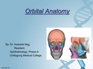
  • 1. Orbital Anatomy By- Dr. Kawshik Nag, Resident, Ophthalmology, Phase-A Chittagong Medical College.
  • 2. Anatomy Of Orbit  Quadrangular truncated pyramidal in shape.  Bounded by- • Superiorly- Anterior cranial fossa • Medially- Nasal cavity and ethmoidal air sinuses • Inferiorly- Maxillary sinus • Laterally- Middle cranial fossa and Temporal fossa.
  • 3. Dimensions  Volume: 30cm3  Rim: Horizontally- 4cm Vertically- 3.5cm  Intra orbital width: 2.5cm  Extra orbital width: 10cm  Depth: Medially- 4.2cm Laterally- 5.0cm  Ratio of vol. of orbit : vol. of globe: 4.5:1
  • 4. Bony Orbit Seven bones make up the bony orbit :  Frontal bone  Zygomatic Bone  Maxillary bone  Ethmoid bone  Sphenoid bone  Lacrimal bone  Palatine bone
  • 5. Walls Of The Orbit  The bony orbit has four walls:  Medial wall  Lateral wall  Roof  Floor
  • 6. Medial Orbital Wall The medial wall is formed from front to back by the:  Frontal process of maxilla  Lacrimal bone  Orbital plate of the ethmoid bone  Body of the sphenoid bone.
  • 7. Medial Orbital Wall Clinical applications: • It is the thinnest wall of the orbit, so it is frequently fragmented as a result of indirect blow out fractures and during orbitotomy operations. • Frequently eroded by chronic inflammatory lesions, neoplasms, cysts. • Medial wall provide alternate access route to the orbit through sinus. • Haemorrhage can occur due to trauma to ethmoidal vessels. • Accidental lateral displacemet of medial wall causes traumatic hypertelorism.
  • 8. Lateral Orbital Wall  Thickest and strongest.  Formed by two bones: • Zygomatic • Greater wing of sphenoid.
  • 9. Lateral Orbital Wall Clinical applications: • The anterior half of globe is not covered by bone on lateral side. Hence, palpation of retrobulbar tumours is easier from the lateral side. • The zygomatico-sphenoid suture is an important landmark in creating the flap in lateral orbitotomy. • It is the strongest portion of the orbit and needs to be sawed open in lateral orbitotomy. • Since lateral wall is almost devoid of foramina, bleeding is less.
  • 10. Roof Of Orbit  Underlies frontal sinus and anterior cranial fossa.  Formed by- • Orbital plate of frontal bone • Lesser wing of sphenoid.  Triangular.  Faces downwards and slightly forwards.
  • 11. Roof Of Orbit Clinical applications: • Thin and periorbita peels away easily. • Objects piercing upper eyelid penetrate roof and damage frontal lobe. • In old age roof may be absorbed so that periorbital and duramater comes into contact. • Any trauma of dura mater and CSF escapes into orbit or nose or both.
  • 12. Floor Of Orbit  Shortest orbital wall.  Formed by: • Maxillary bone- medially • Zygomatic bone- laterally • Palatine bone- posteriorly.  Triangular in shape.  Bordered laterallly by inferior orbital fissure and medially by maxilloethmoidal sinus.  Overlies maxillary sinus.
  • 13. Floor Of Orbit Clinical applications: • Commonly involved in Blow out fractures of the orbit. Infra orbital vessels and nerves almost always involved. • Diplopia is the main symptoms of blow-out fracture. • Easily invaded by tumors of the maxillary antrum. Figure- Mechanism of blow-out fracture from displacement of the globe itself into the orbital walls. The globe is displaced posteriorly, striking the orbital walls and forcing them outward.
  • 14. Base Of Orbit  The anterior open part.  Bounded by four orbital margins- • Superior orbital margin • Inferior orbital margin • Medial orbital margin • Lateral orbital margin.  It gives attachment to the septum orbitale.
  • 15. Apex Of Orbit  Orbital apex is the posterior end of the orbit.  Four orbital walls converge.  Two orifices: • Optic Canal • Superior orbital fissure
  • 16. Optic Canal  It connects the orbit to the middle cranial fossa.  It transmits: • Optic Nerve • Ophthalmic artery.
  • 17. Superior Orbital Fissure  Structure passing:  Upper lateral part: • Lacrimal and frontal nerves • Trochlear nerve • Superior ophthalmic vein • Recurrent branch of ophthalmic artery.  Middle part: • Superior and inferior divisions of occulomotor nerve • Nasociliary branch of ophthalmic division of trigeminal nerve. • Abducent nerve.  Lower medial part: • Inferior ophthalmic vein.
  • 18. Superior Orbital Fissure Clinical applications: • Radiographic enlargement of superior orbital fissure may accompany pathologic processes,  Aneurysm  Meningioma  Choroidoma  Pituitary adenoma  tumours of orbital apex. • When idiopathic inflammation involves the superior orbital fissure, the “Tolosa Hunt syndrome” which is painful ophthalmoplegia results.
  • 19. Periorbita  Periorbita refers to periosteum lining the orbitlal surface of the bones of orbit.  Loosely adherent to the bones.  Fixed firmly at- • Orbital margins • Suture lines • Various fissures and foramina • Lacrimal fossa.  Applied Anatomy- • Surgery in the orbital roof in the areas of fissures and suture lines may be complicated by cerebrospinal fluid leakage.
  • 20. Orbital Fascia  It is a complex interwoven thin connective tissue membrane joining the various intraorbital contents.  Parts- • Fascia bulbi, • Muscular sheaths, • Intermuscular septa, • Membranous expansions of the extraocular muscles, • Ligament of Lockwood.
  • 21. Extraocular Muscles  Voluntary Muscles: • Superior rectus • Inferior rectus • Medial rectus • Lateral rectus • Superior oblique • Inferior oblique • Levator palpebrae superioris.  Involuntary Muscles: • Superior tarsal or Muller’s muscle, • Inferior tarsal muscle.
  • 22. Surgical Spaces In Orbit  Orbit is divided into 4 surgical spaces- • Subperiosteal space • Peripheral orbital space/ Extraconal space • Central orbital space/ Intraconal space • Subtenon’s space
  • 23. Surgical Spaces In Orbit  Importance of these spaces- • Most of the orbital tumours tends to remain with in a space in which they are formed unless they are large or malignant or represents an infiltrative process such as pseudotumour.
  • 24. Subperiosteal Space  Lies between orbital bone and periorbita.  tumours arising from bone separates periorbita from bone.  Here periorbita acts as a effective barrier against spread of tumour to eye.
  • 25. Subperiosteal Space  tumours in this space are- • Dermoids cyst • Epidermoid cyst • Mucocele • Subperiosteal abscess • Osteomatous tumour
  • 26. Peripheral Orbital Space  Known as extraconal space.  Lies between periorbita at periphery, extraocular muscles and their intermuscular septa internally and orbital septum anteriorly.  Posteriorly it merges with central space.  tumours in this space are usually approached by anterior orbitotomy and sometimes by lateral orbitotomy.
  • 27. Peripheral Orbital Space  tumours in this space produce eccentric proptosis.  tumours in this space are- • Malignant Lymphoma • Capillary haemangioma of childhood • Intrinsic neoplasm of lacrimal gland • Pseudotumours.
  • 28. Central Orbital Space  Known as muscle cone/ retro- orbital space/posterior space/ intraconal space.  Bounded by- • Anteriorly tenon’s capsule • Posteriorly by 4 recti and intermuscular septa.  In posterior part, space become continuous with peripheral space.
  • 29. Central Orbital Space  tumours of this space- • Cavernous haemangioma of adults • Solitary neurofibroma • Neurolemoma • Nodular orbital meningiomas • Optic nerve glioma.  Produce axial proptosis.  tumours are approached through lateral orbitotomy.
  • 30. Subtenon’s space  Space around eyeball between sclera and tenon’s capsule.  Pus collection in this space is drained by incision on tenon’s capsule through conjunctiva.
  • 31. Contents Of The Orbit  Eyeball  Fascia: Orbital and bulbar.  Muscles: Extraocular.  Vessels: • Ophthalmic artery • Superior and inferior ophthalmic vein • Lymphatics.  Nerves: Optic,Oculomotor, Trochlear, Abducent, Branches of ophthalmic nerves and sympathetic nerves.  Ciliary ganglion  Lacrimal gland and lacrimal sac  Orbital fat.