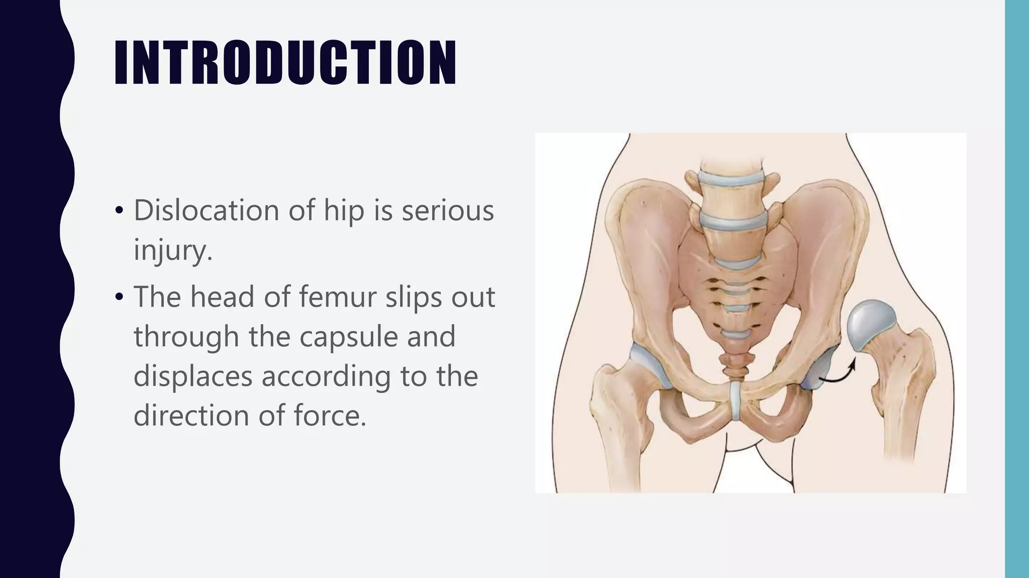
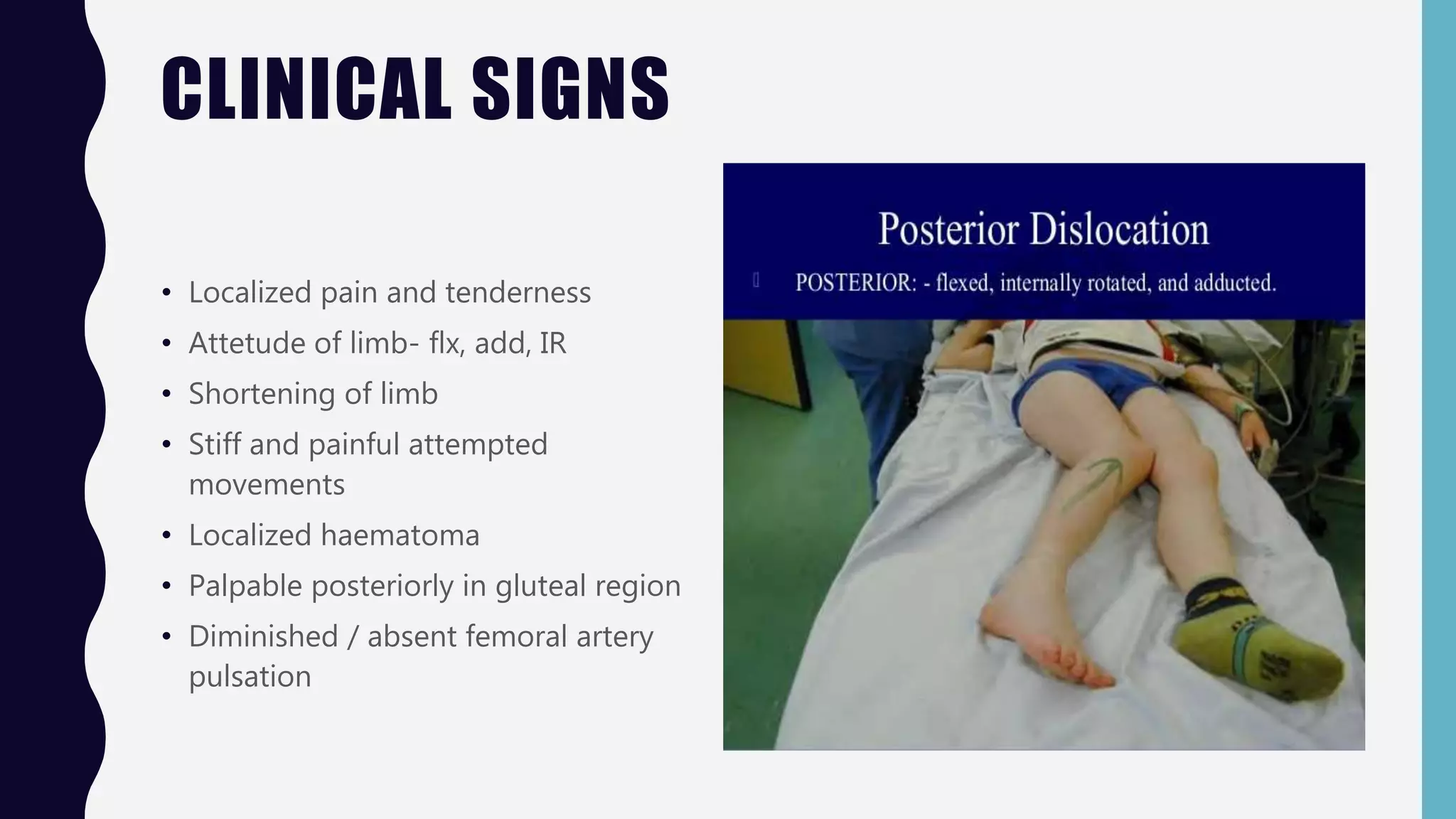
DISLOCATION OF
HIP JOINT
D R . K R U PA R A I T H AT H A
A S S I S T A N T P R O F E S S O R ,
S C H O O L O F P H Y S I O T H E R A P Y , R K U N I V E R S I T Y , R A J K O T , G U J A R A T , I N D I A
INTRODUCTION
• Dislocation of hip is serious
injury.
• The head of femur slips out
through the capsule and
displaces according to the
direction of force.
TYPES
• Posterior Dislocation (the
commonest)
• Anterior Dislocation
• Central Dislocation
POSTERIOR
DISLOCATION
OF HIP
MECHANISM
• Commonest
• 50% of cases , it is associated with a
chip fracture of posterior lip of
acetabulum, in which case it is known
as a fracture- dislocation.
• It occurs when – adducted and flexex
femur is pushed backwards by violent
thrust to the knee.
CLINICAL SIGNS
• Localized pain and tenderness
• Attetude of limb- flx, add, IR
• Shortening of limb
• Stiff and painful attempted
movements
• Localized haematoma
• Palpable posteriorly in gluteal region
• Diminished / absent femoral artery
pulsation
CLASSIFICATION
THOMPSON AND EPSTEIN
CLASSIFICATION
Type Description
I with or without a minor fracture
II
with a large single fracture of the
posterior acetabular rim
III
with comminution of
the acetabular ring
IV with a fracture of the acetabular floor
V with a fracture of the femoral head
CLASSIFICATION
PIPKIN CL ASSIFICATION
Type Description
I
Fracture below the fovea; not involving
weight-bearing surface of the head
II
Fracture above the fovea; involving
weight-bearing surface of the head
III
Type I or II fracture with
associated femoral neck fracture
IV
Type I or II fracture with
associated acetabulum fracture
INVESTIGATIONS
• Head out of acetabulum
• Thigh internally rotated
• Lesser trochanter less prominent
• Shelton’s line is broken.
• Look for any bony chip.
TREATMENT
• Reduction is an emergency- more
chance of being avascular
• Reduced under general
anaesthesia by classical Watson-
jones axial traction technique.
• old dislocation, failure of closed
reduction, intra articular loose
fragment,large acetabular
fragment--- cause of open
reduction.
• immobilization after both- 6-8
weeks.
• Mobilization started after 6-8
weeks.
STIMONS GRAVITY METHOD
HIP SPICA THOMAS SPLINT
COMPLICATIONS
• Injury to sciatic nerve
• Avascular necrosis
• Osteoarthritis
• Myositis ossificans
ANTERIOR
DISLOCATION
OF HIP
MECHANISM
• Rare
• Violent abduction force, with
thigh flexed
• Occurs in RTAs
• Dislocated head of femur lies in
obturator foramen of symphysis
pubis.
• Types: pubic, obturator,
perineal.
CLINICAL PRESENTATION
• Attitude of limb- externally
rotated
• Head palpable in groin
• Localized pain, stiffness,
tenderness
• Inability to bear weight
• Apparent lengthening of
limb
TREATMENT AND COMPLICATIONS ARE SAME.
CENTRAL
FRACTURE-
DISLOCATION OF
HIP
MECHANISM
• Femoral head is driven
through the medial wall of
the acetabulum towards
the pelvic cavity.
• Displacement varies: from
minimal- to as much as
whole head in pelvis.
• Mechanism- Fall on greater
trochanter, or RTAs.
CLINICAL FEATURES
• Severe localized pain and
stiffness
• Hip abduction and rotation
markedly restricted with
severe pain
• Bony mass is palpable in
per rectal examination
• Limb remains in neutral
rotation and short.
INVESTIGATION
TREATMENT
• Aim of treatment in these cases
is to achieve as congruous an
articular surface as possible.
• For that skeletal traction is
applied distally and laterally.
• If fragments fall in place- cont.
for 8-12 weeks; if not- surgical
reconstruction of the acetabular
floor may be necessary.
• Full weight bearing started after
3 months (after radiographic
evidence of consolidation).
QUESTIONS??
THANK YOU…

Dislocation of hip joint

  • 1.
    DISLOCATION OF HIP JOINT DR . K R U PA R A I T H AT H A A S S I S T A N T P R O F E S S O R , S C H O O L O F P H Y S I O T H E R A P Y , R K U N I V E R S I T Y , R A J K O T , G U J A R A T , I N D I A
  • 3.
    INTRODUCTION • Dislocation ofhip is serious injury. • The head of femur slips out through the capsule and displaces according to the direction of force.
  • 4.
    TYPES • Posterior Dislocation(the commonest) • Anterior Dislocation • Central Dislocation
  • 6.
  • 7.
    MECHANISM • Commonest • 50%of cases , it is associated with a chip fracture of posterior lip of acetabulum, in which case it is known as a fracture- dislocation. • It occurs when – adducted and flexex femur is pushed backwards by violent thrust to the knee.
  • 8.
    CLINICAL SIGNS • Localizedpain and tenderness • Attetude of limb- flx, add, IR • Shortening of limb • Stiff and painful attempted movements • Localized haematoma • Palpable posteriorly in gluteal region • Diminished / absent femoral artery pulsation
  • 9.
    CLASSIFICATION THOMPSON AND EPSTEIN CLASSIFICATION TypeDescription I with or without a minor fracture II with a large single fracture of the posterior acetabular rim III with comminution of the acetabular ring IV with a fracture of the acetabular floor V with a fracture of the femoral head
  • 10.
    CLASSIFICATION PIPKIN CL ASSIFICATION TypeDescription I Fracture below the fovea; not involving weight-bearing surface of the head II Fracture above the fovea; involving weight-bearing surface of the head III Type I or II fracture with associated femoral neck fracture IV Type I or II fracture with associated acetabulum fracture
  • 11.
    INVESTIGATIONS • Head outof acetabulum • Thigh internally rotated • Lesser trochanter less prominent • Shelton’s line is broken. • Look for any bony chip.
  • 12.
    TREATMENT • Reduction isan emergency- more chance of being avascular • Reduced under general anaesthesia by classical Watson- jones axial traction technique. • old dislocation, failure of closed reduction, intra articular loose fragment,large acetabular fragment--- cause of open reduction. • immobilization after both- 6-8 weeks. • Mobilization started after 6-8 weeks.
  • 13.
  • 14.
  • 15.
    COMPLICATIONS • Injury tosciatic nerve • Avascular necrosis • Osteoarthritis • Myositis ossificans
  • 16.
  • 17.
    MECHANISM • Rare • Violentabduction force, with thigh flexed • Occurs in RTAs • Dislocated head of femur lies in obturator foramen of symphysis pubis. • Types: pubic, obturator, perineal.
  • 18.
    CLINICAL PRESENTATION • Attitudeof limb- externally rotated • Head palpable in groin • Localized pain, stiffness, tenderness • Inability to bear weight • Apparent lengthening of limb TREATMENT AND COMPLICATIONS ARE SAME.
  • 19.
  • 20.
    MECHANISM • Femoral headis driven through the medial wall of the acetabulum towards the pelvic cavity. • Displacement varies: from minimal- to as much as whole head in pelvis. • Mechanism- Fall on greater trochanter, or RTAs.
  • 21.
    CLINICAL FEATURES • Severelocalized pain and stiffness • Hip abduction and rotation markedly restricted with severe pain • Bony mass is palpable in per rectal examination • Limb remains in neutral rotation and short.
  • 22.
  • 23.
    TREATMENT • Aim oftreatment in these cases is to achieve as congruous an articular surface as possible. • For that skeletal traction is applied distally and laterally. • If fragments fall in place- cont. for 8-12 weeks; if not- surgical reconstruction of the acetabular floor may be necessary. • Full weight bearing started after 3 months (after radiographic evidence of consolidation).
  • 25.
  • 26.