Downloaded 102 times


![Optical properties of biological tissue
Basic properties
• n [–]: index of refraction; e.g., 1.37
• μa [cm–1]: absorption coefficient; e.g., 0.1
• μs [cm–1]: scattering coefficient; e.g., 100
• g [–]: scattering anisotropy, <cosθ>; e.g., 0.9
Derived properties
• μt [cm–1]: total interaction (extinction) coefficient, μa + μs
• lt [cm]: mean free path, 1/ μt; e.g., 0.1 mm
• μs’ [cm–1]: reduced scattering coefficient, μs(1 – g)
• μt’ [cm–1]: transport interaction coefficient, μa + μs’
• lt’ [cm]: transport mean free path, 1/ μt’; e.g., 1 mm
• μeff [cm–1]: effective attenuation coefficient, (3μa μt’)1/2
• δ [cm]: penetration depth, 1/(3μa μt’)1/2; e.g., 5 mm](https://image.slidesharecdn.com/lecture1-141119145945-conversion-gate01/85/Introduction-to-Biomedical-Optics-7-320.jpg)


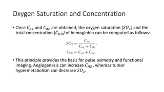

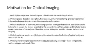
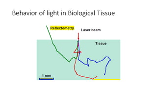
This document describes the syllabus for a biomedical optics course. It covers 13 topics including Monte Carlo modeling of photon transport, the radiative transfer equation, diffuse optical tomography, and ultrasound-modulated optical tomography. The first chapter introduces motivation for optical imaging in biology, general behavior of light in tissue, and basic light-matter interactions. It discusses optical properties of tissue, Beer's law, absorption and scattering origins and coefficients, and exercises calculating light transmission through absorbing and scattering media. Oxygen saturation and concentration are also introduced.