Top down proteomics of soluble and integral membrane proteins
•
1 like•190 views

Mitochondria provide important cellular functions including oxidative phosphorylation, fatty acid biosynthesis, and acting as gatekeepers to apoptosis.

Recommended
More Related Content
What's hot
What's hot (19)
Similar to Top down proteomics of soluble and integral membrane proteins
Similar to Top down proteomics of soluble and integral membrane proteins (20)
More from Expedeon
More from Expedeon (20)
Recently uploaded
Recently uploaded (20)
Top down proteomics of soluble and integral membrane proteins
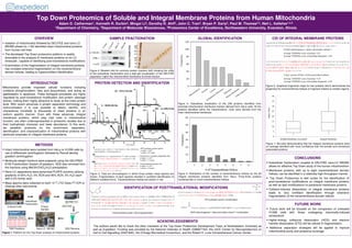
- 1. IDENTIFICATION OF POSTTRANSLATIONAL MODIFICATIONS PROTEIN DETECTION AND IDENTIFICATION GLOBAL IDENTIFICATION INTRODUCTION Mitochondria provide important cellular functions including oxidative phosphorylation, fatty acid biosynthesis, and acting as gatekeepers to apoptosis. These biological processes are highly regulated by post-translational modification and protein cleavage events, making them highly attractive to study at the intact protein level. With recent advances in protein separation technology and instrumentation it is now possible to detect, identify, and characterize hundreds to thousands of intact proteins in an isoform specific manner. Even with these advances, integral membrane proteins, which play vital roles in mitochondrial function, are often underrepresented in proteomic studies due to their hydrophobic character and lower abundance. In this work, we establish protocols for the enrichment, separation, identification, and characterization of mitochondrial proteins with particular emphasis on integral membrane proteins. OVERVIEW Isolation of mitochondria followed by GELFrEE and nano LC- MS/MS allows for >100 identified intact mitochondrial proteins from human cell lines The developed Top Down proteomics platform is readily amenable to the analysis of membrane proteins on an LC timescale , capable of identifying post-translational modifications Examination of the fragmentation of integral membrane proteins has revealed extensive fragmentation on the transmembrane domain helices, leading to hyperconfident identification Top Down Proteomics of Soluble and Integral Membrane Proteins from Human Mitochondria Adam D. Catherman1, Kenneth R. Durbin2, Mingxi Li2, Dorothy R. Ahlf2, John C. Tran3. Bryan P. Early3, Paul M. Thomas2,3, Neil L. Kelleher1,2,3 1Department of Chemistry, 2Department of Molecular Biosciences, 3Proteomics Center of Excellence, Northwestern University, Evanston, Illinois 75 ACKNOWLEDGEMENTS The authors would like to thank the other members of the Top Down Proteomics Development Team at Northwestern University as well as Expedeon. Funding was provided by the National Institutes of Health (GM067193), the UIUC Center for Neuroproteomics on Cell to Cell Signalling (DA01830), the Chicago Biomedical Consortium, and the Robert H. Lurie Comprehensive Cancer Center. CONCLUSIONS Subcellular fractionation coupled to GELFrEE nano-LC MS/MS allows for effective Top Down study of the human mitochondrion Membrane proteins, with up to eight transmembrane domain helices, can be identified in a relatively high-throughput manner Top Down Proteomics is well suited for the identification of post-translational modifications on integral membrane proteins as well as lipid modifications on peripheral membrane proteins Collision-induced dissociation of integral membrane proteins leads to very confident identification through extensive fragmentation of the transmembrane domain helices FUTURE WORK Future work will be focused on the comparison of untreated H1299 cells with those undergoing chemically-induced senescence Higher-energy collisional dissociation (HCD) and electron transfer dissociation (ETD) will be utilized fir fragmentation. Additional separation strategies will be applied to improve mitochondrial purity and proteome coverage METHODS Intact mitochondrial were isolated from HeLa or H1299 cells by use of differential centrifugation followed by Percoll density gradient centrifugation Molecular weight fractions were prepared using the GELFREE 8100 Fractionation System (Expedeon). SDS was removed from the fractions using MeOH/CHCl3/H2O precipitation. Nano-LC separations were performed PLRPS columns utilizing gradients of 05% H2O, 5% ACN and 95% ACN, 5% H2O each with 0.2% formic acid. Mass spectra were collected on both 12 T LTQ Velos FT-ICR or Orbitrap Elite instruments. SAMPLE FRACTIONATION Figure 1: Platform for the Top Down analysis of mitochondrial proteins. CID OF INTEGRAL MEMBRANE PROTEINS Protein Sample GELFrEE SDS RemovalNano-LC MS/MS Database Search Cultured Cells Subcellular Fractionation TMD Prediction Figure 6: Graphical fragments maps for two proteins which demonstrate the propensity for transmembrane helices to fragment relative to soluble regions. Figure 7: Box-plot demonstrating that the integral membrane proteins were on average identified with more confidence than the soluble and membrane associated proteins. Figure 3: Total ion chromatogram in which three protein mass spectra are shown. Fragmentation of each species resulted in confident identification of different modified forms Transmembrane helices are shown in red. 10 15 20 25 37 50 75 100 Figure 2: Western blot for common protein markers (left) showing the utility of the subcellular fractionation and a slab gel visualization of the GELFrEE separation (right) the mitochondrial membrane enriched fraction. Figure 4: Subcellular localization of the 246 proteins identified from enriched mitochondrial membrane fraction derived from HeLa cells. Of the proteins identified within the mitochondrion, most were derived from the inner mitochondrial membrane. Figure 5: Distribution of the number of transmembrane helices for the 83 integral membrane proteins identified from HeLa. Thirty-three proteins contained two or more transmembrane helices.