Antibody oligonucelotide conjugates application note
•
1 like•206 views

Antibody-oligonucleotide (Ab-Oligo) conjugates have been used in numerous applications from diagnostics to therapeutics and were developed through an unmet need for precise and efficient detection of low-abundance proteins.

Recommended
More Related Content
What's hot
What's hot (20)
Similar to Antibody oligonucelotide conjugates application note
Similar to Antibody oligonucelotide conjugates application note (20)
More from Expedeon
More from Expedeon (20)
Recently uploaded
Recently uploaded (20)
Antibody oligonucelotide conjugates application note
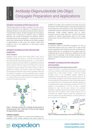
- 1. www.expedeon.com info@expedeon.com ANTIBODY-OLIGONUCLEOTIDE CONJUGATES Antibody-oligonucleotide (Ab-Oligo) conjugates have been used in numerous applications from diagnostics to therapeutics and were developed through an unmet need for precise and efficient detection of low-abundance proteins. Ab-Oligo conjugates have since played a significant role in enhancing an extensive range of biological techniques that include immunological and proteomic research, biomarker discovery, clinical diagnostics – including point-of-care, as well as other novel techniques. Antibodies can be readily conjugated to oligonucleotides via their amino acid residues, making them suitable for most in vitro applications, as they possess several functional groups. ANTIBODY-OLIGONUCLEOTIDE CONJUGATION CHEMISTRY Amine conjugation Antibodies contain several amine groups (NH2), which can be distributed throughout as lysine (K / Lys) side chain epsilon-amine and N-terminal alpha-amine groups (see Figure 1.). These residues are the two that are most often targeted for conjugation as they can easily be modified owing to their steric accessibility. Apart from amine groups, glutamic acid and aspartic acid residues can also be conjugated, as well as carboxylic acid (-COOH) residues via their C- terminal end. However, the numerous amine and carboxylic acid functional groups distributed on the surface of antibodies, means that the method of conjugation used may potentially result in partially active / inactive Ab-Oligo conjugates that may not bind to the antigen, i.e., reduced affinity, due to steric hindrance of conjugated residues located on the antigen binding sites. Figure 1. Schematic structure of an antibody showing location of functional groups (-NH2, -COOH and S–S) that are often targeted for oligonucleotide conjugation. Sulfhydryl conjugation Conjugation is also possible via a reactive thiol (sulfhydryl) group. Antibodies contain oxidized sulfhydryl (-SH) groups present as disulfide (S–S) bridges, which contribute to the tertiary structure of the antibody. These disulfide bridges must be reduced to expose their reactive groups by a reducing agent (e.g., 2-ME / SDS). Antibodies can be selectively cleaved to create either two half antibody molecules or alternatively, smaller antibody fragments such as F(ab')2. Conjugation using the ‘hinge’ region free / reduced -SH group also orientates the attached oligonucleotide away from the antigen binding regions, hence preventing steric hindrance and preserving activity. Carbohydrate conjugation An alternative method of site-directed conjugation can occur at carbohydrate residues, which occur mainly in the Fc region, as they are less susceptible to steric hindrance due to their remoteness from antigen binding sites. For conjugation via this complex method the carbohydrate group must be oxidized to an aldehyde (-CHO) using periodic acid. An advantage is that Ab-Oligos produced via site- specific conjugation techniques, have distinct advantages for in vivo applications. ANTIBODY-OLIGONUCLEOTIDE CONJUGATE APPLICATIONS: Immuno-Polymerase Chain Reaction (immuno-PCR) Immuno-PCR (iPCR) is a powerful technique that is similar to an ELISA, as an antibody is used to detect and quantify a specific antigen (analyte) from a mixed sample. However, in iPCR, the Ab-oligo conjugate binds to the immobilized analytes, followed by amplification of the attached DNA using RT-PCR (see Figure 2.). The use of Ab-Oligo conjugates in iPCR provides a highly sensitive method for protein detection and quantification, which is significantly superior to a standard ELISA as it combines both the detection specificity of an antibody with the nucleic acid detection sensitivity of RT-PCR. The coupling of immunodetection to RT-PCR has been a standard technique for more than 20 years. Owing to the enormous amplification capability, specificity and increased sensitivity given by iPCR, lower limits of detection, which surpass ELISA by 100- to 10,000-fold, are available. Products arising from amplification can then be visualized using gel electrophoresis, so that the presence of bound analytes can be established. Figure 2. Schematic representation of the immuno-PCR approach. Antibody-Oligonucleotide (Ab-Oligo) Conjugate Preparation and Applications APPLICATION NOTE
- 2. www.expedeon.com info@expedeon.com Proximity Ligation Assay (PLA) Proximal Ligation Assay (PLA), developed by Fredriksson et al., is used for localized detection, visualization, and quantification of single protein or protein-protein interactions in adherent cell lines, cytospin preparations or tissue samples (including frozen or paraffin- embedded). This technique utilizes two Ab-Oligo conjugates bound in close proximity (30–40nm apart) to different epitopes of the same protein, or two proteins in a complex. The cells / tissue must be fixed with a fixative that is appropriate for the antibodies used in the protocol. If required, antigen retrieval and antibody-specific blocking must also be performed. Figure 3. Direct and indirect PLA techniques. The direct method uses antibody pairs with primary conjugation and the indirect method uses secondary antibody conjugates Electrochemical Proximity Assay (ECPA) The Electrochemical Proximity Assay (ECPA) is a simple separation / wash free, electrochemical format based on the marriage between the proximity assay concept and electrochemical detection. ECPA produces a direct readout suitable for highly sensitive (low femtomolar (fm) range) and selective quantitation of a wide variety of protein targets. Figure 4. Schematic of the principles of Electrochemical Proximity Assay. EPCA uses the proximity effect of two antibody-oligo conjugate probes bound to a protein target to move an electrochemically active label – methylene blue (MB), closer to a gold electrode with a preassembled thiolated DNA / competitor monolayer (Figure 4.). In the presence of the protein target, the redox current in ECPA is quantified using square wave voltammetry (SWV) and is found to depend directly on the concentration of target. ECPA may be useful for any protein with available antibody pairs. Multiplexed Assay Development Multiplex protein detection aids the simultaneous measurement of multiple analytes of interest across multiple samples, using universal capture technology in a quantitative manner, thereby reducing workflow and sample volume problems. However, this detection method has previously been constrained by factors that include assay specificity, sensitivity, and throughput (see Figure 5.). Figure 5. In a conventional immunoassay, cross-reactivity due to unspecific binding of antibodies, limited the degree of potential multiplexing available. Several methods are typically used for multiplex immunoassays, which loosely fit into two categories: Immobilization on a solid surface in which the assays for each analyte are spatially separated, or immobilization on beads or particles in which the assays for each analyte are on a different bead/particle. Multiplex protein detection technology now takes advantage of the specific binding between two complementary strands of DNA, with one strand (analyte) for each cognate immobilized onto a solid support e.g., a microtiter plate. The Ab-Oligo conjugates are pooled and hybridized to their specific immobilized capture oligos in the well to create an antibody array (see Figure 6.). Figure 6. Cross-reactive binding is not detected as only matched oligo-pairs are detected and reported (e.g., 3A+3B). EXPEDEON TECHNOLOGIES & SERVICES FOR EASY ANTIBODY-OLIGONUCLEOTIDE CONJUGATION Previously, the standard chemistry and methods used to link oligonucleotides to antibodies were difficult, time consuming, and required specialist scientific knowledge of chemical modification techniques. Nowadays, straightforward, efficient and high yielding technologies have been developed for the quick and easy preparation of Ab-Oligo conjugates. Thunder-Link® PLUS Technology Expedeon’s Thunder-Link® PLUS Oligo Conjugation System facilitates the simple and rapid conjugation of antibodies to oligonucleotides, with high recovery of materials and a superior clean- up procedure. The kit is quick and simple to use, overcoming time
- 3. www.expedeon.com info@expedeon.com consuming and lengthy protocols generally associated with standard conjugation methods (see Figure 7.). Thunder-Link® PLUS Features and Benefits: Quick and easy to use, only 30 minutes antibody and oligo activation and 1-hour oligo conjugation, saving you time with no specialist scientific knowledge required Robust and flexible clean-up procedure (works with antibody fragments and other proteins) High levels of antibody and oligo recovery so you save precious reagents Unidirectional chemistry so there’s no risk of cross-linking Covalent bond forms highly stable conjugates Suitable for single-stranded oligos of 10–120 bases, double- stranded oligos up to 80 base pairs therefore covers the majority of sequences Linking chemistry works at both the 5’ and 3’ end providing the ability to combine with other modifications. Figure 7. The Thunder-Link® PLUS conjugation process. The Ab- Oligo conjugation protocol consists of three main steps: activation of both the antibody and the oligonucleotide followed by desalting, after which the two are mixed and left to incubate overnight. If an unbound oligonucleotide removal step is necessary for the end application, this can be performed the next morning. Following this, the conjugate is ready to use. Main applications for Thunder-Link® PLUS: Immuno-PCR (iPCR), Proximity Ligation Assay (PLA), Electrochemical Proximity Assay (ECPA), Lateral Flow, siRNA-antibody delivery in therapeutic applications, pre-targeting cancer therapeutics. Immuno-qPCR Data Prepared with Thunder-Link® PLUS: The top graph plots the number of qPCR cycles undertaken vs. fluorescence intensity generated by SYBR green containing qPCR probes at specific antigen concentrations (see Figure 8.). The bottom graph then converts this data to antigen amount vs. cycle number to permit calculation of a standard curve (see Figure 9.). Figure 8. Number of cycles generated by SYBR green containing qPCR probes at specific antigen concentrations. Figure 9. A mouse monoclonal antibody specific for human CRP (clone C7) was purchased in unconjugated format from HyTest. The unconjugated antibody was conjugated to an oligonucleotide using a Thunder-Link® PLUS kit and was used as detection antibody in a sandwich immuno-PCR assay using a polyclonal anti-CRP antibody as capture reagent. Graph depicts standard curve of amount of antigen versus cycle number. The results show that the assay utilizes 1,000-fold less capture antibody, 100-fold less detection antibody and provides 1,000-fold more sensitivity than the equivalent ELISA. USEFUL TIPS FOR THE DESIGN OF OLIGOS WHEN USING THE EXPEDEON THUNDER-LINK® OLIGO CONJUGATION SYSTEM The amino or thiol modification should be located at either the 5’ or 3’ end of the oligonucleotide. The 5’ end tends to be the preferred location as it results in higher purity oligonucleotides, which in turn slightly increases Ab-oligo conjugation efficiency. Minimize base repetition (homopolymer regions, e.g., CCCCCC). It is best to avoid stretches of more than four consecutive G bases (GGGG) as these sequences can form a G-quadruplex or cruciform structures, which can result in lower hybridization and coupling efficiencies. EXPEDEON’S AB-OLIGO CONJUGATION AND CONJUGATE OPTIMIZATION SERVICES Expedeon also offers Ab-Oligo conjugation and conjugate optimization services, performed in-house by our expert conjugation scientists and tailored to your requirements. If you are looking to outsource your conjugation or would like to enhance the performance of your conjugates, then please contact us and we will put you in touch with one of our experts.
- 4. www.expedeon.com info@expedeon.com EXPEDEON RELATED PRODUCTS: AbSelectTM Antibody Purification Systems Unfortunately, many antibodies are provided in buffers containing additives that are incompatible with labeling technologies, making purification a key consideration prior to carrying out any conjugation reaction. To help you, we have developed a range of purification kits that complement our labeling technologies. These are extremely easy and convenient to use and have been carefully designed to complement our bioconjugation kits. Find out more here. References: Chang, L., Li, J., Wang, L. Immuno-PCR: An ultrasensitive immunoassay for biomolecular detection. Anal Chim Acta. 2016 Mar 3;910:12–24. doi: 10.1016/j.aca.2015.12.039. Epub 2016 Jan 8. Dennler, P., Fischer, E., Scibli, R. Antibody Conjugates: From Heterogeneous Populations to Defined Reagents. Antibodies. 2015;4(3):197–224; doi:10.3390/antib4030197. Available at: http://www.mdpi.com/2073-4468/4/3/197/htm [Accessed September 2018]. Fan, R., Vermesh, O., Srivastava, A., Yen, BK., Qin, L., Ahmad, H., Kwong, GA., Liu, CC., Gould, J., Hood, L., Heath, JR. Integrated barcode chips for rapid, multiplexed analysis of proteins in microliter quantities of blood. Nature Biotechnology. 2008;26:1373–1378. Fredriksson, S., Horecka, J., Brustugun, OT., Schlingemann, J., Koong, A., Tibshirani, R. and Davis, R. Multiplexed Proximity Ligation Assays to Profile Putative Plasma Biomarkers Relevant to Pancreatic and Ovarian Cancer. Clinical Chemistry. 2008;54:582–589. Fredriksson S., Gullberg M., Jarvius J., Olsson C., Pietras K., Gústafsdóttir SM., Östman A., Landegren U. Protein detection using proximity dependent DNA ligation assays. Nature Biotechnology. 2002;20:473–477. Gong, H., Holcomb, I., Ooi, A., Wang, X., Majonis, D., Unger, MA., Ramakrishnan, R. Simple Method to Prepare Oligonucleotide-Conjugated Antibodies and Its Application in Multiplex Protein Detection in Single Cells. Bioconjugate Chem. 2016;27:217−225. doi: 10.1021/acs.bioconjchem.5b00613. Epub 2016 Jan 4. Jiaming, H., Tanyu, W., Joonyul, K., Curtis, S., Christopher, E., Quantitation of Femtomolar Protein Levels via Direct Readout with the Electrochemical Proximity Assay. Journal of the American Chemical Society, 2012;134:7066–7072. Kozlov, IA., Melnyk, PC., Stromsborg, KE., Chee, MS., Barker, .L., Zhao, C., Efficient strategies for the conjugation of oligonucleotides to antibodies enabling highly sensitive protein detection. Biopolymers. 2004;73:621. Kwong, GA., Radu, CG., Hwang, K., Shu, CJ., Ma, C., Koya, RC., Comin- Anduix, B., Hadrup, SR., Bailey, RC., Witte, ON., Schumacher, TN., Ribas, A., Heath, JR. Modular Nucleic Acid Assembled p/MHC Microarrays for Multiplexed Sorting of Antigen-Specific T Cells. J Am Chem Soc. 2009;131(28):9695–9703. doi:10.1021/ja9006707. Noriyuki, K., Kazuhiro, K., Shinobu, S., Yukitaka, Y., Fusanori, Y., Shingo, M., Eisuke, M., Junichi, E. Soluble heparin-binding EGF-like growth factor (HB-EGF) detected by a newly developed immuno-PCR method is a clear-cut serological biomarker for ovarian cancer. American Journal of Translational Research, 2012;4:415–421. Proteinatlas.org. (2018). Learn: method proximity ligation assay - The Human Protein Atlas. [online] Available at: https://www.proteinatlas.org/learn/method/proximity+ligation+assay [Accessed September 2018]. Sano, T., Smith, CL., Cantro, CR. Immuno-PCR: Very Sensitive Antigen Detection by Means of Specific Antibody-DNA Conjugates. Science.1992;258(5079):120–122. Söderberg, O., Gullberg, M., Jarvius, M., Ridderstråle, K., Leuchowius, KJ., Jarvius, J., Landegren, U. Direct observation of individual endogenous protein complexes in situ by proximity ligation. Nature Methods. 2006;3(12):995–1000.