Respiratory System Anatomy & Physiology
•Download as PPT, PDF•
7 likes•2,276 views

Respiratory System Anatomy & Physiology for Nurses

Recommended
More Related Content
What's hot
What's hot (20)
Similar to Respiratory System Anatomy & Physiology
Similar to Respiratory System Anatomy & Physiology (20)
More from Dow University of Health Sciences
More from Dow University of Health Sciences (20)
Recently uploaded
Recently uploaded (19)
Respiratory System Anatomy & Physiology
- 2. Lecture Outline At the end of this unit learners will be able to: 1.Define respiratory system. 2.Define respiration. 3.Describe the structure and function of upper respiratory and lower respiratory tract. 2
- 3. Lecture Outline • 4. Discuss the physiology of respiration by explaining the mechanism of: • Pulmonary Ventilation • External Respiration • Internal Respiration • 5. Discuss nervous control of respiration. • 6. Briefly discuss the lung volumes & capacities. • 7. References 3
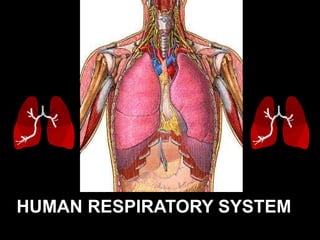
- 4. * It is the system, consisting of tubes and is responsible for the exchange of gases in Humans by filtering incoming air and transporting it into the microscopic alveoli where gases are exchanged * Your respiratory system provides the energy needed by cells of the body to funtion accroding to their designated tasks. THE HUMAN RESPIRATORY SYSTEM 4
- 5. THE HUMAN RESPIRATORY SYSTEM 5
- 6. The organs of the “Respiratory Tract” can be divided into two groups “STRUCTURALLY” ** The Upper Respiratory Tract ** The Lower Respiratory Tract * Nose * Nasal cavity * Sinuses * Pharynx * Larynx * Trachea * Bronchial Tree * Lungs 6
- 7. THE HUMAN RESPIRATORY SYSTEM 7
- 8. The organs of the “Respiratory Tract” can be divided into two groups “FUNCTIONALLY” ** The Conducting Portion - system of interconnecting cavities and tubes that conduct air into the lungs ** The Respiratory Portion - system where the exchange of respiratory gases occurs * Nose * Pharynx * Larynx * Trachea * Bronchi * Respiratory bronchioles * Alveolar Ducts * Alveoli 8
- 10. THE HUMAN RESPIRATORY SYSTEM I. N O S E A. N a s a l C a v i t y B. P a r a n a s a l S i n u s e s II. P H A R Y N X III. L A R Y N X A. E p I g i o t t i s B. V o c a l C o r d s IV. T R A C H E A v. B R O N C H I A. B r o n c h i a l T r e e VI. L U N G S A. L o b e s o f t h e L u n g s B. P l e u r a l C a v i t i e s C. A l v e o l i 10
- 11. THE NOSE 11
- 12. * It provides an entrance for air in which air is filtered by coarse hairs inside the nostrils. * It has 2 portions : the external and internal * The external portion is supported by a framework of bone and cartilage covered with skin and lined with mucous membrane. * The internal portion is a large cavity in the skull, merging with the extrenal nose anteriorly and communicating with the throat posteriorly. THE NOSE 12
- 14. * Interior area of the nose; lined with a sticky mucous membrane and contains tiny, surface hairs, cilia. divided medially by the nasal septum. * Nasal conchae divide the cavity into passageways that are lined with mucous membrane, and help increase the surface area available to warm and filter incoming air. •Particles trapped in the mucus are carried to the pharynx by ciliary action, swallowed, and carried to the stomach where gastric juice destroys any microorganisms in the mucus. The Nasal Cavity 14
- 16. * Sinuses are air-filled spaces within the maxillary, frontal, ethmoid, and sphenoid bones of the skull. * These spaces open to the nasal cavity and are lined with mucus membrane that is continuous with that lining the nasal cavity. * The sinuses reduce the weight of the skull and serve as a resonant chamber to affect the quality of the voice. Paranasal Sinuses 16
- 17. THE PHARYNX 17
- 18. * The “throat” is a funnel shaped tube that lies posterior to the nasal cavity, oral cavity and larynx; and anteriorly to the cervical vertebra. * It is composed of: Nasopharynx – uppermost portion Oropharynx – middle portion Laryngopharynx – lowermost portion * It is a common passageway for air and food and it provides a resonating chamber for speech sounds THE PHARYNX 18
- 19. THE LARYNX 19
- 20. * It is an enlargement in the airway superior to the trachea and inferior to the pharynx. * It helps keep particles from entering the trachea and also houses the vocal cords. * It is composed of a framework of muscles and cartilage bound by elastic tissue THE LARYNX 20
- 22. * It is a large leaf-shaped piece of cartilage. * A flap of cartilage that prevents food from entering the trachea (or windpipe). * During swallowing, there is elevation of the larynx The Epiglottis 22
- 24. * Inside the larynx, 2 pairs of folds of muscle and connective tissues covered with mucous membrane make up the vocal cords. a. The upper pair is the false vocal cords. b. The lower pair is the true vocal cords. c. Changing tension on the vocal cords controls pitch, while increasing the loudness depends upon increasing the force of air vibrating the vocal cords. The Vocal Cords 24
- 25. * During normal breathing, the vocal cords are relaxed and the glottis is a triangular opning. * During swallowing, the false vocal cords and epiglottis close off the glottis. The Vocal Cords 25
- 26. THE TRACHEA 26
- 27. * It is a tubular passageway for air, located anterior to the esophagus * It extends from the larynx to the 5th thoracic vertebra where it divides into the right and left bronchi. THE TRACHEA 27
- 28. THE TRACHEA 28
- 29. * The inner wall of the trachea is lined with ciliated mucous membrane with many goblet cells that serve to trap incoming particles. * The tracheal wall is supported by 20 incomplete cartilaginous rings. THE TRACHEA 29
- 30. BRONCHI 30
- 31. * The Bronchi are the two main air passages into the lungs. * They are composed of the: ** “Right Primary Bronchus” - leading to the right lung. ** “Left Primary Bronchus” - leading to the left lung. BRONCHI 31
- 33. * The bronchial tree consists of branched tubes leading from the trachea to the alveoli. * The bronchial tree begins with the two primary bronchi, each leading to a lung. * The branches of the bronchial tree from the trachea are right and left primary bronchi; these further subdivide until bronchioles give rise to alveolar ducts which terminate in alveoli. * It is through the thin epithelial cells of the alveoli that gas exchange between the blood and air occurs. The Bronchial Tree 33
- 34. THE LUNGS 34
- 35. •The paired soft, spongy, cone-shaped lungs, separated medially by the mediastinum and are enclosed by the diaphragm and thoracic cage. •2 layers of serous membrane, collectively known as pleural membrane, enclose and protect each lung. ** Parietal Pleura - outer layer attached to the thoracic cavity ** Visceral Pleura - inner layer covering the lung itself THE LUNGS 35
- 36. Right-3 lobes Left-2 lobes THE LUNGS trachea 36
- 37. * The two organs that extract oxygen from inhaled air and expel carbon dioxide in exhaled air. * This is the main and primary organ of the Respiratory System. * The bronchus and large blood vessels enter each lung. THE LUNGS 37
- 38. Lobes of the Lungs 38
- 39. * The right lung has three lobes. * The left lung has two lobes. * Each lobe is composed of lobules that contain air passages, alveoli, nerves, blood vessels, lymphatic vessels, and connective tissues. Lobes of the Lungs 39
- 41. * A layer of serous membrane, between the visceral pleura and the parietal pleura. * It contains a lubricating fluid secreted by the membranes that prevents friction between the membranes and allows their easy movement on one another during breathing. The Pleural Cavities 41
- 42. The Alveoli 42
- 43. * They are cup-shaped out pouching lined by epithelium and supported by a thin elastic basement membrane. •With that you can imagine having bunch of grapes with each grape indicating and alveolus. * Alveolar sacs are 2 or more alveoli that share a common opening. * This is where the primary exchange of gases occur. The Alveoli 43
- 44. Respiratory Centers of the CNS • The primary portions of the brainstem that control ventilation are the medulla oblongata and the pons. 44
- 46. 46 Two respiratory nuclei in medulla oblongata Expiratory center (ventral respiratory group, VRG) •involved in forced expiration Inspiratory center (dorsal respiratory group, DRG) • more frequently they fire, more deeply you inhale • longer duration they fire, breath is prolonged, slow rate
- 47. 47 Respiratory Centers in Pons Apneusticcenter (lower pons) •Sends continual inhibitory impulses to inspiratory center of the medulla oblongata, •As impulse frequency rises, breathe faster and shallower •Stimulation causes apneusis •Integrates inspiratory cutoff information Pneumotaxic center (upper pons)
- 48. Lung Volumes and Capacities • The tidal volume (TV), about 500 mL, is the amount of air inspired during normal, relaxed breathing. 48
- 49. The inspiratory reserve volume (IRV) • About 3,100 mL, is the additional air that can be forcibly inhaled after the inspiration of a normal tidal volume. 49
- 50. expiratory reserve volume (ERV) • about 1,200 mL, is the additional air that can be forcibly exhaled after the expiration of a normal tidal volume. 50
- 51. Residual volume (RV) • about 1,200 mL, is the volume of air still remaining in the lungs after the expiratory reserve volume is exhaled. 51
- 52. Total Lung Capacity (TLC), • about 6,000 mL, is the maximum amount of air that can fill the lungs (TLC = TV + IRV + ERV + RV). • The vital capacity (VC), about 4,800 mL, is the total amount of air that can be expired after fully inhaling (VC = TV + IRV + ERV = approximately 80 percent TLC). The value varies according to age and body size. 52
- 53. inspiratory capacity (IC) • The), about 3,600 mL, is the maximum amount of air that can be inspired (IC = TV + IRV). • The functional residual capacity (FRC), about 2,400 mL, is the amount of air remaining in the lungs after a normal expiration (FRC = RV + ERV). 53
- 54. Dead Space • Some of the air in the lungs does not participate in gas exchange. Such air is located in the anatomical dead space within bronchi and bronchioles—that is, outside the alveoli. 54
- 55. STRUCTURE FUNCTION nose / nasal cavity warms, moistens, & filters air as it is inhaled pharynx (throat) passageway for air, leads to trachea larynx the voice box, where vocal chords are located trachea (windpipe) tube from pharynx to bronchi rings of cartilage provide structure, keeps the windpipe "open" trachea is lined with fine hairs called cilia which filter air before it reaches the lungs bronchi two branches at the end of the trachea, each lead to a lung bronchioles a network of smaller branches leading from the bronchi into the lung tissue & ultimately to air sacs alveoli the functional respiratory units in the lung where gases (oxygen & carbon dioxide) are exchanged (enter & exit the blood stream) Summary 55
- 56. References • Ross, & Wilson. (2000) Anatomy & Physiology in Health & Illness.Edinburgh: Churchill. Eight Edition. • Tortora, G. J. (2000). Principles of Human Anatomy and Physiology (3rd ed). New York: Happer & Row. 56