Lymphatic system
•
6 likes•438 views

types of circulatory system, function of lymphatic system, components of lymphatic system, and explaination of these components, formation of lymph and factors contributing ti n the drainage of lymph, types of lymphoid organ, blood supply of lymphoid organ clinical aspect

Recommended
More Related Content
What's hot
What's hot (20)
Similar to Lymphatic system
Similar to Lymphatic system (20)
More from Dr. sana yaseen
More from Dr. sana yaseen (20)
Recently uploaded
Recently uploaded (20)
Lymphatic system
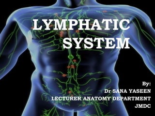
- 1. LYMPHATIC SYSTEM By: Dr SANA YASEEN LECTURER ANATOMY DEPARTMENT JMDC
- 2. INTRODUCTION Second Circulatory system of body Also referred as Drainage system of coarse type Venous system is called as Drainage system of fine type
- 4. FUNCTIONS 1. Drains excess tissue fluid to venous system 2. Absorbs fat and fat soluble vitamins, A,D, E, K from intestine & transport it to blood 3. Helps to provide immunological defense against disease causing agents (particular microbes)
- 5. LYMPH LYMPHCAPILLARIES LYMPH VESSELS LYMPH TRUNKS & DUCTS LYMPH ORGAN a. Lymph nodes b. Spleen c. Thymus d. Bone marrow Epithelio-lymphoid system COMPONENTS
- 6. LYMPH Is the tissue fluid that enters the lymph capillaries It is clear watery fluid similar in composition to plasma with exception of plasma protein.
- 7. Composition of lymph Lyphocytes – 1000-2000/mm3 Plasma cells Electrolytes same as plasma Urea, aminoacis, creatinine same as plasma Protein, calcium phosphorus lower than plasma Chloride and glucose more than plasma Clottin factors, plasma enzumes and antibodies
- 9. Lymph capillaries in intestine is called LACTEALS. Lymph from small intestine is milky white called CHYLE as it contains fat droplets.
- 10. LYMPH CAPILLARIES Are microscopic blind ended sacs of smallest lymph vessels which begin in inter cellular space. Walls of lymph capillaries are similar to blood capillaries, but are more permeable. Abundant: skin, glands, mucous, serous membrane. Absent: epidermis, hari, nail, cornea, articular cartilage, brain & spinal cord, bone marrow, splenic pulp.
- 12. FLOW OF LYMMPH INTO THE LYMPHATICS CAPILLARIES
- 14. FACTORS INFLUENCE FORMATION OF LYMPH Increase hydrostatic pressure Increase capillary surface Increase capillary permeability (hypoxia, bradykinin, histamin) Increase functional activity
- 16. Difference between blood and lymph capillaries LYMPH CAPILLARIES BLOOD CAPILLARIES Colorless, difficult to observe Reddish, easy to observe Blind ends Joins arterioles & venules Wider lumen Narrower lumen Wall consist of thin endothelium & poorly developed basement membrane Wall consist of normal endothelium & well developed basement membrane Lymph-colorless Blood-red Low pressure Relatively high pressure Absorbs tissue fluid from inter cellular space Add and absorbs tissue fluid
- 17. LYMPH VESSELS Union of lymph capillaries Thin walled vessels, Dia 0.5-1mm They posses numerous valves within them- beaded appearance The flow of lymph is in uni-direction towards large veins. Types of lymph vessels: Superficial: in superficial fascia, drains into deep lymph vassels Deep : in deep fascia & accompany the blood vessels
- 18. Before lymph is drained into venous system it passes through number of filter stations/ barriers called LYMPH NODES. These nodes purify the lymph
- 19. As lymph vessels exist lymph nodes in particular region of body they unite to form lymph trunks. The principal trunks are: Lumbar, intestinal, bronchomediastinal, subclavian and jugular trunks.
- 20. LYMPHATIC DUCTS There are principally two large lymphatic trunks draining the lymph from whole body: 1. THORACIC DUCT 2. RIGHT- LYMPHATIC DUCT
- 21. THORACIC DUCT Is largest lymphatic duct (45cm long) in the body Origin: sac like dilated lymph channel called CYSTERNA CHYLI. Terminate: angle between L-internal jugular vein & left subclavian vein Area drains: Abdomen L-thoracic region L- upper limb L side of head and neck
- 23. RIGHT- LYMPHATIC DUCT Is dilated vessel of 1 cm long Lies in root of neck on right side & is formed by union of 3 vessels: 1. R- jugular trunk 2. R- subclavian trunk 3. R – broncho-mediastinal trunk Terminates: open into angle between Right brachicephalic vein & Right sbclavian vein Area drain: right upper limb Right side of head and neck Right thoracic region
- 25. FACTORS CONTRIBUTING IN DRAINAGE OF LYMPH Muscle tissues in the walls of large lymph vessels has intrinsic ability to contract rhythmically & so called “lymphatic pump” Pulsation of arteries lying near lymph vessels Massing action from contraction of surrounding muscles Filteration pressure in tissue spaces generated by filteration of fluid from blood capillaries Respiratory movement Negative pressure in brachio-cephalic trunk Valves within the lumen of vessels
- 26. LYMPHOID ORGANS Are composed of specialised connective tissue cells called Lymphatic tissue: Composed of : Reticular framework Lymphocytes, plasma cells, macrophages
- 27. TYPES OF LYMPHOID ORGANS PRIMARY/ CENTRAL: Eg, bone marrow, thymus Involve in the production of lymphocytes SECONDARY/ PERIPHERAL: Eg , lymphnodes, spleen, epithelio lymphoid tissue. Activation of lymphocytes
- 28. LYMPH NODE Oval shaped 1-25mm long Along course of lymph vessels Occur in cluster/groups in specific region. Ex. Cervical LN – neck Axillary LN- upper limb Mediastinal – thorax Aortic & mesenteric LN- abdomen Iliac LN – pelvis Popliteal & inguinal LN- lower limb
- 29. INTERNAL STRUCTURE OF LYMPH NODE Has depression on one side called hilum Composed of Outer fibrous capsule Inner ground substance (parenchyma) Outer cortex Inner medulla
- 31. CAPSULE: Is fibrous structure From its deep surface a number of trabeculae extend into ground substance Trabeculae are accompanied by para trabecular spaces which are continuous with sub capsular sinus. SUBCAPSULAR SINUS: Is subcapsular space that separates the capsule from gland substance Surrounds node except the hilum
- 33. CORTEX: On basis of distribution of B & T lymphocytes it is divided into: Outer cortex: immature B-lymphocytes B-lymphocytes forms variable number of densely packed lymphoid follicles/nodules Many of these nodules shows less denser area called GERMINAL CENTRES. Are areas of rapid lymphocytes division & occupied by lymphoblasts & plasmoblasts INNER PARACORTEX: contain T-lymphocytes (thymic dependent zone)
- 35. PRIMARY LYMPHOID NODULE: follicles without germinal centre SECONDARY LYMPHOID NODULE: follicles with germinal centre MEDDULA: Central part of lymphnode Contain network of anastomosing cords of cells called Medullary cords Cells present are: B-lymphocytes (mature) plasma cells macrophages
- 39. BLOOD FLOW THROUGH LYMPHNODES
- 40. CLINICAL ASPECTS LYMPHADINITIS:inflammation of lymphnodes LYMPHANGITIS: inflammation of lymph vessels
- 41. ELEPHANTIASIS CAUSATIVE ORGANISM: filarial parasite Mechanism: lives in lymphatics which may become blocked leading to solid edema in peripheral area of drainage
- 43. LYMPHOMA Blood tumor occuring in lymphatic system Types: Hodgkin’s lymphoma Non hodgkin’s lymphoma