Signal Sorting by G Protein Linked Receptors
•Download as PPT, PDF•
0 likes•130 views

By Bayer Darmel

Recommended
More Related Content
What's hot
What's hot (19)
Similar to Signal Sorting by G Protein Linked Receptors
Similar to Signal Sorting by G Protein Linked Receptors (20)
More from Bayer Darmel
More from Bayer Darmel (11)
Recently uploaded
Recently uploaded (20)
Signal Sorting by G Protein Linked Receptors
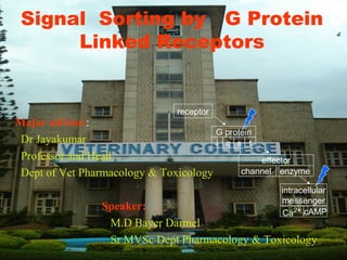
- 1. Signal Sorting by G Protein Linked Receptors Major advisor: Dr Jayakumar Professor and Head Dept of Vet Pharmacology & Toxicology Speaker: M.D Bayer Darmel Sr MVSc Dept Pharmacology & Toxicology receptor tsqi G protein cAMPCa2+ intracellular messenger enzymechannel effector
- 2. History • Refers to a “receptive substance” describing the cellular sites of interaction of drugs curare/nicotine and atropine/pilocarpine in neuromuscularjunctions. (Langley. 1909) • 1969: proposition of an intermediate transducer to link distinct receptors to common effector adenylyl effector, cyclase, and identification of the heterotrimeric G‐ protein,Gs.(John C. Foreman,.2003) • 1983: rhodopsin was the first GPCR to be cloned • The classical G protein signaling pathway that was identified very early on was the activation of the adenylyl cyclase‐cAMP pathway by G s (Gilman, 1987). • Rodbell and Gilman were jointly awarded the Nobel Prize in 1994. (John C. Foreman,.2003) • 2000: first crystal structure of a GPCR(John C. Foreman,.2003)
- 3. Noble Prize for G PCR Rodbell and Gilman were jointly awarded the Nobel Prize in 1994 for their discovery of G protein couple receptors and the role of these protein in signal transduction in cell Martin Rodbell, 1925–1998 USA Alfred Goodman Gilman USA
- 4. G protein couple receptors • G protein-coupled receptors (GPCRs), also known as seven transmembrane domain receptors, 7TM receptors, heptahelical receptors, and G protein- linked receptors (GPLR) .(David L et al,.2005) • Also called metabotropic receptors and serpentine receptors.(Miligan.1995 )
- 5. Importance G protein-coupled receptors are involved in many diseases, and are also the target of around half of all modern medicinal drugs.(Hardman et al,.2001)(Marchese et al.) G protein-coupled receptors are found only in eukaryotes, including yeast(Saccharomyces cerevisiae)Saccharomyces cerevisiae), plants, choanoflagellates, and animals Pathways involving these receptors are the targets of hundreds of drugs, including antihistamines, neuroleptics, antidepressants, and antihypertensives(.(Miligan.1995 Ad.Ph V32) • All GPCRs signal via the use of G-proteins
- 6. GPCRs are receptors for: • Light, odours and gustative molecules • Biogenic amines; dopamine, histamine, serotonin • Eicosanoids . • opioids, • amino acids such as GABA, and many other peptide and protein ligands • Peptide and protein hormones Panacreatic hormones Gastrointestinal Thyroid (Hardman,.2001)
- 7. Classification(I) • Muscarinic acetylcholine receptors (several types) • Catecholamine receptors • Serotonin receptors 5-HT1,2,4,6 • GABA receptor • Metabotropic’ glutamate receptors (11 subtypes) • Purine receptors (P2Y): Adenosine, AMP, ADP, ATP • Peptide hormone receptors(Michael ,.2005)
- 8. Classification(II) • Class A (or 1) (Rhodopsin-like) • Class B (or 2) (Secretin receptor family) • Class C (or 3) (Metabotropic glutamate/pheromone) • Class D (or 4) (Fungal mating pheromone receptors) • Class E (or 5) (Cyclic AMP receptors) • Class F (or 6) (John C. 2000
- 10. The number of sequences in each subfamily
- 11. G-Protein-Coupled Receptors John C. Foreman,et al,.2003
- 12. Physiological roles The visual sense, sense of smell and pheromones (vomeronasal receptors), Behavioral and mood regulation: receptors in the mammalian brain ( serotonin, dopamine) Regulation of immune system activity and inflammation (chemokines) Autonomic nervous system transmission: both the sympathetic and parasympathetic nervous systems (blood pressure, heart rate and digestive processes) The G-Protein-Coupled Receptor GCR1 Regulates DNA Synthesis GPCRs comprise the largest family of cell surface receptors. In mice, there are 1000 different receptors involved in smell alone GPCR are able to regulate the rate of second messenger production or degration GPLR regulate ion flux through a battery oion chenall by direct GP regulation or via second messenger. • The human genome encodes morethan 1,000 members of this family of receptors, specialized for transducing messages as diverse as light, smells, tastes, and hormones (.(David L et alt,.2005)
- 13. Ligand • It is a signal triggering molecule binding to a site on a target protein, by intermolecular forces such as ionic bonds, hydrogen bonds and Van der Waals forces. • Ligand can be selective for receptor or non selective . • Ligands include substrates, inhibitors, activators, and neurotransmitters. • The affinity of ligand belongs to intermolecular force • Can work as agonist or antagonist.(Miligan,1995)
- 14. Physiological role of GPCR Wiki free encyclopedia
- 15. Cell-to-cell communication by extracellular signaling usually involves six steps • (1) synthesis of the signaling molecule by the signaling cell • (2) release of the signaling molecule by the signaling cell • (3) transport of the signal to the target cell • (4) detection of the signal by a specific receptor protein – receptor-ligand specificity • (5) a change in cellular metabolism, function, or development = cellular respons. • (6) removal of the signal, which usually terminates the cellular response – degredation of ligand
- 17. Structure of GPCR • Order of segments are known – N-terminus.. – Helix – Intracellular loop – Extracellular loop – C-Terminus
- 18. Gether & Koblikas, 1998. JBC 273
- 19. GPCR cellular domains • Extracellular domain • By definition, a receptor's main function is to recognize and respond to a specific ligand, for example, a neurotransmitter or hormone • Transmembrane domain • Intracellular domain • Adenylate Cyclase (AC) is a transmembrane protein, with cytosolic domains forming the catalytic site.
- 20. Coupling to G protein • Intracellular loop I3 – Main point of interaction – 12 amino acids near N terminal of I3 mediates specificity (G protein subtype) – Amino acids near C terminal of I3 mediate efficiency – Varies in size between receptor subtypes • Intracellular loop I2 (from TM3 to TM4) • Mediates specificity and efficacy • C terminal tail – Determines efficiency • Neurotransmitter interacting with amino acids in TM5 and TM6 transmit conformation change to area of I3
- 21. G proteins(molecular switches) • short for guanine nucleotide-binding proteins, • G-proteins are heterotrimeric proteins composed of α (45 KDa), β (37 KDa), and γ (9 KDa) subunits (David L et alt,.2005) • G-proteins interact with a receptor comprised of 7-membrane spanning α-helices. Ligand binding induces. (Michael ,.2005) Alpha • Binds to guanosine nucleotides: GDP or GT • four main families exist for Gα subunits: Gαs , Gαi , Gαq/11 , and Gα12/13 . (modified by attachment of fatty acid chain) • Gαs stimulates the production of cAMP from ATP. • Gαi inhibits the production of cAMP from ATP • Gαq/11 stimulates membrane-bound phospholipase C beta, which then cleaves PIP2 • Gα12/13 are involved in Rho family GTPase signaling
- 22. G Proteins Beta and Gamma(CAAX) • Five members of Beta subunit are identified(B1-B5) • Binds to alpha subunit • Stabilizes G protein in membrane • Blocks alpha from interacting with effector • Can be effectors • The α and γ subunits have covalently attached lipid anchors, that insert into the plasma membrane, binding a G-protein to the cytosolic surface of the plasma membrane • There is a larger family of small GTP-binding switch proteins, • initiation & elongation factors (protein synthesis) • Ras (growth factor signal cascades) • Rab (membrane vesicle targeting and fusion) • ARF (formation of vesicle coatomer coats) • Ran (transport of proteins into & out of the nucleus) • Rho (regulation of actin cytoskeleton
- 23. Lüllmann, Color Atlas of Pharmacology © 2000 Thieme
- 24. Types of G Protein Actions • Indirect action – G subunit activates enzyme – Wide spatial extent due to diffusible second messenger – Examples • Adenylate cyclase • Phospholipase C • Phospholipase A2 • Phosphodiesterase – Activates intracellular signalling pathways
- 25. Types of G Protein Actions • Direct action – G subunit directly gates channel – Limited spatial extent – Usually Gβγ – Examples • Stimulation of GIRK potassium channels • Inhibition of calcium channels • Regulation of Na-K pump Lüllmann, Color Atlas of Pharmacology © 2000 Thieme
- 26. G-protein linked receptors coupled to ion channels • Acetylcholine (muscarinic) • Adenosine & adenine nucleotides • Adrenaline & noradrenaline • Angiotensin • Bombesin • Bradykinin • Calcitonin • Cannabinoid • Chemokine • Cholecystokinin & gastrin • Dopamine • Endothelin • Galinin • GABA (GABAB) • Glutamate (quisqualate) • Histamine • 5-Hydroxytryptamine (1,2) • Leukotriene • Melatonin • Neuropeptide Y • Neurotensin • Odorant peptides • Opioid peptides • Platelet-activating factor • Prostanoid • Protease-activated • Tachykinins • Taste receptors • VIP • Vasopressin and oxytocin
- 27. G-protein activation 1. Initially the G-protein α subunit has bound GDP, and the α, β, & γ subunits are complexed together. Gβ,γ , the complex of β & γ subunits, inhibits Gα 2. When the ligand binds to the GPCR it Altering the conformation of the alpha subunit allows it to exchange GDP for GTP. 3. Substitution of GTP for GDP causes another conformational change in Gα . Gα -GTP dissociates from the inhibitory βγ subunit complex, and can now bind to and activate Adenylate Cyclase 4. Adenylate Cyclase, activated by the stimulatory Gα - GTP, catalyzes synthesis of cAMP 5. Protein Kinase A (cAMP-Dependent Protein Kinase) catalyzes transfer of phosphate from ATP to serine or threonine residues of various cellular proteins, altering their activity. 6. The complex of Gβ,γ that is released when Gα binds GTP is itself an effector that binds to and activates or inhibits several other proteins. For example, Gβ,γ inhibits one of several isoforms of Adenylate Cyclase, contributing to rapid signal turnoff in cells that Gprotn.gif
- 28. G protein Inactivation 1. Gα hydrolyzes GTP to GDP + Pi (GTPase). The presence of GDP on Gα causes it to rebind to the inhibitory βγ complex. AdenylateCyclase is no longer activated. 2. Phosphodiesterases catalyze hydrolysis of cAMP to AMP. 3. Receptor desensitization varies with the hormone. In some cases the activated receptor is phosphorylated via a G-protein Receptor Kinase. The phosphorylated receptor then may bind to a protein β-arrestin **GTPase activating proteins (GAPs), when bound to the alpha subunit, enhance the GTPase activity tremendously. GAPs are critical negative regulators of G proteins.
- 29. The role of G Protein
- 30. There are three basic types of secondary messenger molecules: • Hydrophobic molecules: like diacylglycerol, IP3 , and phosphatidylinositols, which are membrane- associated and diffuse from the plasma membrane into the space where they can reach and regulate membrane-associated effector proteins • Hydrophilic molecules: like cAMP, cGMP, and Ca2+ , that are located within the cytosol • Gases: nitric oxide (NO) and carbon monoxide (CO), which can diffuse both through cytosol and across cellular membranes.
- 31. Lüllmann, Color Atlas of Pharmacology © 2000 Thieme The Stimulation of G Protein-Linked Signal Transduction Pathways by α- and β-Adrenergic Receptors
- 32. Production of cAMP 1. Adenylyl cyclase produces cAMP by removing two phosphate groups from ATP. 2. The phosphates are removed as pyrophosphate (P-P). 3. Along with the removal of pyrophosphate the molecule is cyclized. 4. cAMP phosphodiesterase then hydrolyzes cAMP to AMP. Cholera toxin: inactivates the GTPase activity of the Gs alpha subunit, thereby keeping it active. This causes oversecretion of chloride ions and water into the gut (severe diahrrhea). Pertussis toxin: this toxin inactivates the alpha subunit of Gi. This blocks its ability to negatively regulate its targets (whooping cough).
- 33. Most effects of cAMP are mediated by protein kinase A (PKA) 1. PKA is activated by cAMP and mediates the majority of cAMP effects. 2. There are many different substrates of PKA in different cells, which may explain why rises in cAMP in different cell types result in very different responses. 3. The inactive form of PKA is a heterotetramer of two catalytic (kinase) subunits and two regulatory subunits. 4. The binding of cAMP to the regulatory subunits causes a conformational change that releases the catalytic subunits. 5. The release of the catalytic subunits activates them, allowing them to phosphorylate their substrates on serine and threonine residues. 33 Alberts 11-31 © Garland serine kinase phosphatase Residue in target protein N H CHC CH2 O O -O OP O O N H CHC CH2 O O OH
- 34. Rapid and Slow responses to PKA activation **Some of the effects of PKA activation are rapid. Example: the stimulation of glycogen breakdown to glucose in muscle cells. This occurs by the direct phosphorylation of proteins involved in glycogen metabolism. This provides glucose for energy production in muscle cells within seconds. **Some of the effects of PKA are slower. Example: the activation of gene expression, such as the somatostatin hormone. • Activated PKA can translocate into the nucleus. • There it phosphorylates the transcription factor CREB (cAMP response element binding protein). 3. When CREB is phosphorylated it binds to the cAMP response element (CRE). 4. CREB-binding protein (CBP) binds to phosphorylated CREB and activates transcription of genes that contain CRE sequences, such as the somatostatin gene. 5. Many cAMP-induced genes contain CRE sequences and are regulated by CREB and CBP.
- 35. Production of inositol phospholipids **Phosphatylinositol (PI) 4-phosphate and PI 4,5 bisphosphate are produced by the sequential actions of PI kinase and PIP kinase, respectively. **PIs exist in the inner leaflet of the plasma membrane. **PI 4,5 bisphosphate is especially important because its breakdown produces two different second messengers. **PI 4,5 bisphosphate is the least abundant of the PIs, and accounts for only 1% of total phospholipids. (Joyce J. Diwan.. 2008)
- 36. Phospholipase C-β is critical for GPCR signaling 1. The G protein q (Gq) alpha subunit activates the enzyme phospholipase C-β (PLC-β) in a manner similar to how Gs activates adenylyl cyclase. 2. Activated PLC-β cleaves PI 4,5 bisphosphate to produce diacylglycerol (DAG) and inositol 1,4,5-trisphosphate (IP3). 3. Importantly, both DAG and IP3 are second messengers that activate distinct intracellular signaling molecules. 4. IP3 is a small, water-soluble molecule that readily diffuses through the cytosol. 5. DAG remains embedded in the plasma membrane, but like other PM lipids, can diffuse laterally through the membrane.
- 37. The targets of IP3 and DAG IP3: When IP3 diffuses to the membrane of the ER it binds to IP3-gated calcium release channels (IP3Rs) in the membrane, triggering their opening. **IP3Rs release calcium from the ER into the cytosol, rapidly increasing the concentration of calcium in the cytosol. Ca2+ is perhaps the most common second messenger in cells. **Calcium levels are quickly reduced by channels that pump it out of the cell, and by the inactivation of IP3 by dephosphorylation and other means. DAG: DAG has two signaling functions. First, it can be further cleaved to arachidonic acid, which can initiate a complex cascade of lipid messengers. Second, DAG can activate a serine/threonine kinase called protein kinase C (PKC). **PKC requires both Ca2+ and DAG, along with membrane phospholipids, to be activated. **PKC has numerous protein substrates that are unique from PKA.
- 38. Calmodulin 1. Calmodulin is a Ca2+ binding protein that has 4 high affinity binding sites for Ca2+ . 2. Calmodulin is extremely abundant in cells and accounts for as much as 1% of total protein. 3. Binding of calcium causes a conformational change in calmodulin. 4. At least two or more Ca2+ must bind before calmodulin changes conformations, making it behave like a switch to increasing concentrations of calcium. 5. Calmodulin has no enzymatic function, and instead binds to target proteins and alters their confirmation (as well as its own). 6. One of the most important group of calmodulin targets is the Ca2+ /calmodulin-dependent protein kinases (CaM-kinases).
- 39. CaM-kinase II 1. CaM-kinase II is composed of a large complex of about 12 subunits of CaM- kinase II. For simplicity, only one is shown here. 2. Upon Ca2+ /calmodulin binding, CaMKII changes conformation and is activated. 3. Upon activation, CaMKII autophosphorylates itself on an autoinhibitory domain. This phosphorylation event sustains CaMKII activity without Ca2+ /calmodulin being present in two ways. First, it locks Ca2+ /calmodulin binding to it such that it will not dissociate without the prolonged return to normal calcium levels. Second, it converts the enzyme to a calcium independent form. 4. After this occurs, CaMKII can only be inactivated if all of the subunits are dephosphorylated by phosphatases (overriding the CaMKII kinase activity).
- 40. GPCR desensitization **Cells desensitize, or adapt, when exposed to high levels of ligand for a long period of time. There are 3 mechanisms of desensitization at the level of the GPCR: 1. Receptor inactivation: The GPCR becomes modified such that it can no longer interact with its G protein. 2. Receptor sequestration: The GPCR can be internalized and transported to an interior compartment of the cell such that it no longer is exposed to ligand. 3. Receptor downregulation: The receptor can be degraded by lysosomes after it is internalized. G-protein-linked receptor kinases (GRKs): phosphorylate GPCRs upon their activation on multiple serines and threonines. This phosphorylation leads to binding of arrestin to the active GPCR. Arrestin triggers desensitization by 1). inhibiting the binding of the G protein to the GPCR and 2). by acting as an adaptor protein for the internalization of the receptor. Whether the receptor is sequestered or degraded depends upon may factors.
- 41. Conclusion ■ A large family of plasma membrane receptors with seven transmembrane segments act through heterotrimeric G proteins. On ligand binding, these receptors catalyze the exchange of GTP for GDP bound to an associated G protein, forcing dissociation of the subunit of the G protein. This subunit stimulates or inhibits the activity of a nearby membrane-bound enzyme, changing the level of its second messenger product.
- 42. Contin.. ■ The cAMP produced by adenylyl cyclase is an intracellular second messenger that stimulates cAMP-dependent protein kinase,which mediates
- 43. Cont.. ■ The cascade of events in which a single molecule of hormone activates a catalyst that in turn activates another catalyst, and so on,results in large signal amplification; this is characteristic of most hormone activated systems. • Some receptors stimulate adenylyl cyclase through Gs; others inhibit it through Gi. Thus cellular [cAMP] reflects the integrated input of two (or more) signals
- 44. Cont.. ■ Cyclic AMP is eventually eliminated by cAMP phosphodiesterase, and Gs turns itself off by hydrolysis of its bound GTP to GDP. When the epinephrine signal persists, -adrenergic receptor–specific protein kinase and arrestin 2 temporarily desensitize the receptor and cause it to move into intracellular vesicles. In some cases, arrestin also acts as a scaffold protein, bringing together protein components of a signaling pathway such as the MAPK cascade
- 45. Cont.. ■ Some serpentine receptors are coupled to a plasma membrane phospholipase C that cleaves PIP2 to diacylglycerol and IP3. By opening Ca2 channels in the endoplasmic reticulum, IP3 raises cytosolic [Ca2]. Diacylglycerol and Ca2 act together to activate protein kinase C, which phosphorylates and changes the activity of specific cellular proteins. Cellular [Ca2] also regulates a number of other enzymes, often through calmodulin. T H E E N DT H E E N D