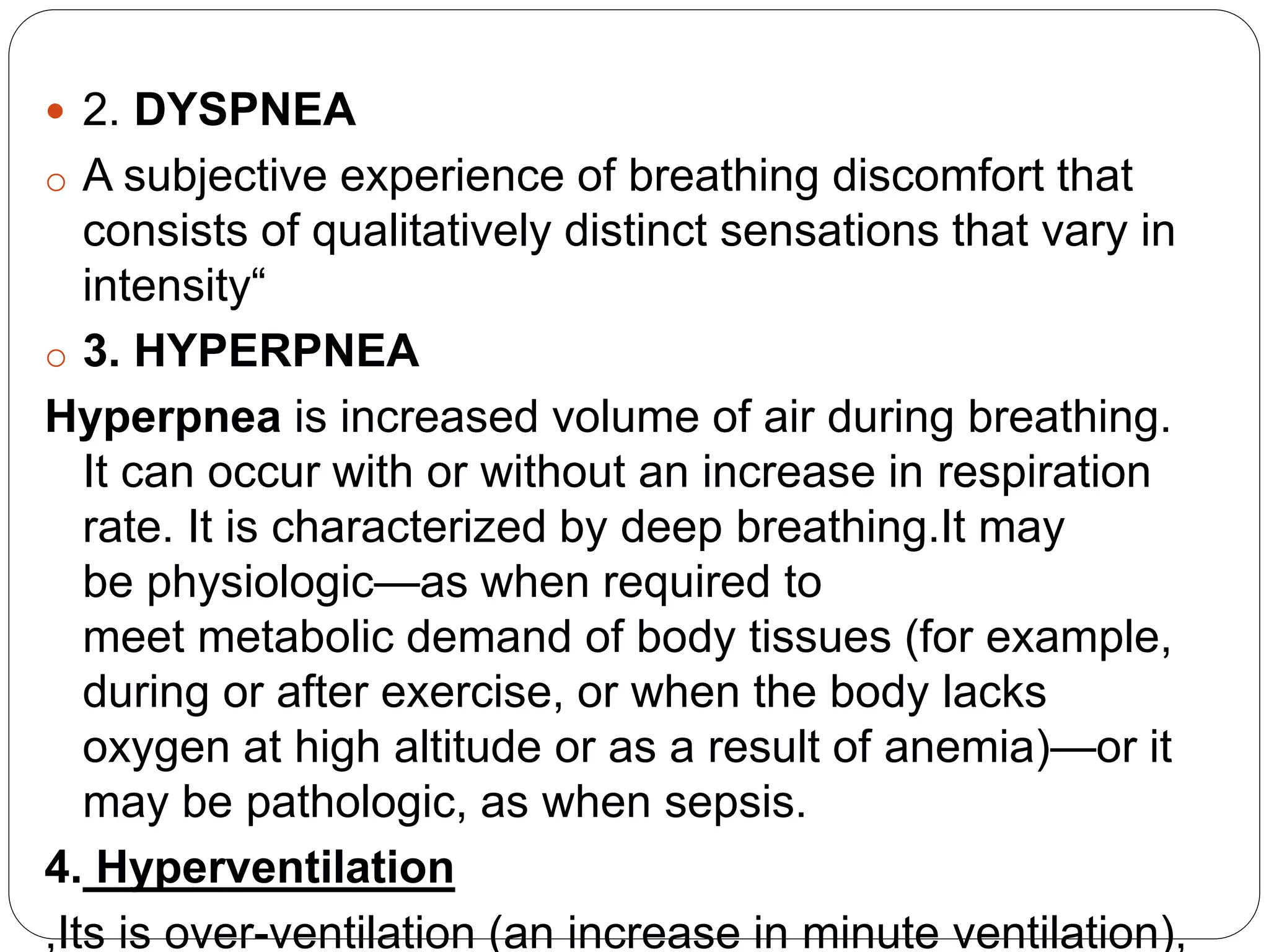
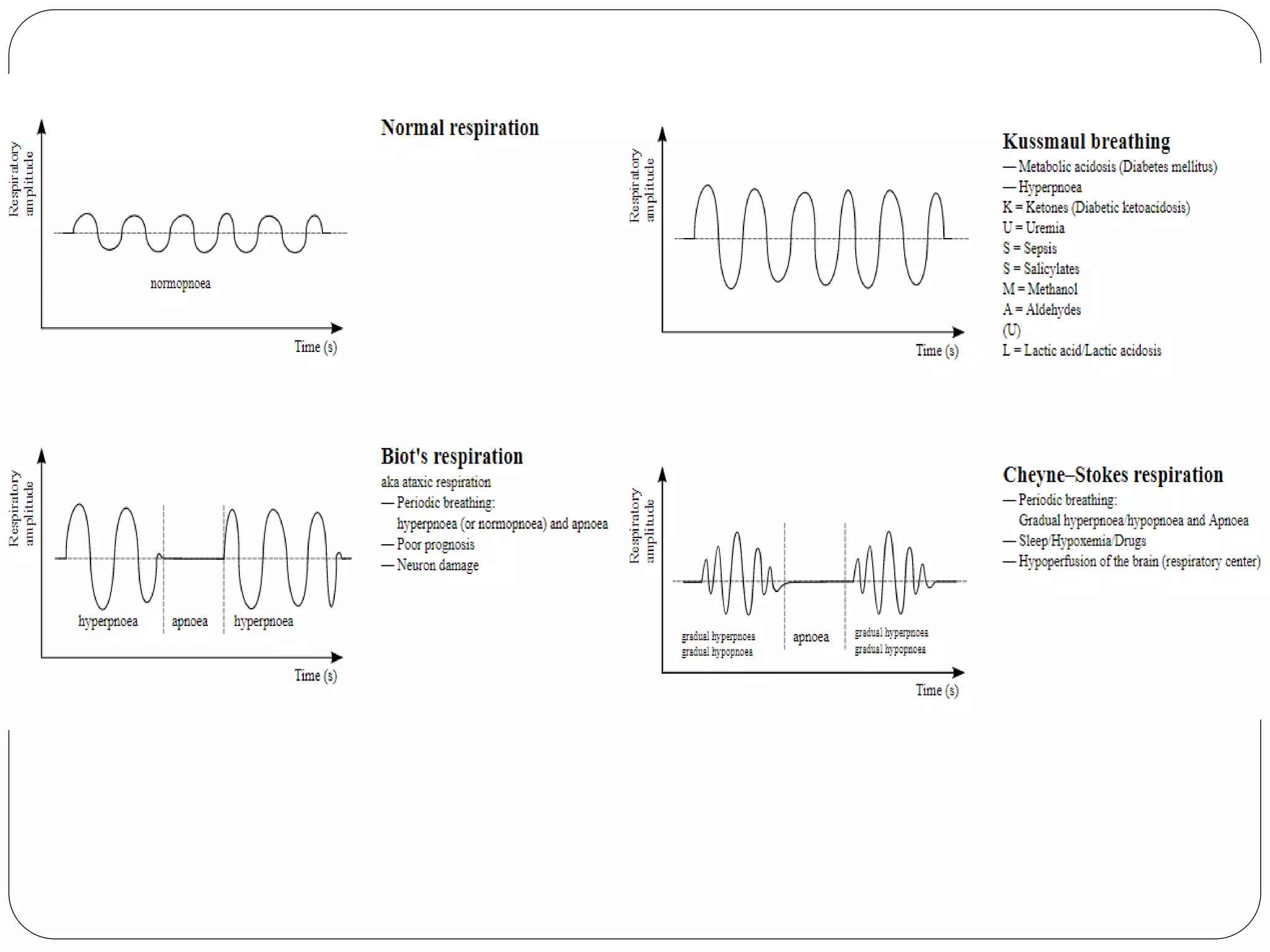
2. The respiratory rate is the rate at
which breathing occurs. This is usually measured
in breaths per minute and is set and controlled by
the respiratory centre.
The pattern, effort,and volume of respiration may
be more indicative of altered respiratory
physiology .
Several prehospital and hospital-based illness or
injury severity scores feature the RR as a cardinal
value .
5. The respiratory centre is responsible for generating
and maintaining the rhythm of respiration, and also of
adjusting this in homeostatic response to
physiological changes
The respiratory center is located in the medulla
oblongata and pons
The respiratory center is made up of three major
respiratory groups of neurons, two in the medulla
and one in the pons.
In the medulla they are the dorsal respiratory
group, and the ventral respiratory group.
In the pons, the pontine respiratory group includes
two areas known as the pneumotaxic centre and the
apneustic centre.
6. The respiratory center receives input
from chemoreceptors, mechanoreceptors the cerebral
cortex, and the hypothalamus in order to regulate the rate
and depth of breathing.
Input is stimulated by altered levels of oxygen, carbon
dioxide, and blood pH, by hormonal changes relating to
stress and anxiety from the hypothalamus, and also by
signals from the cerebral cortex to give a conscious
control of respiration.
Dorsal respiratory group
-initiating inspiration (inhalation)
• Ventral respiratory group
-exhalation(expiratory) area of respiratory control.
-The VRG contains both inspiratory and expiratory
neurons.[6The VRG of neurons are active in forceful
breathing and inactive during quiet, restful
respirations.[ The VRG sends inhibitory impulses to the
apneustic center.
7. Pneumotaxic center
o The pneumotaxic center is located in the upper part of
the pons
o The pneumotaxic center controls both the rate and
the pattern of breathing.
o Responsible for limiting inspiration, providing
an inspiratory off-switch.
o Absence of the center results in an increase in depth
of respiration and a decrease in respiratory rate.
o Apneustic center
o The apneustic center sends signals to the dorsal
group in the medulla to delay the 'switch off',
the inspiratory off switch.
8. Normal range
Average resting respiratory rates by age are
birth to 6 weeks: 30–40 breaths per minute
6 months: 25–40 breaths per minute
3 years: 20–30 breaths per minute
6 years: 18–25 breaths per minute
10 years: 17–23 breaths per minute
Adults: 12–18 breaths per minute
Elderly ≥ 65 years old: 12–28 breaths per
minute.
Elderly ≥ 80 years old: 10-30 breaths per
minute.
9. IMPORATANCE OF RR
The respiratory status of both adults and children
plays acrucial role in determining the overall
assessment of illness.Although it is a sensitive yet
nonspecific indicator of respiratory dysfunction.
Using tachypnea alone as a predictor of
pulmonary pathology, infants with an RR higher
than 60 breaths/min are found to be hypoxic 80%
of the time.
An RR higher than 25 breaths/min in prehospital
trauma patients was associated with increased
mortality.
Increased RRs may be seen in patients with a
variety of pulmonary or cardiac diseases, and
10. Procedure
Sit down and try to relax.
It's best to take your respiratory rate while sitting
up in a chair or in bed.
Measure your breathing rate by counting the
number of times your chest or abdomen rises over
the course of one minute.
It is common to measure respirations over 15
seconds and multiply by 4, but this can
significantly alter the true RR per minute .
11. Respiratory Pattern
1 .Apnea
It is the cessation of breathing. During apnea, there is
no movement of the muscles of inhalation, and the
volume of the lungs initially remains unchanged.
Voluntarily doing this is called holding one's breath.
causes : drug-induced (such as by opiate toxicity),
mechanically induced ( strangulation or choking),
consequence of neurological disease or trauma
During sleep in patients who are suffering from sleep
apnea
12. 2. DYSPNEA
o A subjective experience of breathing discomfort that
consists of qualitatively distinct sensations that vary in
intensity“
o 3. HYPERPNEA
Hyperpnea is increased volume of air during breathing.
It can occur with or without an increase in respiration
rate. It is characterized by deep breathing.It may
be physiologic—as when required to
meet metabolic demand of body tissues (for example,
during or after exercise, or when the body lacks
oxygen at high altitude or as a result of anemia)—or it
may be pathologic, as when sepsis.
4. Hyperventilation
,Its is over-ventilation (an increase in minute ventilation),
13. 5. Tachypnea
it is a respiration rate greater than normal, resulting
in abnormally rapid breathing.
6. Hypopnea
Hypopnea is overly shallow breathing or an
abnormally low respiratory rate. Hypopnea is
defined by some to be less severe than apnea .
7. Orthopnea
shortness of breath (dyspnea) that occurs
when lying flat causing the person to have to
sleep propped up in bed
8. Platypnea
shortness of breath(dyspnea) that is relieved
when lying down and worsens
14. Biot's respiration
It is an abnormal pattern of breathing
characterized by groups of regular deep
inspirations followed by regular or irregular
periods of apnea.
Biot's respiration is caused by damage to
the pons due to strokes or trauma or by pressure
on the pons due to uncal or tentorial herniation.
It can be caused by opioid use
It is distinguished from ataxic respirations by
having more regularity and similar-sized
inspirations, whereas ataxic respirations are
characterized by completely irregular breaths and
15. Cheyne–Stokes respiration
an abnormal pattern of breathingcharacterized by
progressively deeper, and sometimes faster,
breathing followed by a gradual decrease that
results in a temporary stop in breathing called
an apnea.
The pattern repeats, with each cycle usually taking
30 seconds to 2 minutes.
It is an oscillation of ventilation between apnea
and hyperpnea with a crescendo-diminuendo
pattern, and is
associatedwithchanging serum partialpressures of
oxygen and carbon dioxide
It may be caused by damage to respiratory center
Apnea leading to increased CO2 which causes
excessive compensatory hyperventilation, in turn
16. Kussmaul breathing
- It is a form of hyperventilation, which is any
breathing pattern that reduces carbon dioxide in
the blood due to increased rate or depth of
respiration.
- In metabolic acidosis, breathing is first rapid and
shallow[1] but as acidosis worsens, breathing
gradually becomes deep, labored and gasping.,
- Kussmaul breathing is respiratory compensation
for a metabolic acidosis, most commonly
occurring in diabetics in diabetic ketoacidosis .