Fluorometry is an analytical technique that uses fluorescence to identify and characterize small amounts of substances. It involves exciting a sample with ultraviolet or visible light, which causes certain molecules to absorb energy and reach an excited electronic state. The molecules then emit light of a longer wavelength as they fall back to the ground state, and the intensity and composition of this fluorescent light can be measured. Fluorometric methods have applications in pharmaceutical analysis to measure compounds like riboflavin, thiamine, and reserpine in drug formulations.
Fluorescence spectroscopy becomes a widely used tool at the interface of biology, chemistry and physics, because of its precise sensitivity and recent technical advancements. The measurements can provide information on a wide range of molecular processes including the interactions of solvent molecules with fluorophores, rotational diffraction of biomolecules, distance between sites of biomolecules, conformational changes and binding interactions. These advances in fluorescence technology are decreasing the cost and complexity of previously complex processes. Fluorescence spectroscopy is a highly developed and non-invasive technique that enables the on-line measurements of substrate and product concentrations or the identification of characteristic process states.
Fluorescence spectroscopy involves three main processes: excitation, where a molecule absorbs a photon and reaches an excited state; internal conversion and vibrational relaxation in the excited state; and fluorescence emission, where the molecule returns to the ground state and emits a photon. It has many applications including structural elucidation of molecules, monitoring molecular interactions and conformational changes, and tracking ions and biomolecules in cells. Specifically, intrinsic protein fluorescence relies on tryptophan residues, while extrinsic labels are often used for non-fluorescent compounds. Fluorescence resonance energy transfer (FRET) also allows measuring distances between fluorophores to study biomolecular interactions and conformational dynamics.
This document provides an overview of infrared (IR) spectroscopy. It discusses the principle behind IR spectroscopy, the different modes of molecular vibration, instrumentation including sources, detectors and monochromators. It also covers sample handling techniques, factors that affect vibrational frequencies and applications of IR spectroscopy such as structure elucidation.
This document presents an overview of fluorimetry. It discusses that fluorimetry is the measurement of emitted fluorescence light. When certain substances are exposed to light, they emit visible light or radiation, known as fluorescence. Fluorescence occurs immediately after light absorption and stops when the light is removed. Phosphorescence is delayed fluorescence that continues after light removal. Fluorimetry works by exciting substances from their singlet ground state to a singlet excited state, then measuring the wavelength of light emitted as they return to the ground state.
This document provides an overview of Fourier Transform Infrared (FT-IR) Spectroscopy. It explains that FT-IR spectroscopy uses an interferometer to measure all infrared frequencies simultaneously, whereas dispersive infrared spectroscopy measures them sequentially. This allows FT-IR to produce spectra much faster. The document also outlines the key components of an FT-IR system, including the Michelson interferometer, beam splitter, fixed and moving mirrors, and how a Fourier transform is used to convert the interferogram signal into an infrared spectrum. Finally, some advantages of FT-IR are noted, such as improved sensitivity and ability to analyze a wide range of sample types.
This document provides an overview of the principles of UV-visible spectroscopy. It discusses how UV-visible spectroscopy involves exciting electrons from lower to higher orbital energies using electromagnetic radiation between 200-800nm. The absorption of radiation is dependent on the structure of the compound and type of electron transition. The main types of electron transitions are σ->σ*, n->π*, π->π*, and n->σ*. Selection rules determine which transitions are allowed. UV-visible spectroscopy is used in pharmaceutical analysis for qualitative, quantitative, and structural analysis of compounds in solution.
This document discusses different types of luminescence including photoluminescence, chemiluminescence, and electroluminescence. It then focuses on fluorescence spectroscopy, describing how it works, common instrumentation used, and parameters that influence fluorescence spectra such as excitation wavelength and concentration. Applications of fluorescence spectroscopy are outlined as well as techniques like steady-state fluorescence, time-resolved fluorescence, fluorescence anisotropy, and quenching of fluorescence.
Flourescence spectroscopy- instrumentation and applicationssinghsnehi01
This document discusses fluorescence and phosphorescence. It defines fluorescence as the emission of light that starts immediately upon absorption of light and stops when the light is removed. Phosphorescence is defined as delayed fluorescence where light continues to be emitted even after the absorbed light is removed. It discusses factors that affect fluorescence like concentration, quantum yield, incident light intensity, oxygen, pH, temperature, viscosity, photodecomposition, and quenchers. Instrumentation for fluorescence includes light sources, filters, sample cells, monochromators, and detectors like photomultiplier tubes. Applications include determination of metals in alloys and fluorescence-based assays.
This document provides an overview of NMR spectroscopy. It begins by explaining the fundamental principles, including that NMR spectroscopy detects the absorption of radio waves by atomic nuclei placed in a magnetic field. It then discusses various aspects of interpreting NMR spectra such as chemical shifts, spin-spin coupling and integrals. The document also covers NMR techniques including Fourier transformation, 2D NMR, and relaxation processes. In summary, the document serves as an introduction to NMR spectroscopy and the principles behind analyzing NMR spectral data.
Nuclear magnetic resonance (NMR) spectroscopy uses the NMR phenomenon to study the physical, chemical, and biological properties of matter. NMR occurs when atomic nuclei are placed in a magnetic field and exposed to a second oscillating field. Only certain atomic nuclei experience NMR, depending on whether they have a quantum property called spin. NMR spectroscopy is valuable in chemistry for determining molecular structure. It is commonly used to map the carbon-hydrogen framework of organic molecules. More advanced NMR techniques also study protein structure and dynamics in biological chemistry.
This document discusses the theory, instrumentation, and applications of dispersive and Fourier transform infrared (FTIR) spectroscopy. It begins with an introduction to IR spectroscopy and the IR region. It then covers dispersive IR instrumentation, which uses prism or grating monochromators to separate wavelengths, and has limitations like slow scan speeds and limited resolution. The document introduces FTIR instrumentation, which uses an interferometer to simultaneously measure all wavelengths and overcomes the limitations of dispersive IR. It concludes that FTIR provides faster, more accurate and sensitive analysis compared to dispersive IR.
Fluorescence spectroscopy analyzes the fluorescent properties of molecules. It works by exciting a molecule to a higher electronic state using a photon, causing it to emit a photon of lower energy as it returns to the ground state. The difference in wavelengths allows detection of emission photons. Key aspects covered include the principles of absorption and emission, instrumentation used, and different types of data that can be recorded such as fluorescence measurements, steady state techniques, and fluorescence anisotropy/polarization.
The document discusses the Jablonski diagram, which was developed by Alexander Jablonski to illustrate electronic transitions in molecules. It shows energy levels and the transitions between them that occur during absorption and emission of light. Absorption involves excitation of electrons to higher energy levels via straight arrows. Emission can occur through fluorescence, phosphorescence, or non-radiative pathways like internal conversion and intersystem crossing, represented by curved arrows. The diagram provides insight into the timescales of the different transition processes.
1) IR spectroscopy uses infrared radiation to identify chemical substances by their absorption patterns.
2) The main components of an IR spectrometer are a radiation source, monochromator, sample cells, detectors, and recorder.
3) Common radiation sources are Nernst glowers, globar sources, and incandescent wires, which emit IR radiation that is focused through the sample.
1. Absorption spectroscopy measures the absorption of light by a sample as it transitions between energy levels. The amount of light absorbed is dependent on characteristics of the sample like concentration and path length.
2. A spectrophotometer directs light from a source through a wavelength selector and sample cell, and a detector measures the intensity of light transmitted. Double beam instruments separately measure light passing through a reference and sample for improved accuracy.
3. Beer's law states absorbance is directly proportional to concentration, path length, and a proportionality constant. Spectrophotometers allow determination of unknown concentrations by measuring absorbance.
This document discusses phosphorescence spectroscopy and provides information about molecular luminescence, including fluorescence and phosphorescence. It describes the basic principles, including how molecules are excited to higher energy states and then emit light as they relax to lower energy states. Singlet and triplet states are defined, along with electronic and vibrational energy levels. Electron transitions like internal conversion, intersystem crossing, and vibrational relaxation are explained. Instrumentation for measuring phosphorescence is also summarized, including components like light sources, monochromators, sample cells, and detectors. Some applications of phosphorescence are mentioned, such as in television screens, pigments, and glow-in-the-dark toys.
Fluorescence spectroscopy is a technique that uses fluorescence from molecules to analyze samples. Certain molecules emit light at longer wavelengths after absorbing ultraviolet or visible light (fluorescence). This technique is highly sensitive and can detect fluorescent compounds even when present at low concentrations. It has various applications like determining drugs in formulations, studying drug-protein binding, and bioanalysis. Factors like temperature, pH, concentration, and molecular structure can influence fluorescence intensity. Fluorometers contain a light source, wavelength selection devices, and photodetectors to measure fluorescence from samples.
Fluorescence spectroscopy involves using ultraviolet light to excite electrons in molecules, causing them to emit visible light. The emitted light has a longer wavelength than the absorbed light. Fluorimeters are used to measure fluorescence, exciting samples at an absorption wavelength and measuring emission at a longer fluorescence wavelength. Fluorescence spectroscopy is useful for applications like determining fluorescent drugs in formulations, carrying out limit tests for fluorescent impurities, and studying drug-protein binding in bioanalysis.
This document discusses overtones and Fermi resonance in infrared spectroscopy. It defines overtones as absorptions that occur at integral multiples of the fundamental frequency, such as a band at 1000 cm-1 accompanying a fundamental at 500 cm-1. Fermi resonance occurs when a fundamental and overtone band have similar energies, causing them to interact and shift in intensity and frequency. This can result in a "Fermi doublet" with one band increasing while the other decreases in energy. The document provides examples of overtones and Fermi resonance in infrared spectra.
This document discusses the principles and applications of fluorimetry. It defines fluorescence as the emission of light from excited electrons returning to ground state. Fluorimetry involves using ultraviolet light to excite sample molecules, which then emit light of lower energy. Factors that influence fluorescence intensity are discussed. Common instrumentation includes light sources, monochromators, cuvettes, detectors, and data analyzers. Applications include determining inorganic substances, proteins, and steroids. Two case studies on analyzing glucose in milk and silver in food/water samples using fluorimetry are presented.
IR spectroscopy analyzes the vibrational frequencies of bonds in molecules to determine their structure. It works by measuring the absorption of IR radiation by molecular bonds. Different functional groups absorb at characteristic frequencies, producing a molecular "fingerprint". IR spectroscopy is useful for identification of unknown compounds, analyzing purity, and monitoring chemical reactions through changes in bond absorption. It is a nondestructive technique applied in various fields such as pharmaceutical analysis, biomedical research, forensic science, and atmospheric studies.
Spectrofluorimetry uses fluorescence to analyze samples. Fluorescence occurs when molecules absorb ultraviolet or visible light then emit light at a higher wavelength. A spectrofluorimeter contains a light source, monochromators to isolate wavelengths, and a detector. It can quantify substances like vitamins, drugs, proteins, and more down to attogram levels. Though specific, fluorescence can be impacted by environmental factors and some compounds may not fluoresce. Areas of application include chemistry, biochemistry, medicine, and more.
This document discusses fluorimetry and phosphorimetry. It defines them as measurement techniques, with fluorimetry measuring fluorescence intensity at a particular wavelength, and phosphorimetry measuring phosphorescence in conjunction with pulsed radiation. It describes the principles behind photoluminescence, including fluorescence and phosphorescence. Factors affecting these processes and instrumentation used are summarized, including light sources, filters, monochromators, and detectors. Applications in pharmaceutical, clinical, environmental, and entertainment fields are also briefly outlined.
Infrared spectroscopy (IR spectroscopy) is the spectroscopy that deals with the infrared
region of the electromagnetic spectrum, that is light with a longer wavelength and
lower frequency than visible light.
Infrared Spectroscopy is the analysis of infrared light interacting with a molecule.
This document discusses instrumentation methods of fluorimetry. It describes the key components of a fluorimeter including light sources like mercury vapor lamps and xenon arc lamps, filters and monochromators to select wavelengths of light, sample cells to hold liquid samples, and detectors like photomultiplier tubes and photovoltaic cells. Common types of fluorimeters are single beam, double beam, and spectrofluorimeters. Applications include determination of inorganic substances, proteins, and drugs.
Spectrofluorimetry involves the absorption of UV or visible radiation by a molecule, exciting it to a higher energy state. The molecule then relaxes and emits light of a longer wavelength. It is a sensitive technique that can detect low concentrations of organic and inorganic substances. Factors like conjugation, substituents, temperature, and oxygen presence affect fluorescence intensity. Instrumentation includes a light source, filters, sample cell, and detector. Applications include analysis of foods, pharmaceuticals, clinical samples, and natural products.
This document discusses atomic absorption spectrophotometry and flame emission spectrophotometry. It begins by explaining the basic principles of atomic absorption spectrophotometry, which measures the concentration of elements by detecting the absorption of light by atoms. It then describes the parts and working of atomic absorption spectrophotometry instruments. Next, it covers flame emission spectrophotometry, which measures the radiation emitted by excited atoms to determine concentrations. The document concludes by comparing the two techniques and discussing some sources of interference.
Introduction, theoretical principle, quantum efficiency of fluorescence, molecular structure of
fluorescence, instrumentation, factors influencing the intensity of fluorescence, comparison of fluorometry with spectrophotometry, application of fluorometry in pharmaceutical analysis
Introduction, theoretical principle, quantum efficiency of fluorescence, molecular structure of
fluorescence, instrumentation, factors influencing the intensity of fluorescence, comparison of
fluorometry with spectrophotometry, application of fluorometry in pharmaceutical analysis
Fluorimetry, principle, Concept of singlet,doublet,and triplet electronic sta...Vandana Devesh Sharma
This document discusses the principles and factors affecting fluorescence and fluorimetry. It begins by defining fluorescence as the emission of light by a substance that has absorbed light or other electromagnetic radiation. It then discusses various processes that can occur in excited molecules including fluorescence, phosphorescence, internal conversion, intersystem crossing, and collisional deactivation. The document also summarizes several factors that can influence fluorescence intensity, including molecular structure, temperature, viscosity, oxygen content, and pH. Structural factors discussed include conjugation, substituent groups, and molecular rigidity.
Fluorimetry.pptx by Saloni Kadam Nanded talukauser621767
The document discusses fluorimetry and provides details about:
- Luminescence processes including fluorescence and phosphorescence
- Factors that affect fluorescence like pH, temperature, and concentration
- Instrumentation used for fluorimetry including radiation sources, monochromators, sample holders, and photomultiplier tube detectors
- Quenching processes that can decrease fluorescence intensity
Introduction, theoretical principle, quantum efficiency of fluorescence, molecular structure of
fluorescence, instrumentation, factors influencing the intensity of fluorescence, comparison of
fluorometry with spectrophotometry, application of fluorometry in pharmaceutical analysis
This document is a student's report on luminescence spectroscopy submitted to their professor. It defines fluorescence and phosphorescence, explaining the principles using the Jablonski diagram. Fluorescence occurs from the first excited singlet state and involves emission of a photon within nanoseconds of absorbing light. Phosphorescence involves intersystem crossing to the triplet state, with emission of a photon over micro- to milliseconds. The key differences are that fluorescence stops immediately upon removing excitation, while phosphorescence emission persists afterwards due to the longer-lived triplet state.
Fluorescence is a type of luminescence where molecules emit light from electronically excited states created by light absorption. The fluorescence process involves three steps: 1) excitation of a molecule to an excited electronic state, 2) vibrational relaxation to the lowest excited vibrational level, and 3) emission of a photon and return to the ground state. Phosphorescence also involves light emission from an excited state, but occurs from a longer-lived triplet excited state following intersystem crossing. The absorption and fluorescence emission spectra of molecules generally overlap but the fluorescence peaks are at slightly longer wavelengths due to a Stokes shift.
1. Fluorescence spectrophotometry measures the intensity of light emitted by a substance that has absorbed ultraviolet or visible light.
2. After light absorption, molecules can deactivate through radiationless processes like internal conversion or intersystem crossing, or through emission of a photon during fluorescence or phosphorescence.
3. Factors like a molecule's structure, solvent, temperature, and pH can affect its fluorescence quantum yield by changing rates of radiationless relaxation versus light emission.
Fluorescence and phosphorescence are forms of luminescence that involve the emission of light from a substance that has absorbed radiation or light. Fluorescence involves emission of light from singlet excited states, while phosphorescence involves emission from triplet excited states. Factors like temperature, concentration, and molecular structure can influence the intensity of fluorescence. Fluorescence and phosphorescence find applications in areas like analytical chemistry, microscopy, lighting, and more. Instrumentation used to study these phenomena include filter fluorimeters and modern fluorescence spectrophotometers.
The document discusses the theory of fluorimetry. It begins by defining luminescence as light emission from a substance when an electron returns to the ground state from an excited state. It then describes the three types of luminescence - fluorescence, phosphorescence, and chemiluminescence. Fluorescence occurs immediately when light is absorbed, while phosphorescence occurs more slowly after light is removed. Fluorimetry is the measurement of fluorescence, involving excitation and emission spectra. The document goes on to discuss singlet and triplet electronic states, Stokes shift, lifetime, quantum yield, and references in the field of fluorimetry.
The document discusses photoluminescence, which is the emission of light from a material when it absorbs photons. There are three main steps in the photoluminescence process: excitation, relaxation, and emission. Excitation occurs when photons are absorbed and electrons are lifted to a higher energy state. Relaxation follows as electrons lose energy non-radiatively. Emission is the radiative decay of electrons as they return to the ground state, emitting photons of lower energy than those absorbed. The two main types of photoluminescence are fluorescence, which is a rapid emission, and phosphorescence, which is a slower emission.
This document discusses photoluminescence, which is the emission of light from a material upon exposure to light or other electromagnetic radiation. It begins by classifying luminescence and describing photoluminescence as a specific type involving absorption of photons and emission of photons as electrons return to lower energy states. The key processes of photoluminescence are excitation, relaxation, and emission. It then distinguishes between two types of photoluminescence - fluorescence, which is a rapid emission, and phosphorescence, which involves a spin-forbidden state and longer-lasting emission. The document concludes by outlining applications of photoluminescence spectroscopy for materials characterization and explaining the differences between photoluminescence and
What is Fluorescence Electrons in an atom or a m.pdfapnashop1
What is Fluorescence? Electrons in an atom or a molecule can absorb the energy in
the electromagnetic radiation and thereby excite to an upper energy state. This upper energy state
is unstable; therefore, electron likes to come back to the ground state. When coming back, it
emits the absorbed wavelength. In this relaxation process, they emit excess energy as photons.
This relaxation process is known as fluorescence. Fluorescence takes place much more rapidly.
Generally, it completes in about 10-5 s or less time from the time of excitation. In atomic
fluorescence, gaseous atoms fluoresce when they are exposed to radiation with a wavelength that
exactly matches one of the absorption lines of the element. For example, gaseous sodium atoms
absorb and excite by absorbing 589 nm radiations. Relaxation takes place after this by
reemission of fluorescent radiation of the identical wavelength. Because of this, we can use
fluorescence to identify different elements. When excitation and reemission wavelengths are the
same, the resulting emission is called resonance fluorescence. Other than fluorescence, there are
other mechanisms by which an excited atom or molecule can give up its excess energy and relax
to its ground state. Nonradiative relaxation and fluorescence emissions are two such important
mechanisms. Because of many mechanisms, the lifetime of an excited state is brief. The relative
number of molecules that fluoresce is small because fluorescence requires structural features that
slow the rate of the nonradiative relaxation and enhance the rate of fluorescence. In most
molecules, these features are not there; therefore, they undergo nonradiative relaxation, and
fluorescence does not occur. Molecular fluorescence bands are made up of a large number of
closely spaced lines; therefore, usually it is hard to resolve. What is Phosphorescence? When
molecules absorb light and go to the excited state they have two options. They can either release
energy and come back to the ground state immediately or undergo other non-radiative processes.
If the excited molecule undergoes a non radiative process, it emits some energy and come to a
triplet state where the energy is somewhat lesser than the energy of the exited state, but it is
higher than the ground state energy. Molecules can stay a bit longer in this less energy triplet
state. This state is known as the metastable state. Then metastable state (triplet state) can slowly
decay by emitting photons, and come back to the ground state (singlet state). When this happens
it is known as phosphorescence. What is the difference between Fluorescence and
Phosphorescence? • When light is supplied to a sample of molecules, we immediately see the
fluorescence. Fluorescence stops as soon as we take away the light source. But phosphorescence
tends to stay little longer even after the irradiating light source is removed. • Fluorescence takes
place when excited energy is released, and the molecule comes back to the gro.
describes the complete history, mechanisms, instrumentation(jablonski diagram), types, comparision and factors affecting, applications of fluorescence and phosphorescence and describes about quenching and stokes shift.
Fluorescence spectroscopy is based on the principle of fluorescence emission that occurs when a molecule absorbs light and is excited to a higher electronic state. The excited molecule then relaxes to the ground state via vibrational relaxation and emission of a photon. The emitted light has a longer wavelength than the absorbed light due to energy losses in vibrational relaxation, following Stokes' rule. Fluorescence spectroscopy can provide information about molecular structure and interactions through analysis of fluorescence emission spectra.
1. Photochemistry involves using light as a chemical reagent to promote molecules to electronically excited states or as a chemical product when excited states return to the ground state.
2. Fluorescence occurs when a molecule in an excited singlet state returns to the ground state and emits light. It is a spin-allowed process that results in emission at a longer wavelength than the absorbed light.
3. The excitation and fluorescence emission spectra of a compound are often approximately mirror images of each other, though there are exceptions when the excited and ground states differ in geometry.
3.2 molecular fluorescence and phosphorescence spectroscopyGaneshBhagure2
This document discusses molecular fluorescence and phosphorescence spectroscopy. It begins with an introduction to the principles and terms, explaining that fluorescence occurs when emission takes place within 10-8 seconds of absorption, while phosphorescence occurs after more than 10-8 seconds. The document then covers electronic transitions, factors that affect fluorescence and phosphorescence like temperature, pH, and solvent, and instrumentation including components of fluorimeters.
1. Fluorimetry is the measurement of fluorescence intensity using a fluorimeter or spectrofluorimeter. It involves exciting a molecule with specific wavelengths of light which causes electrons to get promoted to excited states, then relax emitting light of longer wavelengths.
2. Key components of fluorimeters include light sources like mercury lamps, filters to select wavelengths, sample cells, and detectors like photomultiplier tubes. Spectrofluorimeters use monochromators instead of filters to isolate wavelengths.
3. Fluorescence can be quenched by factors like oxygen, pH, temperature. Applications include determining inorganic/organic substances, proteins, pigments in nanogram amounts.
The document discusses the basics of lasers. It explains that lasers work via the process of stimulated emission, where photons stimulate excited electrons to emit additional photons of the same frequency and direction. This leads to coherent, highly directional light that is monochromatic and has high intensity and brightness. The key aspects that enable lasers are population inversion, where more atoms are in excited states than ground states, and stimulated emission, where incident photons cause excited electrons to emit additional photons coherently.
This document provides an overview of spectrofluorimetry. It begins with an introduction that defines fluorescence and phosphorescence as types of photoluminescence that occur when electrons return to the ground state from an excited state. It then discusses the principle, theory, instrumentation, factors affecting fluorescence, and applications of spectrofluorimetry. The instrumentation section describes the main components, including a light source, excitation and emission monochromators, sample holder, detector, and readout device. Common factors that can affect fluorescence intensity are concentration, incident light intensity, quantum yield, absorption, pH, oxygen, temperature, viscosity, and scatter. Applications include chemical modification of compounds, identification of compounds based on excitation and emission spectra, and assays of vitamins
About Healthcare system of Bangladesh: Health care delivery is a daunting challenge area of the Bangladesh’s healthcare systems. The Health
care system in Bangladesh falls under the control of the Ministry of Health and Family Planning. The
government is responsible for building health facilities in urban and rural areas.
The document discusses tests used to identify carbohydrates from unknown sources, including the Molisch test, Benedict's test, Barfoed's test, and Seliwanoff's test. It also describes how to distinguish between glucose and fructose using bromine water oxidation and lists pharmaceutical uses of glucose and fructose such as in foods, medicines, and supplements.
Glycosides are compounds that contain a sugar component (glycone) bonded to a non-sugar component (aglycone). Upon hydrolysis, glycosides break down into these components. Glycosides are widely found in plants and some are used medicinally as laxatives, cardiotonics, or expectorants. Examples discussed include senna, rhubarb, cascara sagrada, and aloe, which contain anthraquinone glycosides that act as stimulant laxatives. Glycyrrhiza (licorice) contains saponin glycosides and is used as a demulcent, expectorant, and tonic.
Raw materials used in capsule shells include water, colorants, preservatives, gelatin, opacifiers, and plasticizers. Gelatin is the main component and is derived from animal bones, hide portions, or pork skin. It is soluble, film-forming, and provides strength to the capsule shell. Colorants like dyes and pigments are used for identification and attractiveness. Preservatives prevent microbial growth. Opacifiers like titanium dioxide make the shell opaque. Plasticizers are added to soft gelatin capsules to decrease the viscosity of the gelatin.
NSAIDs are a class of drugs that reduce pain, fever, and inflammation by blocking prostaglandin production. They include aspirin, ibuprofen, and naproxen which are available over-the-counter. NSAIDs work by inhibiting the COX enzymes involved in prostaglandin synthesis. They can cause side effects like ulcers and bleeds from their effects on COX-1. COX-2 selective NSAIDs aim to reduce inflammation while sparing the stomach lining by selectively inhibiting COX-2. Paracetamol is considered separately due to its different mechanism of action, providing analgesia and antipyresis through central COX inhibition in the brain.
This document provides an introduction to the field of pharmacology. It defines pharmacology as the study of drug action, including their origins, properties, and interactions with living organisms. The document then discusses several key areas within pharmacology, including clinical pharmacology, neuropharmacology, psychopharmacology, and pharmacokinetics. It also defines important terms like drugs, medicines, pro-drugs, and the four main processes involved in pharmacokinetics - absorption, distribution, metabolism, and excretion (ADME).
The male reproductive system produces and transports sperm and reproductive hormones. It includes internal organs like the testes, which produce sperm and testosterone, and the epididymis, seminal vesicles, and prostate gland, which produce fluids that nourish and transport sperm. Externally, it includes the penis, which delivers sperm during intercourse, and the scrotum, which houses the testes and maintains the temperature needed for sperm production. The testes contain seminiferous tubules that produce sperm and Leydig cells that secrete testosterone, both of which are essential for male fertility and sexual function.
- Video recording of this lecture in English language: https://youtu.be/Pt1nA32sdHQ
- Video recording of this lecture in Arabic language: https://youtu.be/uFdc9F0rlP0
- Link to download the book free: https://nephrotube.blogspot.com/p/nephrotube-nephrology-books.html
- Link to NephroTube website: www.NephroTube.com
- Link to NephroTube social media accounts: https://nephrotube.blogspot.com/p/join-nephrotube-on-social-media.html
5-hydroxytryptamine or 5-HT or Serotonin is a neurotransmitter that serves a range of roles in the human body. It is sometimes referred to as the happy chemical since it promotes overall well-being and happiness.
It is mostly found in the brain, intestines, and blood platelets.
5-HT is utilised to transport messages between nerve cells, is known to be involved in smooth muscle contraction, and adds to overall well-being and pleasure, among other benefits. 5-HT regulates the body's sleep-wake cycles and internal clock by acting as a precursor to melatonin.
It is hypothesised to regulate hunger, emotions, motor, cognitive, and autonomic processes.
Breast cancer: Post menopausal endocrine therapyDr. Sumit KUMAR
Breast cancer in postmenopausal women with hormone receptor-positive (HR+) status is a common and complex condition that necessitates a multifaceted approach to management. HR+ breast cancer means that the cancer cells grow in response to hormones such as estrogen and progesterone. This subtype is prevalent among postmenopausal women and typically exhibits a more indolent course compared to other forms of breast cancer, which allows for a variety of treatment options.
Diagnosis and Staging
The diagnosis of HR+ breast cancer begins with clinical evaluation, imaging, and biopsy. Imaging modalities such as mammography, ultrasound, and MRI help in assessing the extent of the disease. Histopathological examination and immunohistochemical staining of the biopsy sample confirm the diagnosis and hormone receptor status by identifying the presence of estrogen receptors (ER) and progesterone receptors (PR) on the tumor cells.
Staging involves determining the size of the tumor (T), the involvement of regional lymph nodes (N), and the presence of distant metastasis (M). The American Joint Committee on Cancer (AJCC) staging system is commonly used. Accurate staging is critical as it guides treatment decisions.
Treatment Options
Endocrine Therapy
Endocrine therapy is the cornerstone of treatment for HR+ breast cancer in postmenopausal women. The primary goal is to reduce the levels of estrogen or block its effects on cancer cells. Commonly used agents include:
Selective Estrogen Receptor Modulators (SERMs): Tamoxifen is a SERM that binds to estrogen receptors, blocking estrogen from stimulating breast cancer cells. It is effective but may have side effects such as increased risk of endometrial cancer and thromboembolic events.
Aromatase Inhibitors (AIs): These drugs, including anastrozole, letrozole, and exemestane, lower estrogen levels by inhibiting the aromatase enzyme, which converts androgens to estrogen in peripheral tissues. AIs are generally preferred in postmenopausal women due to their efficacy and safety profile compared to tamoxifen.
Selective Estrogen Receptor Downregulators (SERDs): Fulvestrant is a SERD that degrades estrogen receptors and is used in cases where resistance to other endocrine therapies develops.
Combination Therapies
Combining endocrine therapy with other treatments enhances efficacy. Examples include:
Endocrine Therapy with CDK4/6 Inhibitors: Palbociclib, ribociclib, and abemaciclib are CDK4/6 inhibitors that, when combined with endocrine therapy, significantly improve progression-free survival in advanced HR+ breast cancer.
Endocrine Therapy with mTOR Inhibitors: Everolimus, an mTOR inhibitor, can be added to endocrine therapy for patients who have developed resistance to aromatase inhibitors.
Chemotherapy
Chemotherapy is generally reserved for patients with high-risk features, such as large tumor size, high-grade histology, or extensive lymph node involvement. Regimens often include anthracyclines and taxanes.
Travel vaccination in Manchester offers comprehensive immunization services for individuals planning international trips. Expert healthcare providers administer vaccines tailored to your destination, ensuring you stay protected against various diseases. Conveniently located clinics and flexible appointment options make it easy to get the necessary shots before your journey. Stay healthy and travel with confidence by getting vaccinated in Manchester. Visit us: www.nxhealthcare.co.uk
STUDIES IN SUPPORT OF SPECIAL POPULATIONS: GERIATRICS E7shruti jagirdar
Unit 4: MRA 103T Regulatory affairs
This guideline is directed principally toward new Molecular Entities that are
likely to have significant use in the elderly, either because the disease intended
to be treated is characteristically a disease of aging ( e.g., Alzheimer's disease) or
because the population to be treated is known to include substantial numbers of
geriatric patients (e.g., hypertension).
Are you looking for a long-lasting solution to your missing tooth?
Dental implants are the most common type of method for replacing the missing tooth. Unlike dentures or bridges, implants are surgically placed in the jawbone. In layman’s terms, a dental implant is similar to the natural root of the tooth. It offers a stable foundation for the artificial tooth giving it the look, feel, and function similar to the natural tooth.
Spontaneous Bacterial Peritonitis - Pathogenesis , Clinical Features & Manage...Jim Jacob Roy
In this presentation , SBP ( spontaneous bacterial peritonitis ) , which is a common complication in patients with cirrhosis and ascites is described in detail.
The reference for this presentation is Sleisenger and Fordtran's Gastrointestinal and Liver Disease Textbook ( 11th edition ).
Computer in pharmaceutical research and development-Mpharm(Pharmaceutics)MuskanShingari
Statistics- Statistics is the science of collecting, organizing, presenting, analyzing and interpreting numerical data to assist in making more effective decisions.
A statistics is a measure which is used to estimate the population parameter
Parameters-It is used to describe the properties of an entire population.
Examples-Measures of central tendency Dispersion, Variance, Standard Deviation (SD), Absolute Error, Mean Absolute Error (MAE), Eigen Value
PGx Analysis in VarSeq: A User’s PerspectiveGolden Helix
Since our release of the PGx capabilities in VarSeq, we’ve had a few months to gather some insights from various use cases. Some users approach PGx workflows by means of array genotyping or what seems to be a growing trend of adding the star allele calling to the existing NGS pipeline for whole genome data. Luckily, both approaches are supported with the VarSeq software platform. The genotyping method being used will also dictate what the scope of the tertiary analysis will be. For example, are your PGx reports a standalone pipeline or would your lab’s goal be to handle a dual-purpose workflow and report on PGx + Diagnostic findings.
The purpose of this webcast is to:
Discuss and demonstrate the approaches with array and NGS genotyping methods for star allele calling to prep for downstream analysis.
Following genotyping, explore alternative tertiary workflow concepts in VarSeq to handle PGx reporting.
Moreover, we will include insights users will need to consider when validating their PGx workflow for all possible star alleles and options you have for automating your PGx analysis for large number of samples. Please join us for a session dedicated to the application of star allele genotyping and subsequent PGx workflows in our VarSeq software.
Know the difference between Endodontics and Orthodontics.Gokuldas Hospital
Your smile is beautiful.
Let’s be honest. Maintaining that beautiful smile is not an easy task. It is more than brushing and flossing. Sometimes, you might encounter dental issues that need special dental care. These issues can range anywhere from misalignment of the jaw to pain in the root of teeth.
Know the difference between Endodontics and Orthodontics.
Fluorometry
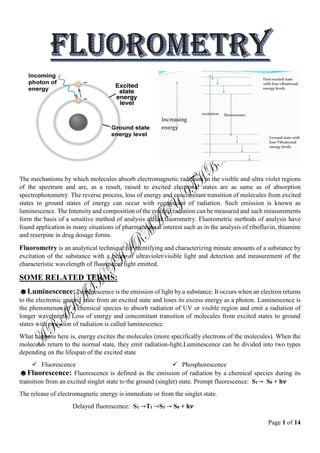
1. Page 1 of 14
The mechanisms by which molecules absorb electromagnetic radiation in the visible and ultra violet regions
of the spectrum and are, as a result, raised to excited electronic states are as same as of absorption
spectrophotometry. The reverse process, loss of energy and concomitant transition of molecules from excited
states to ground states of energy can occur with reemission of radiation. Such emission is known as
luminescence. The Intensity and composition of the emitted radiation can be measured and such measurements
form the basis of a sensitive method of analysis called fluorometry. Fluorometric methods of analysis have
found application in many situations of pharmaceutical interest such as in the analysis of riboflavin, thiamine
and reserpine in drug dosage forms.
Fluorometry is an analytical technique for identifying and characterizing minute amounts of a substance by
excitation of the substance with a beam of ultraviolet/visible light and detection and measurement of the
characteristic wavelength of fluorescent light emitted.
SOME RELATED TERMS:
☻Luminescence: Luminescence is the emission of light by a substance. It occurs when an electron returns
to the electronic ground state from an excited state and loses its excess energy as a photon. Luminescence is
the phenomenon of a chemical species to absorb radiation of UV or visible region and emit a radiation of
longer wavelength. Loss of energy and concomitant transition of molecules from excited states to ground
states with emission of radiation is called luminescence.
What happens here is, energy excites the molecules (more specifically electrons of the molecules). When the
molecules return to the normal state, they emit radiation-light.Luminescence can be divided into two types
depending on the lifespan of the excited state
Fluorescence Phosphorescence
☻Fluorescence: Fluorescence is defined as the emission of radiation by a chemical species during its
transition from an excited singlet state to the ground (singlet) state. Prompt fluorescence: S1→ S0 + h𝝂
The release of electromagnetic energy is immediate or from the singlet state.
Delayed fluorescence: S1 →T1 →S1 → S0 + h𝝂
2. Page 2 of 14
This results from two intersystem crossings, first from the singlet to the triplet, then from the triplet to the
singlet.
Fluorescence is a kind of a luminescence, which is the emission of photons from electronically excited states.
Fluorescence occurs when the electron is transferred from a lower energy state into an "excited" higher energy
state. The electron will remain in this state for 10⁻⁸ sec. then the electron returns to the lower energy state and
it releases the energy in form of fluorescence. In ultraviolet absorption spectroscopy when molecule absorbs
UV radiation at one wavelength and it’s immediately re-emission, usually in a longer wavelength. Some
molecules fluoresce naturally and others can be modified to make fluorescent compounds.
The phenomenon of radiation emission during
transition from the lowest vibrational energy level of
the excited singlet state to the ground state is called
fluorescence.
☻Fluorometry: It is measurement of
fluorescence intensity at a particular wavelength
with the help of a filter fluorimeter or a
spectrofluorimete. It’s measured by a fluorometer or
fluorimeter.
☻Phosphorescence: Phosphorescence is defined as the emission of radiation by a chemical species during
its transition from the excited triplet state to the singlet ground state. The triplet state of a molecule has a lower
energy than its associated singlet state so that transitions back to the ground state are accompanied with the
emission of light of lower energy than from the singlet state. Therefore, we would typically expect
phosphorescence to occur at longer wavelengths than fluorescence. Phosphorescence is often characterized
by an afterglow because of the long life of the triplet state, 10-4
-10 seconds.
An important feature of phosphorescence is
afterglow. Light is emitted from phosphorescent
molecules after radiation energy source is removed.
This is because the luminescence continues for 10-4
seconds to 10 seconds as the triplet state has greater
longevity. In phosphorescence, similar to the
fluorescence, vibrational relaxation must occur.
So, Phosphorescence may be defined as emission of
radiation resulting from transition of molecule from
excited triplet state to ground state.
☻Singlet state (SS): Singlet state
is the state in which all of the electrons
are paired and in each pair the two
electrons spin about their own axis in
opposite directions.
At the ground state, the molecular orbitals are occupied by two electrons. The spins of the two
electrons in the same orbital must be antiparallel.This implies that the total spin, S, of the molecule
in the ground state is zero [½ + (-½)]. This energy state is called “singlet state” and is labeled as S0.
3. Page 3 of 14
☻Excited singlet state: When two electrons of the singlet state are goes to the excited state it is called
excited singlet state. In excited singlet state electrons remain as in exciting position. In the ground state of a
molecule, the two electrons responsible for bonding lie in the bonding molecular orbital in opposite spins.
Now when energy is applied to excite the molecule, one of the electrons will transit to the excited state i.e. the
anti-bonding molecular orbital. If the excited electron in the anti-bonding orbital has spin opposite to the
electron present in the bonding orbital of ground sate, then the excited state is called excited singlet state.
☻Triplet state: Triplet state is a state lying at an energy level intermediate between ground and excited
state and characterized by an unpairing of two electrons. In contrast to the singlet state, there is a spin reversal
involving one electron of the pair and the pair of two electrons spins about their axis in the same direction.
The life time of the molecule in the triplet state in 10-4
to 10-2
seconds.
Basically the triplet state is the excited state between the ground state and the excited singlet state and electron
in this state spins in the same direction as that of ground state.
☻Chemiluminescence: Chemiluminescence is another phenomenon that falls in the category of
luminescence. This refers to the emission of radiation during a chemical reaction.
However, in such cases theexcited state is not a result of absorption of electromagnetic radiation. The oxidation
of luminol (3-aminophthalhydrazide) in an alkaline solution is an example of chemiluminescence.
☻Bioluminescence: Bioluminescence is the production and emission of light by a living organism.
Bioluminescence occurs widely in marine vertebrates and invertebrates, as well as in some fungi,
microorganisms including some bioluminescent bacteria, and terrestrial arthropods such as fireflies.
☻Fluorescein: Fluorescein is a fluorescence label that absorbs light at 490 nm and releases this energy at
520 nm.
Theoretical consideration /Basic concept / Principal:
4. Page 4 of 14
Fluorescence spectroscopy aka fluorometry or spectrofluorometry is an analytical technique for identifying
and characterizing minute amounts of a fluorescent substance by excitation of the substance with a beam of
ultraviolet light and detection & measurement of the characteristic wavelength of the fluorescent light emitted.
It is a spectrochemical method. These terms are explained with the illustration above (Theory of Fluorescence
and Phosphorescence)
When energy is applied to certain molecules in the form of UV or visible electromagnetic radiation, the
molecules temporally transit to an excited singlet state where the excited electron is in paired condition with
the ground electron. In the excited state, the molecules lose energy in radiationless manner to descend to the
lowest vibrational energy level of the excited state. The excited state lasts only 10-8
to 10-4
seconds and then
the excited molecule will return to ground state by losing energy through emitting radiation. This is termed
fluorescence and the emitted radiation is of longer wavelength. By measuring the emitted wavelength we can
determine the presence and amount of a compound in a sample.
1. Absorption of radiant energy: The absorption of UV-Vis radiation is necessary to excite molecules
from the ground state to one of the excited states. When a molecule absorbs radiant energy it is got promoted
from the ground state to the excited state and gets distributed in the various vibrational energy levels mostly
to the excited singlet state. Average time of molecule stay in excited state is 10-8
sec.
2. Radiationless vibrational relaxation (RVR): Absorption of radiation will excite molecules to
different vibrational levels of the excited state.This process is usually followed by successive vibrational
relaxations (VR) as well as internal conversion to lower excited states. Molecules initially undergo a more
rapid process, a radiationless loss of vibrational energy (loss energy by emitting photons) and so quickly falls
to the lowest vibrational energy level of the excited state, known as vibrational relaxation.
3. Radiationless internal conversion: From the lowest vibrational energy level of the excited singlet state,
a molecule can return to the ground state by photoemission or by radiationless process followed by vibrational
relaxation. When an excited molecule undergo a radiationless loss of vibrational energy, sufficient to drop to
the ground state then it is termed internal conversion
4. Fluorescence: After vibrational relaxation to first excited electronic level takes place, a molecule can
return to the ground state by emission of a photon, called fluorescence (FL).The radiation emitted in the
transition of a molecule from a singlet excited state to a singlet ground state is called fluorescence.The
radiation emitted as fluorescence is of lower energy and therefore of longer wavelength than that originally
absorbed.The fluorescence lifetime is much greater than the absorption time and occurs in the range from10-7
to 10-9
s.All absorbed energy will not emitted as fluorescence.
5. Intersystem crossing: Molecule in the lowest vibrational energy level of the excited singlet state converts
to a triplet state (the state lying at an energy level intermediate between ground state and excited).This process
is called intersystem crossing. Here molecules do not losses energy.
6. Radiationless vibrational relaxation: Once intersystem crossing has occurred, a molecule so quickly
falls to the lowest vibrational energy level of the excited triplet state by vibrational relaxation. The lifetime of
molecule in the triplet state is 10-4
to 10-2
seconds (Longer than corresponding singlet state)
7. Radiationless internal conversion: It is a process whereby excited molecules lose their energy due to
collisions with other molecules or by transfer of their energy to solvent or other unexcited molecules.After
RVR molecule goes to the ground state from triplet state.Energy is released here in the form of heat radiation.
8. Phosphorescence: Electrons crossing the singlet state to the triplet state can follow one of three choices
including:
returning to the singlet state
5. Page 5 of 14
relax to ground state by internal or/and external conversion
lose their energy as a photon (phosphorescence) and relax to ground state
It requires a much longer time than fluorescence (10-4
s to up to few s).
Instrumentation for fluorescence spectroscopy:
Figure: A diagrammatic representation of an instrument
used to measure intensity of fluorescence.
In contrast to spectrophotometry, the intensity of light transmitted by a sample is not of direct concern in
fluorometry. Rather, it is the intensity of radiation that is emitted as fluorescence that is measured and related
to the concentration of fluorescing species. The components of instruments which are used in fluorometry are,
however, quite similar in design and function to those employed in spectrophotometers and colorimeters. A
diagrammatic representation of such a device is shown in figure above. The chief components are:
☻Light sources: The lamp or light source provides the energy that excites the compound of interest by
emitting light. The light from an excitation source passes through a filter or monochromator, and strikes the
sample. The light source must be intense and stable. Light sources include:
a. Gas discharge lamps:
Xenon arc lamp (all wave length)
b. Incandescent lamps: Tungsten wire filament lamp
c. Laser: tunable dye laser
6. Page 6 of 14
High pressure mercury vapor lamp d. X-ray source for X-ray fluorescence
The emission of a mercury lamp is concentrated in several very intense bands. Among those having a
wavelength of 254-365 nm are of a great value as excitation radiation is evenly distributed over a wide range
of wavelengths.
☻Excitation Filter (or monochromator): The excitation filter is used to screen out the wavelengths of
unabsorbed light by the compound being measured. It filters the source light and isolates the band of exciting
light that is to be passed to the sample holder. If the instrument uses coarse monochromator then the instrument
is called fluorometer.If grating or prism monochromator is used then the instrument is called
spectrofluorometer, spectrophotofluorometer or florescence spectrometers. Usually glass filters are used.
☻Sample compartment: The sample cell or cuvette holds the sample. The cuvette material must allow the
compound's absorption and emission light energy to pass through. Also, the size of the sample cell affects the
measurement. The greater the path length (or diameter) of the cell, the lower the concentration that can be
read. Glass cells (300nm) are used for most analysis. If measurement is to be under 320nm wavelength then
quartz cells (200-800nm) fused silica are used.
☻Emission filter/monochromator: It selects the band of fluorescence which is to be detected. It is usually
placed at right angle (90º) to the beam of exciting (transmitting) light but other arrangements are possible.
Stray light such as Rayleigh and Raman scatter is also emitted from the sample.
☻Detectors: The detector is placed at a right angle to the direction of travel of beam of exciting light. The
light detector is most often a photomultiplier tube or photoconductive target vidicon or return beam vidicon
or intensified target vidico though photodiodes are increasingly being used. The light passing through the
emission filter is detected by the photomultiplier or photodiode. The light intensity, which is directly
proportional (linear) to the compound's concentration, is registered as a digital readout.
☻Recorder & amplifier: The output of the detector is connected to a meter, a digital display or a recorder.
Recorder (indicates the detector response) gives the intensity of radiation in terms of electrical signal produced
by the detector.
Amplifier is used to amplify the detector response before recording.
Some most common types of fluorimeter are:
Single beam (filter) fluorimeter: It contains tungsten lamp as a source of light and has an optical system
consists of primary filter. The emitted radiations is measured at 90° by using a secondary filter and detector.
Primary filter absorbs visible radiation and transmit UV radiation which excites the molecule present in sample
cell. Single beam instruments are simple in construction cheaper and easy to operate.
Double beam (filter) fluorimeter: it is similar to single beam except that the two incident beams from
a single light source pass through primary filters separately and fall on the another reference solution. Then
the emitted radiations from the sample or reference sample pass separately through secondary filter and
produce response combinly on a detector.
Spectrofluorimeter (double beam): In this primary filter in double beam fluorimeter is replaced by
excitation monochromator and the secondary filter is replaced by emission monochromator. Incident beam is
split into sample and reference beam by using beam splitter.
Structural factors affecting fluorescence:
☻Fluorescence is expected in molecules that are aromatic or multiple conjugated double bonds with a high
degree of resonance stability. Conjugation is necessary for fluorescence.This is because mobile π electrons
7. Page 7 of 14
are responsible for UV-Vis absorption characteristics of compounds.Thus cyclohexane (saturated, no π
electron) is not fluorescent, benzene is weakly fluorescent and anthracene is highly fluorescent.
☻Fluorescence is also expected in polycyclic aromatic systems.
Simple heterocyclic do not exhibit
fluorescence.
Fusion of heterocyclic nucleus to benzene ring
increases fluorescence.
☻Substituents such as –NH3, –OH, –F, –OCH3, –NHCH3, & –N(CH3)2 groups, often enhance fluorescence.
☻On the other hand, these groups decrease or quench fluorescence completely: –Cl, –Br, –I, –NHCOCH3, –
NO2, –COOH.
☻Changes in the system pH, if it affects the charge status of chromophore, may influence fluorescence.
☻Many compounds show fluorescence at ionized state. But this is dependent upon pH of the solution.
☻The higher the rigidity the greater is the
fluorescence intensity. This is because, rigidity and
planarity will prevent vibration and free rotation of
aromatic rings hence less energy is dissipated in
radiationless manner.
☻Substances fluoresce more brightly in a glassy state or viscous solution.
☻Formation of chelates with metal ions also promotes fluorescence. Complexation increases rigidity and
minimizes internal vibration hence fluorescence intensity is increased.
e.g. Tetracycline has a weak native fluorescence but
complexes of the antibiotic with Ca2+
and a
barbiturate fluorescence quiet intensely.
8. Page 8 of 14
Environmental factors affecting fluorescence:
☻Temperature:
A rise in temperature is almost always accompanied by a decrease in fluorescence.
The change in temperature causes the viscosity of the medium to change which in turn changes the n
umber of collisions of the molecules of the fluorophore with solvent molecules.
The increase in the number of collisions between molecules in turn increases the probability for deac
tivation by internal conversion and vibrational relaxation.
Temperature of the reaction must be regulated to within +/- 0.1℃.
In general, a 1⁰C rise in temperature results in a decrease of fluorescence intensity by 1%.
☻PH
:
Relatively small changes in pH can sometimes cause substantial changes in the fluorescence intensity
and spectral characteristics of fluorescence.Example: Serotonin shows a shift in fluorescence emission
maximum from 330 nm at neutral pH to 550 nm in strong acid without any change in the absorption
spectrum.
In the molecules containing acidic or basic functional groups, the changes in pH of the medium change
the degree of ionisation of the functional groups. This in turn may affect the extent of conjugation or
the aromaticity of the molecule which affects its fluorescence. Example: Aniline shows fluorescence
while in acid solution it does not show fluorescence due to the formation of anilinium ion.
Therefore, pH control is essential while working with such molecules and suitable buffers should be
employed for the purpose.
☻Dissolved oxygen:
Dissolved oxygen often decreases fluorescence dramatically and is an interference in many
fluorometric methods.
The paramagnetic substances like dissolved oxygen and many transition metals with unpaired
electrons dramatically decrease fluorescence and cause interference in fluorimetric determinations.
The paramagnetic nature of molecular oxygen promotes intersystem crossing from singlet to triplet
states in other molecules.
The longer lifetimes of the triplet states increases the opportunity for radiationless deactivation to
occur.
Presence of dissolved oxygen influences phosphorescence too and causes a large decrease in the
phosphorescence intensity.
It is due to the fact that oxygen at the ground state gets the energy from an electron in the triplet state
and gets excited.
This is actually the oxygen emission and not the phosphorescence. Therefore, it is advisable to make
phosphorescence measurement in the absence of dissolved oxygen.
Other paramagnetic substances, including most transition metals, exhibit this same effect.
☻Solvents:
Solvents affect fluorescence through their ability to stabilize ground and excited states differently,
thereby changing the probability and the energy of both absorption and emission.
The changes in the “polarity” or hydrogen bonding ability of the solvent may also significantly affect
the fluorescent behaviour of the analyte.
The difference in the effect of solvent on the fluorescence is attributed to the difference in their ability
to stabilize the ground and excited states of the fluorescent molecule.
9. Page 9 of 14
Besides solvent polarity, solvent viscosity and solvents with heavy atoms also affect fluorescence and
phosphorescence.
Increased viscosity increases fluorescence as the deactivation due to collisions is lowered.
A higher fluorescence is observed when the solvents do not contain heavy atoms while
phosphorescence increases due to the presence of heavy atoms in the solvent.
Some solvents e.g Ethanol, also cause appreciable fluorescence.
Other sample matrix, e.g. proteins and bilirubin are more serious contributors to unwanted
fluorescence.
Quantum efficiency of fluorescence:
Quantum efficiency is defined as the ratio of number of light quanta emitted and the number of light quanta
absorbed. It is denoted by ∅.
∅ =
emitted light quanta
absorbed light quanta
Its significance is that, it is an indicator of how fluorescent a molecule is.
if ∅ is near 1, the molecule is highly fluorescent molecule
if ∅ is near 0, the molecule is a very low fluorescent molecule
Q-How you can chemically convert a non-fluorescence compound to fluorescence compound?
1. Complexation: Tetracycline is weak fluorescence but complexes with Ca2+
and barbiturate makes it
fluorescence.
2. Acid treatment: Hydrocortisone usually not fluorescence but they form strongly fluorescence compound
in sulphuric acid in presence of ethanol.
3. Oxidation: By oxidation and hydroxylation epinephrine forms strongly fluorescing compound.
Mirror image rule:
10. Page 10 of 14
Vibrational levels in the excited states and ground states are similar.
An absorption spectrum reflects the vibrational levels of the electronically excited state.
An emission spectrum reflects the vibrational levels of the electronic ground state.
Fluorescence emission spectrum is mirror image of absorption spectrum.
Q-Why absorbed energy and emitted energy is not identical?
rearrangement of atoms in different energy levels is not similar
triplet state
When an electron absorbs energy either from light (photons) or heat (phonons), it receives that incident
quantum of energy. But transitions are only allowed between discrete energy levels such as the two shown
above. This leads to emission lines and absorption lines.
Emitted energy: Photon is emitted and electron drops to lower quantum energy state. Emission spectra always
involve electrons going down in energy level.
Absorbed energy: Photon is absorbed and excites electron to higher quantum energy state. Absorption spectra
always involve atoms going up in energy level.
Common problems of fluorescence measurements:
Reference materials and sample:
Reference materials is as fluorescent as the sample Contaminating substances, Raman scattering,
Rayleigh scattering.
Fluorescence reading is not stable:
Fogging of the cuvette when the contents are much colder than the ambient temperature.
Drops of liquid on the external faces of the cuvette.
Light passing through the meniscus of the sample.
Bubbles' forming in the solution as it warms.
Self-quenching:
It results when luminescing molecule collide and lose their excitation energy by radiationless due to
presence of impurities like molecular oxygen.
Inadequate sensitivity:
Fluorometry is significantly more sensitive as an analytical tool
Absorption of radiant energy:
Absorption either of the exciting or of the luminescent radiation reduces the luminescent signal.
11. Page 11 of 14
Self-absorption:
Attenuation of the exciting radiation as it passes through the cell can be caused by too concentrated an
analyte.
Excimer formation:
Formation of a complex between the excited-state molecule and another molecule in the ground state,
called an excimer, causes a problem when it dissociates with the emission of luminescent radiation at
longer wavelengths than the normal luminescence.
Applications of fluorometry:
☻Application in Chemistry:
Fluorometry is used in chemistry for –
Determination of metal ions: Complexes of metals ions may give strong fluorescence which is utilized
for this purpose.
Separation and identification: In many cases, after separation, chemicals are identified using
fluorometry. e.g. aminocrine.
☻Application in Biopharmaceutics:
Measurement of drug in blood, urine and other body fluids.
Study of the rate and mechanism of drug absorption, metabolism and excretion.
Selection of toxic compounds.
12. Page 12 of 14
☻Pharmaceutical applications:
Fluorometry is used for quantitative analysis of
Hormones: Adrenaline, aldosterone, testosterone
Alkaloids:
a.Opioids: Morphine, codeine etc.
b. Rauwolfia alkaloids: Reserpine
c.Others: Atropine, emetine etc.
Vitamin: Riboflavin and thiamine are indicated for fluorometric assay by USP and BP.
Antibiotics: tetracycline, sulfonamide etc.
Cardiac glycosides: Such as digoxin, digitoxin, etc.
Fluerometry is widely used in the analysis of drugs in systems (physiological systems) other than
dosage forms. The sensitivity of the method of analysis is applied for a large number of
pharmacological, biochemical, toxicological, pharmacokinetic (ADME) & biopharmaceutical studies
for the analysis of amount of drugs in biological fluids and tissues.
Advantages of fluorometry:
Sensitivity: In case of Fluorescence, detectability to parts per billion or even parts per trillion is
common for most analytes.
Specificity: Fluorometers are highly specific and less susceptible to interferences because fewer
materials absorb and also emit light (fluoresce).
Wide Concentration Range: Fluorescence output is linear to sample concentration over a very broad
range.
Simplicity: Fluorometry is a relatively simple analytical technique.
Low Cost: Reagent and instrumentation costs are low when compared to many other analytical
techniques, such as gas chromatography and HPLC.
Limitations of Fluorometry:
The extent of applicability of this technique is limited, because of the fact that all elements and
compounds are unable to exhibit fluorescence
The method is not suited for determination of major constituents of a sample, because the accuracy is
very less for large amounts
Careful buffering is necessary as fluorescence intensity may be strongly dependent
The presence of dissolved oxygen may cause increased photochemical destruction
Selection criteria:
Fluorometry only for fluorescence compound
Conversion to fluorescence.
Fluorometry low conc
Electronic transition
Chemistry
Fluorometry is more sensitive tool:
Fluorometer directly measures intensity of light
Measures at low con
More sensitive
More specific
Amplifier is used
13. Page 13 of 14
☻Comparison of fluorometry with spectroscopy:
Fluorometry Spectrophotometry
More sensitive Less sensitive
Fluorometer directly measures intensity of light Cannot directly measures intensity of light
Intensity can be amplified Intensity cannot be amplified
More specific Less specific
Large numbers of variables can be controlled Small numbers of variables can be controlled
Temperature must be controlled Temperature don’t need to be controlled
Intensity of light maintain constant Intensity of light don’t maintain constant
Extraneous solutes affects Extraneous solutes don’t affects
Influence of pH is complex Influence of pH is not complex
☻Difference between absorption spectroscopy & fluorescent spectroscopy:
Features Absorption spectroscopy Fluoroscence spectroscopy
Theoretical consideration Measurement of amount of light
absorbed
Measurement of intensity of
fluorescence
Wavelength of light used Which gives maximum
absorption
Which gives maximum
fluorescence
Instruments Determines only the absorption
of light
Determines absorption of light as
well as emission of radiation
Light source Tungsten, H2-discharge lamp Mercury arc lamp, Xenon arc
lamp
Cell used Silica cell Glass and metal cells
Detector Phototube or photo multiplier is
used to detect the radiation
absorbed
Emission filter is used to separate
the emitted light from the
transmitted light
Concentration Concentration depends on the
molar absorptivity.
Concentration depends on the
characteristics of the instrument
Electrical transition Applicable for both π→π* &
n→π* transition.
Not applicable for the compound
containing n→π* transition
Experimental variables
(temperature & Extraneous solution)
Not so restricted Highly restricted
Sensitivity & selectivity Less sensitive & less specific. More sensitive & highly specific.
☻Differences between fluorescence & phosphorescence:
Property Fluorescence Phosphorescence
Transition Molecule transits from excited
singlet state to ground state.
Molecule transits from excited
triplet state to ground state
Lifespan Fluorescence is continued for only
10-8
to 10-4
seconds.
Phosphorescence continues for
10-4
seconds to 10-2
seconds.
Afterglow Not present. Occurs and luminescence slowly
fades.
Analytical application Yes No