
Recommended
More Related Content
Similar to Investigations for Chest trauma
Similar to Investigations for Chest trauma (20)
Recently uploaded
Recently uploaded (20)
Investigations for Chest trauma
- 2. Investigations: ● Investigation of chest trauma is based on clinical examination, supplemented by chest radiography. Ultrasound – eFAST. Underwater Chest drain. Chest Radiograph.
- 3. ● Ultrasound- Extended Focused Assessment with Sonar for Trauma (eFAST). ● Ultrasound can be used to differentiate between contusion and the actual presence of blood. ● This technique uses sonar assessment in the chest, looking for a cardiac tamponade or free blood and air in the hemithorax on each side, ● Assessment for blood in the abdominal cavity, in sub-diaphragmatic spaces and pelvis.
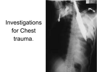
- 5. Chest Radiograph. ● If the patient is haemodynamically unstable or the spine is at risk, an anteroposterior (AP) supine chest radiograph is done which will provide information regarding tracheal deviation, lung and mediastinal pathology, as well as skeletal injury. ● In penetrating injury, chest radiograph is performed in the erect position, as this will best reveal a small pneumothorax, fluid meniscus, air–fluid level or the presence of free gas under the diaphragm. ● The presence of thoracic skeletal injury might also injure the abdominal viscera.
- 6. ● Rupture of the thoracic aorta can be seen in fractures of the first and second rib, bilateral clavicular fracture, fracture of the sternum, thoracic spine or scapula. ● Fracture of the lower ribs can injure the liver or spleen. ● Fracture of ribs can injure the lung parenchyma or thoracic wall vasculature, causing pneumothorax, haemothorax or lung contusion.
- 10. Computed tomography scan. ● Principal and most reliable examination for major injury in chest trauma. ● In blunt chest trauma, the CT scan identifies fractures, haematomas, pneumothorax and pulmonary contusion. In penetrating chest trauma, the scan show the track or presence of the missile and allow the proper planning of definitive surgery.
- 11. ● Also detects the migration of abdominal contents into the chest in case of rupture of diaphragm. ● It is the diagnostic modality of choice for the assessment of the thoracic aorta and mediastinal vessels.
- 12. Pneumothorax
- 14. Reference: Bailey and Love’s Short Practice of Surgery – 27th edition SRB’s Manual of Surgery – 5th edition
- 15. Thank you.