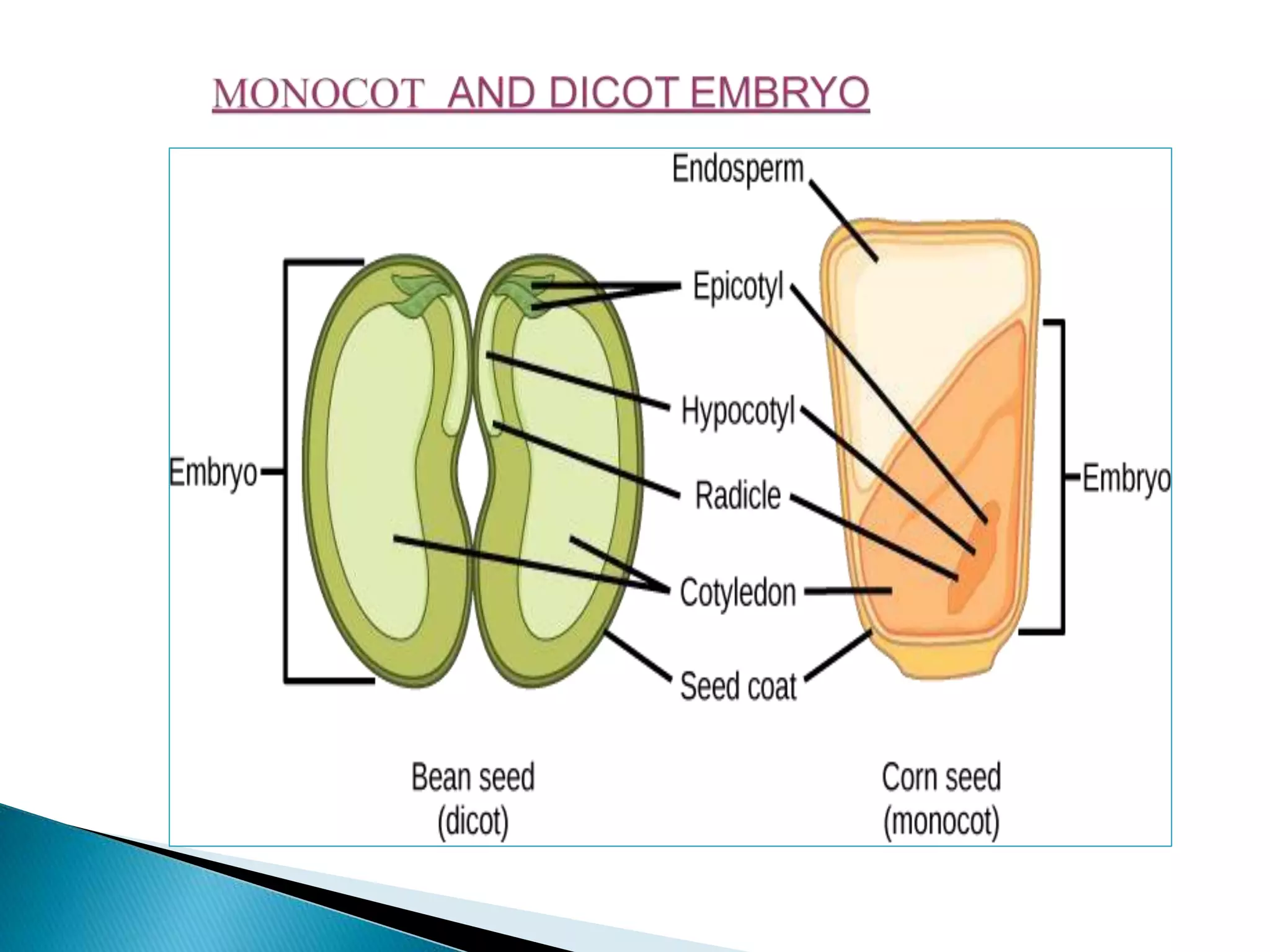
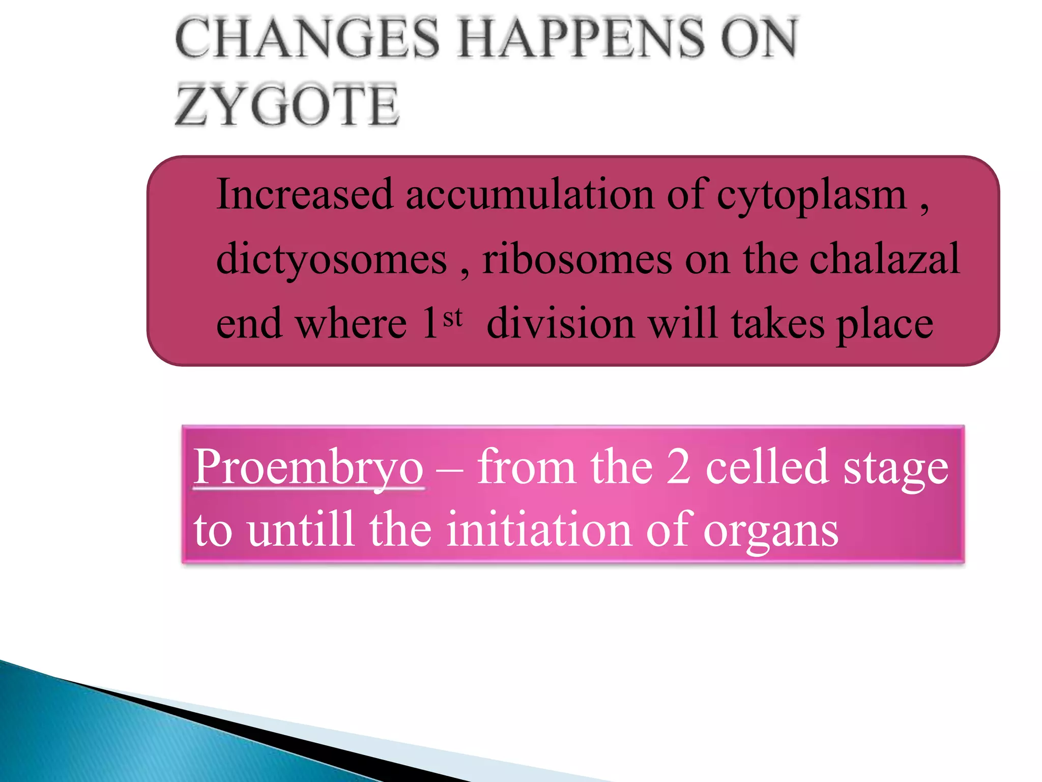
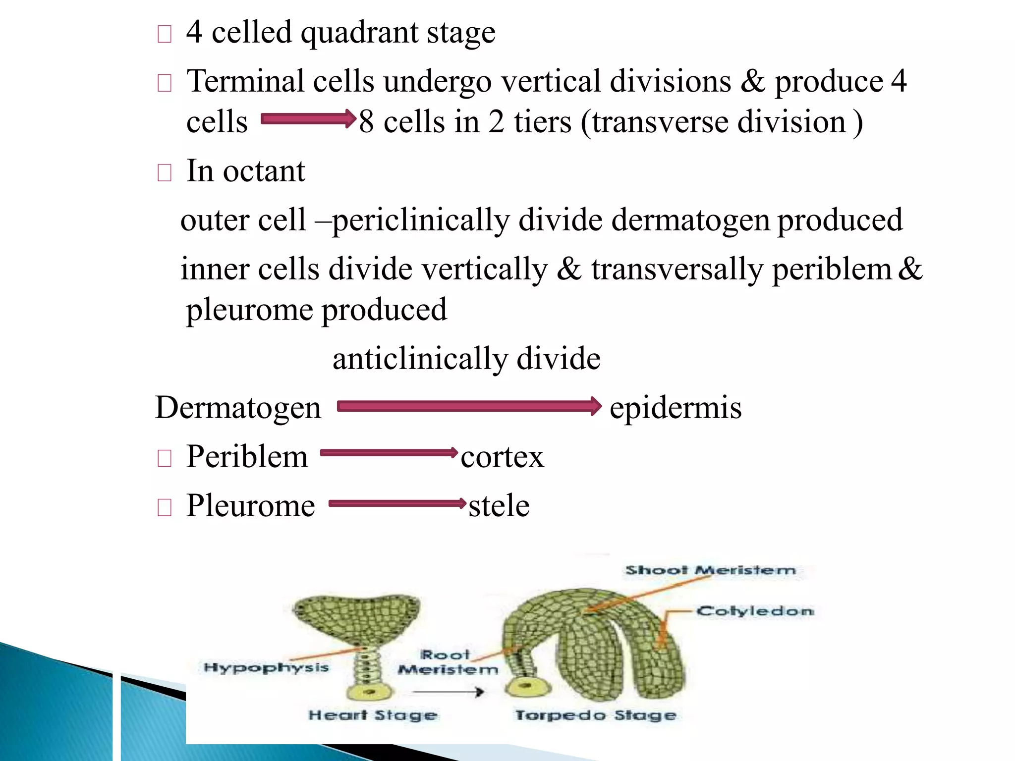
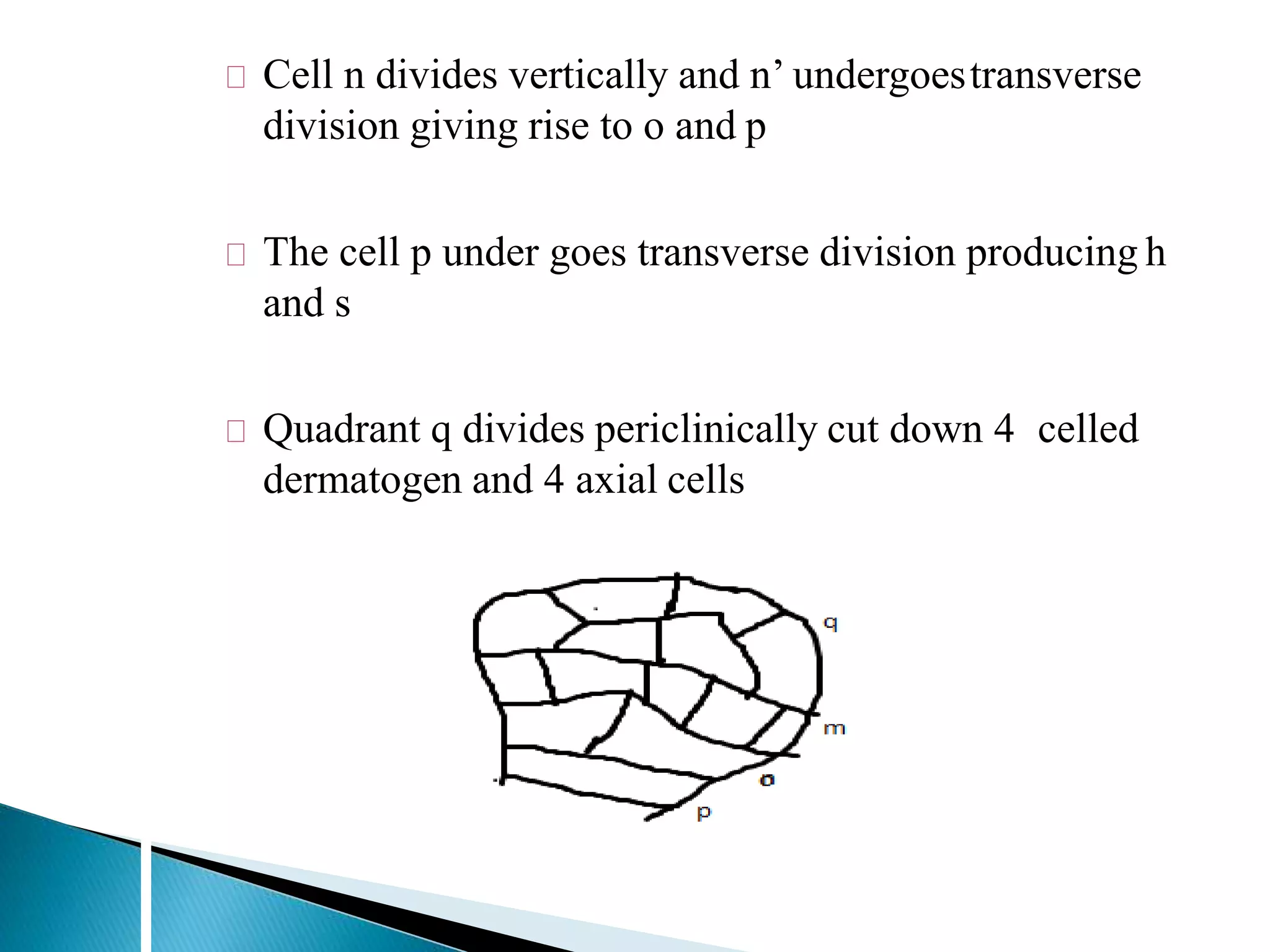
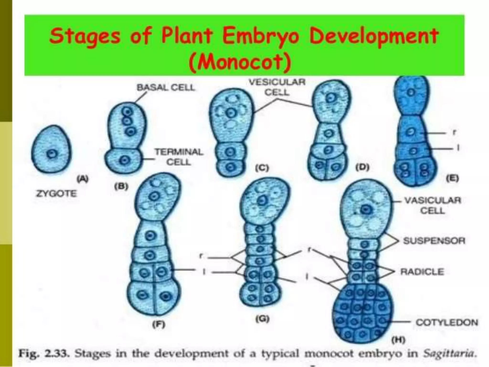
SUBMITTED BY:
A.MOHAMED MANSUR ALI
ROLL.NO: 19PA7763
Zygote
Embryo
Plant
Region of embryonal axis above cotyledons – epicotyl
Epicotyl terminates in plumule (embryonic shoot),
covered by coleorhiza
Region below of embryonal axis below cotyledons
hypocotyl
Hypocotyl terminates in radicle (embryonal root ),
covered by coleoptile
Monocot cotyledon is called scutellum
Increased accumulation of cytoplasm ,
dictyosomes , ribosomes on the chalazal
end where 1st division will takes place
Proembryo – from the 2 celled stage
to untill the initiation of organs
Apical cell of pro
embryo divides
transversely
Basal cell role
minor or none
Solanad type caryophyllad
type
Apical & basal
cell divides
Chenopodiad
type
Johansen said 6th type – piperad type – loranthaceae
, piperaceae , first division of zygote is vertical
ONAGRAD TYPE – capsellabursa pastoris
brassicaceae / cruciferaceae
4 celled quadrant stage
Hanstein in 1870
4 celled quadrant stage
Terminal cells undergo vertical divisions & produce 4
cells 8 cells in 2 tiers (transverse division )
In octant
outer cell –periclinically divide dermatogen produced
inner cells divide vertically & transversally periblem&
pleurome produced
anticlinically divide
Dermatogen epidermis
Periblem cortex
Pleurome stele
Filament tube structure
The basal cells divides transversly
& produce 6- 10 cells
The terminal cells of suspensor
enlarges and form haustoria( help
to get the nutrition for the
developing embryo)
The lower most cell in the
suspensor called hypophysis
The hypophysis undergo 2 vertical
divisions & produce 8 cells in 2
tiers
Then the upper tier produces root
cap & root epidermis , the lower
tier produces root cortex
Illustrated by swami & lakshman in 1962
Complete embryo from apical cell
Basal cell to haustoria
Apical cell divides transversally to form c and d
D divides transversally form m and ci- linear proembryo
formed
2 vertical divisions in the sense c and
m lead to the formation of two tiers of 4
cells each q & m
In the mean time ci divides transversly
to give rise to n and n’
Cell n divides vertically and n’ undergoestransverse
division giving rise to o and p
The cell p under goes transverse division producing h
and s
Quadrant q divides periclinically cut down 4 celled
dermatogen and 4 axial cells
The rapidly growing portion of the tier q forms the single
cotyledons
And the slow growing tissue derived from 4 th axial cells
give rise to initials of epicotyl
The radicle is organised from the derivatives of n
The cells in the tier m divides by vertical & transverse
division and become 2 tiered , at this stage proembryo is
slightly spherical
Transverse division of m and n tiers - makes
proembryo elongated & become oval
Central core of cells of q,m, &n become pleurome
initials
3 of axial cells divide fastly than 4th one
So symmetry changes and the embryo become notched
Deveopment of embryo in monocot and dicot

Deveopment of embryo in monocot and dicot

  • 1.
    SUBMITTED BY: A.MOHAMED MANSURALI ROLL.NO: 19PA7763
  • 2.
  • 4.
    Region of embryonalaxis above cotyledons – epicotyl Epicotyl terminates in plumule (embryonic shoot), covered by coleorhiza Region below of embryonal axis below cotyledons hypocotyl Hypocotyl terminates in radicle (embryonal root ), covered by coleoptile Monocot cotyledon is called scutellum
  • 5.
    Increased accumulation ofcytoplasm , dictyosomes , ribosomes on the chalazal end where 1st division will takes place Proembryo – from the 2 celled stage to untill the initiation of organs
  • 6.
    Apical cell ofpro embryo divides transversely Basal cell role minor or none Solanad type caryophyllad type Apical & basal cell divides Chenopodiad type
  • 7.
    Johansen said 6thtype – piperad type – loranthaceae , piperaceae , first division of zygote is vertical
  • 8.
    ONAGRAD TYPE –capsellabursa pastoris brassicaceae / cruciferaceae 4 celled quadrant stage Hanstein in 1870
  • 9.
    4 celled quadrantstage Terminal cells undergo vertical divisions & produce 4 cells 8 cells in 2 tiers (transverse division ) In octant outer cell –periclinically divide dermatogen produced inner cells divide vertically & transversally periblem& pleurome produced anticlinically divide Dermatogen epidermis Periblem cortex Pleurome stele
  • 11.
    Filament tube structure Thebasal cells divides transversly & produce 6- 10 cells The terminal cells of suspensor enlarges and form haustoria( help to get the nutrition for the developing embryo) The lower most cell in the suspensor called hypophysis The hypophysis undergo 2 vertical divisions & produce 8 cells in 2 tiers Then the upper tier produces root cap & root epidermis , the lower tier produces root cortex
  • 12.
    Illustrated by swami& lakshman in 1962 Complete embryo from apical cell Basal cell to haustoria Apical cell divides transversally to form c and d D divides transversally form m and ci- linear proembryo formed
  • 13.
    2 vertical divisionsin the sense c and m lead to the formation of two tiers of 4 cells each q & m In the mean time ci divides transversly to give rise to n and n’
  • 14.
    Cell n dividesvertically and n’ undergoestransverse division giving rise to o and p The cell p under goes transverse division producing h and s Quadrant q divides periclinically cut down 4 celled dermatogen and 4 axial cells
  • 15.
    The rapidly growingportion of the tier q forms the single cotyledons And the slow growing tissue derived from 4 th axial cells give rise to initials of epicotyl The radicle is organised from the derivatives of n
  • 16.
    The cells inthe tier m divides by vertical & transverse division and become 2 tiered , at this stage proembryo is slightly spherical Transverse division of m and n tiers - makes proembryo elongated & become oval Central core of cells of q,m, &n become pleurome initials 3 of axial cells divide fastly than 4th one So symmetry changes and the embryo become notched