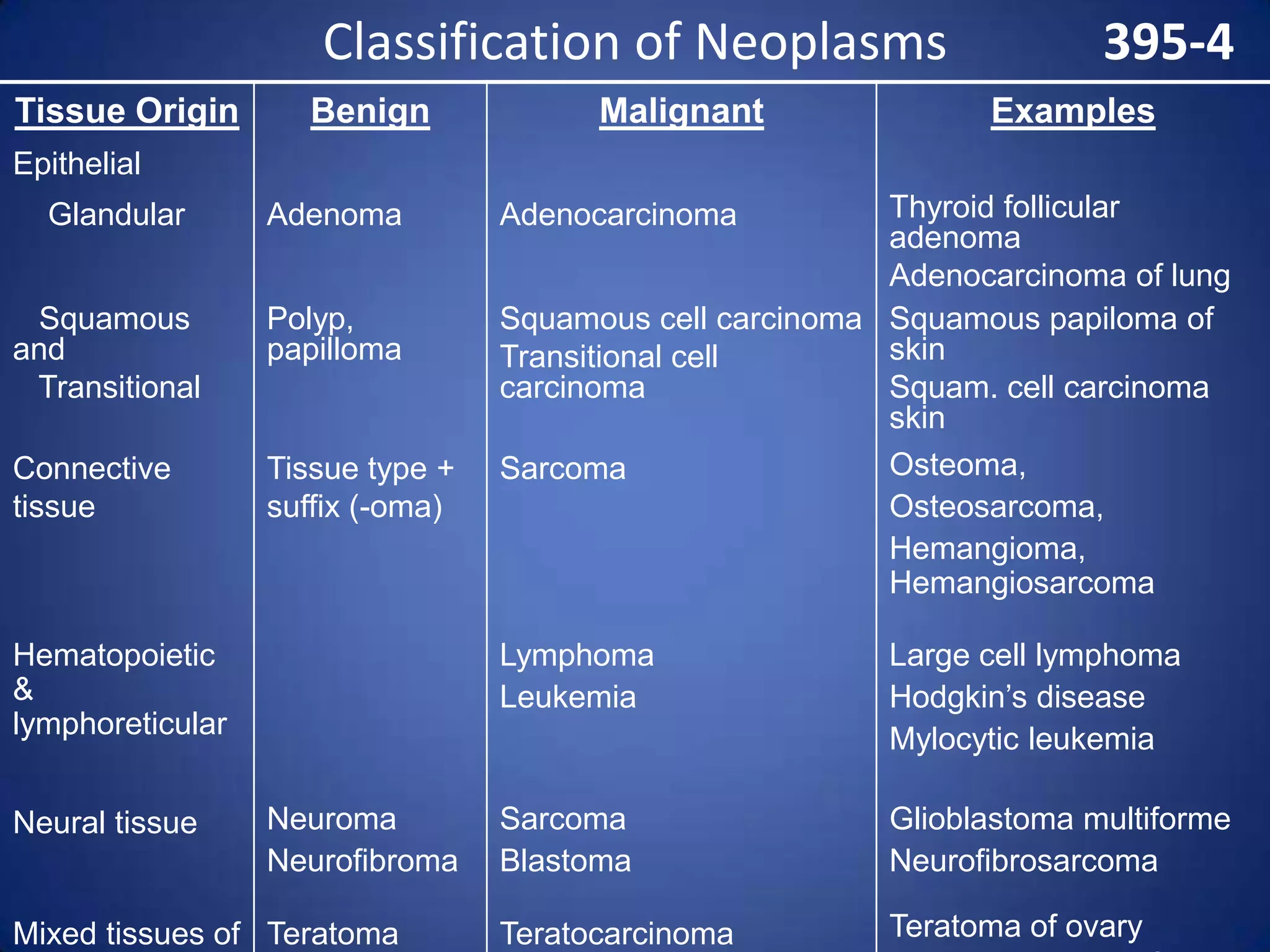
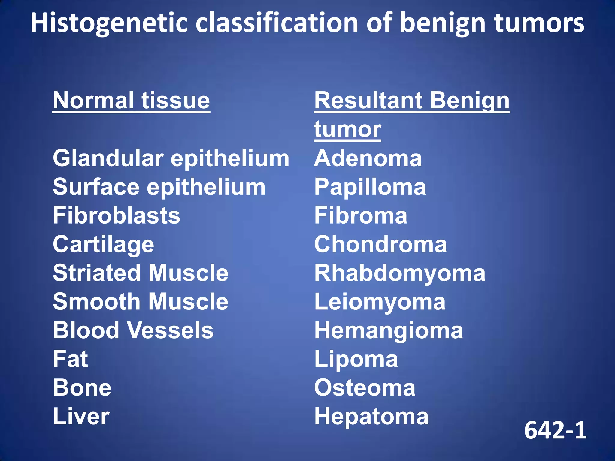
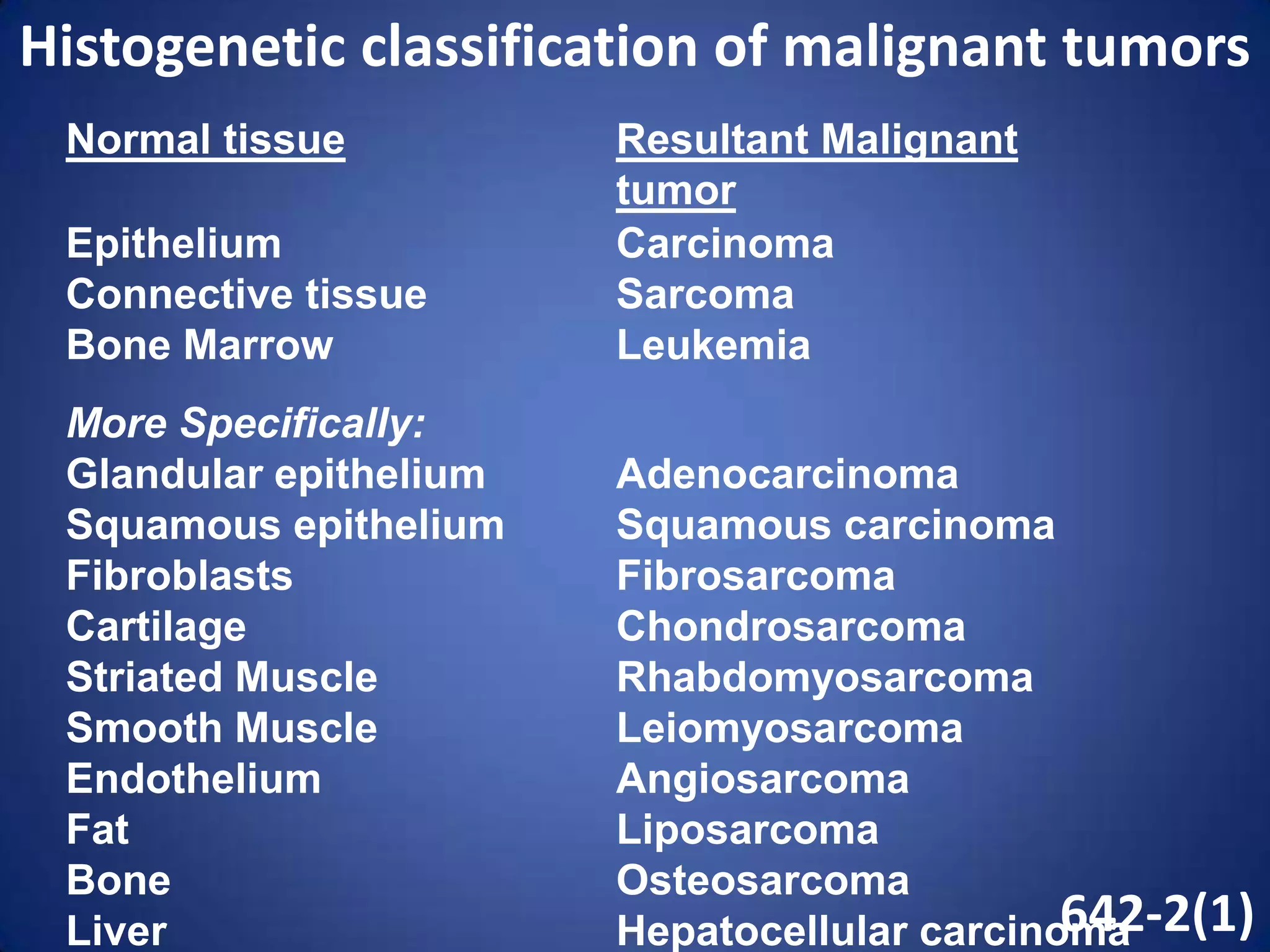
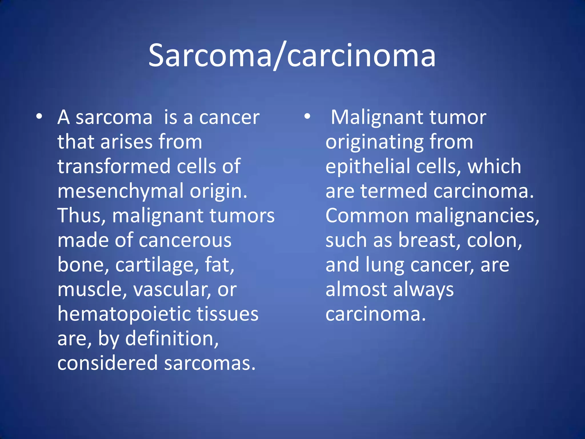
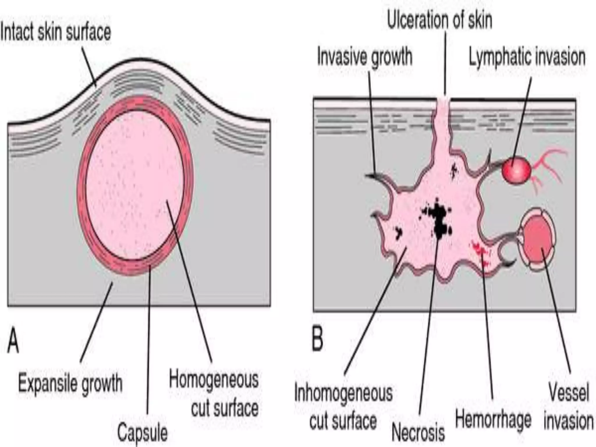
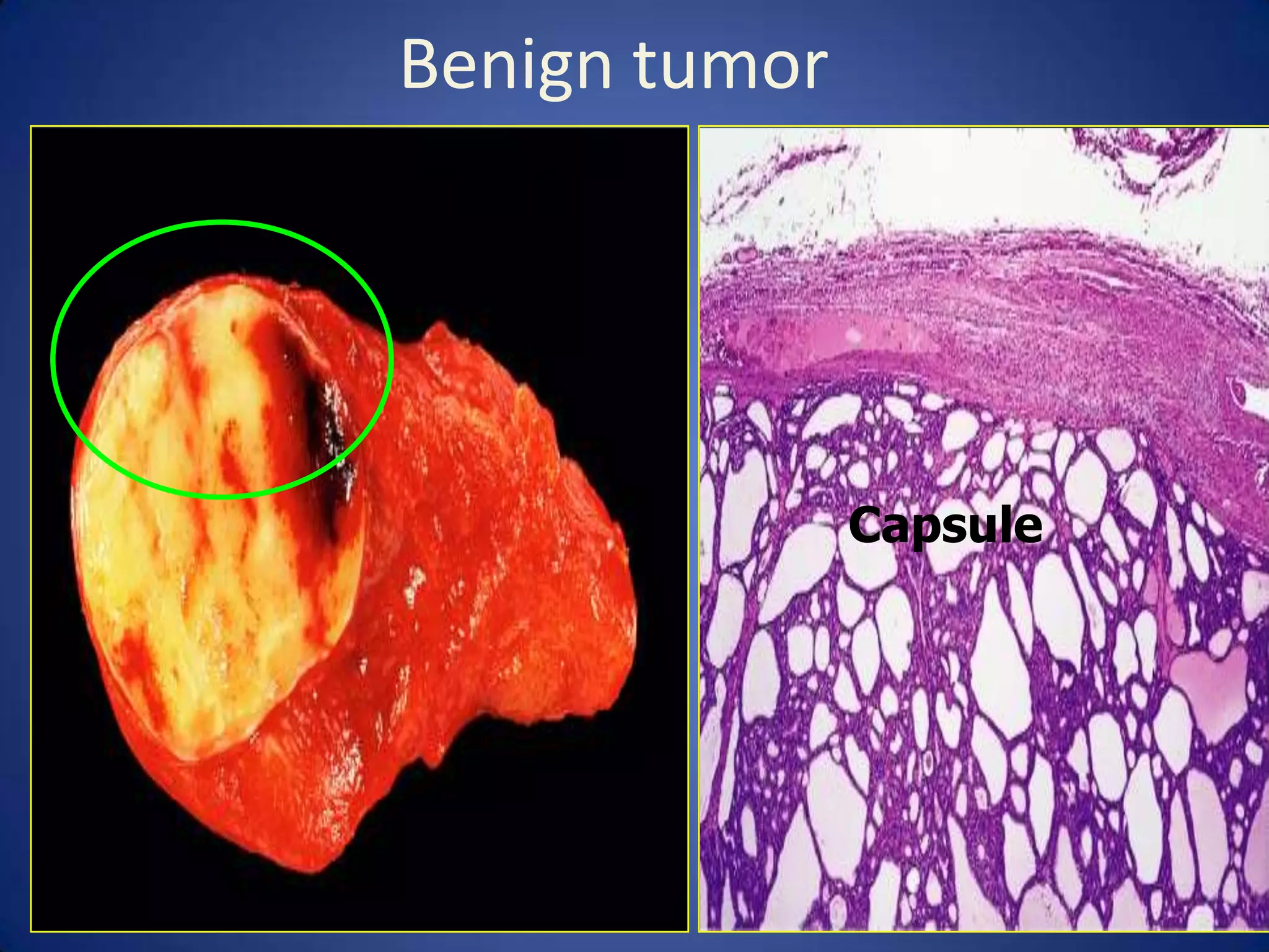
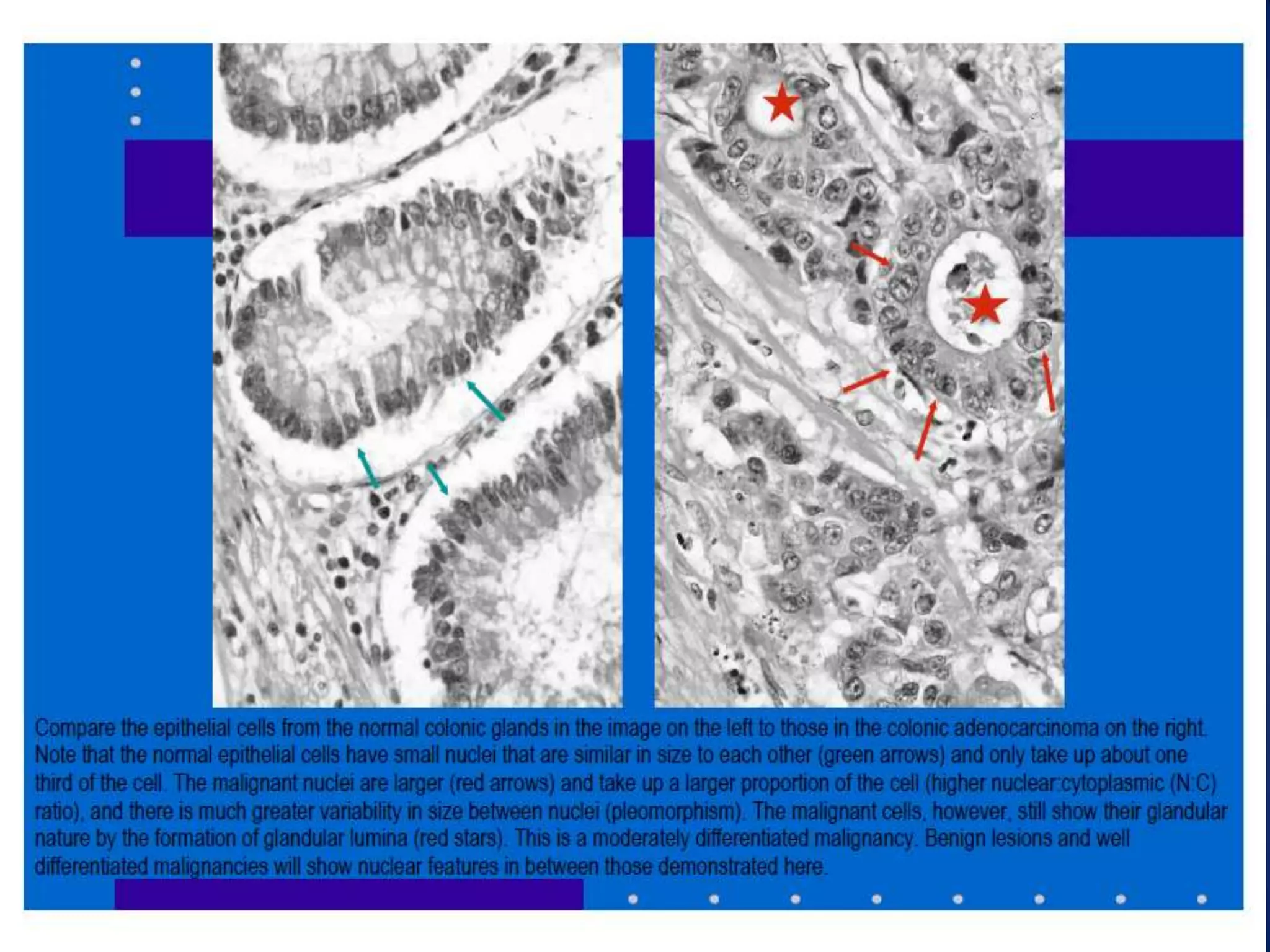
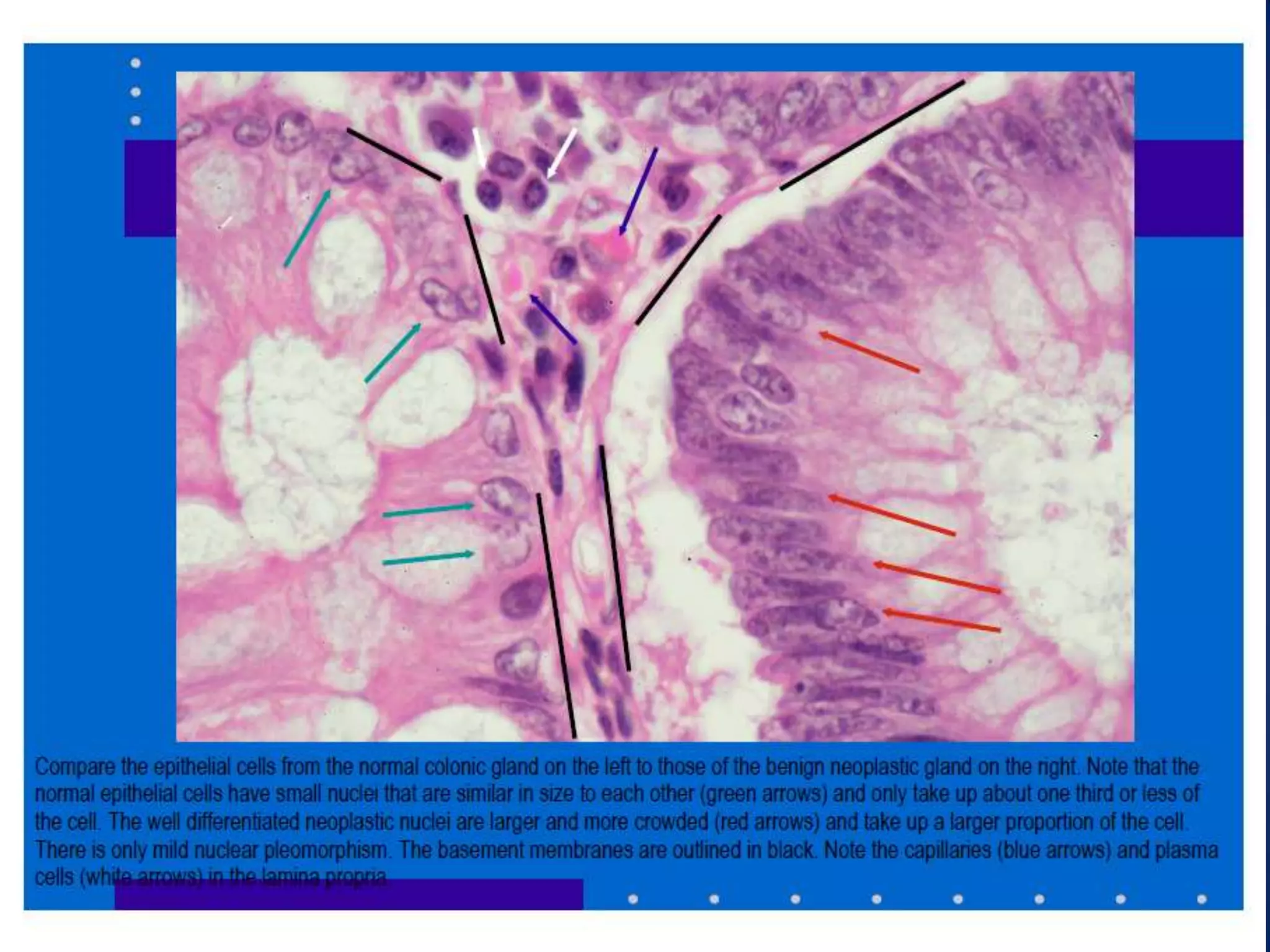
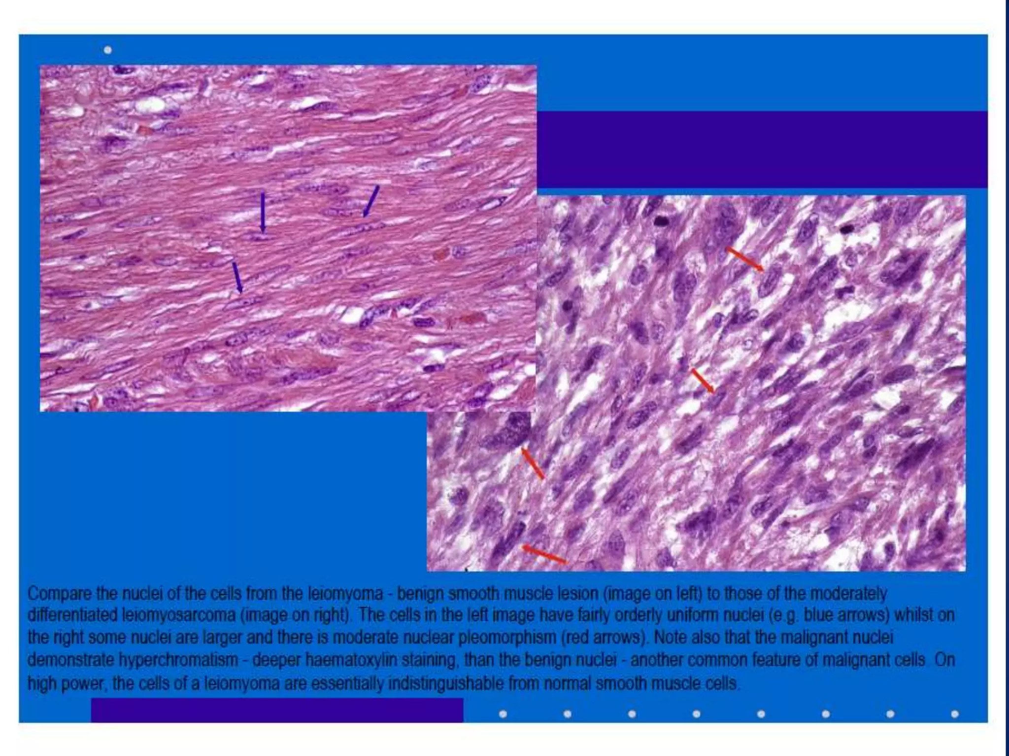
Oncology
• The term oncology literally means a branch of
science that deals with tumours and cancers.
The word “onco” means bulk, mass, or tumor
while “-logy” means study.
• Neoplasm (from ancient Greek neo-, "new" +
plasma, "formation", "creation") or tumor is
an abnormal mass of tissue as a result of
abnormal growth or division of cells.
Classification of Neoplasms
Tissue Origin

Benign

Malignant

395-4
Examples

Epithelial
Glandular

Adenoma

Squamous
and
Transitional

Polyp,
papilloma

Connective
tissue

Tissue type +
suffix (-oma)

Hematopoietic
&
lymphoreticular
Neural tissue

Thyroid follicular
adenoma
Adenocarcinoma of lung
Squamous cell carcinoma Squamous papiloma of
skin
Transitional cell
carcinoma
Squam. cell carcinoma
skin
Osteoma,
Sarcoma
Osteosarcoma,
Hemangioma,
Hemangiosarcoma
Adenocarcinoma

Lymphoma
Leukemia

Neuroma
Neurofibroma

Mixed tissues of Teratoma

Large cell lymphoma
Hodgkin’s disease
Mylocytic leukemia

Sarcoma
Blastoma

Glioblastoma multiforme
Neurofibrosarcoma

Teratocarcinoma

Teratoma of ovary
Histogenetic classification of benign tumors
Normal tissue

Resultant Benign
tumor
Glandular epithelium Adenoma
Surface epithelium
Papilloma
Fibroblasts
Fibroma
Cartilage
Chondroma
Striated Muscle
Rhabdomyoma
Smooth Muscle
Leiomyoma
Blood Vessels
Hemangioma
Fat
Lipoma
Bone
Osteoma
Liver
Hepatoma

642-1
Histogenetic classification of malignant tumors
Normal tissue
Epithelium
Connective tissue
Bone Marrow

Resultant Malignant
tumor
Carcinoma
Sarcoma
Leukemia

More Specifically:
Glandular epithelium
Squamous epithelium
Fibroblasts
Cartilage
Striated Muscle
Smooth Muscle
Endothelium
Fat
Bone
Liver

Adenocarcinoma
Squamous carcinoma
Fibrosarcoma
Chondrosarcoma
Rhabdomyosarcoma
Leiomyosarcoma
Angiosarcoma
Liposarcoma
Osteosarcoma
642-2(1)
Hepatocellular carcinoma
Sarcoma/carcinoma
• A sarcoma is a cancer
that arises from
transformed cells of
mesenchymal origin.
Thus, malignant tumors
made of cancerous
bone, cartilage, fat,
muscle, vascular, or
hematopoietic tissues
are, by definition,
considered sarcomas.

• Malignant tumor
originating from
epithelial cells, which
are termed carcinoma.
Common malignancies,
such as breast, colon,
and lung cancer, are
almost always
carcinoma.
Charateristics of neoplastic cells
 Abnormal size and irregular shape of cells.
 Nuclei
Nuclei increase in size
 Nucleolus (a) often prominent (b) sometimes multiple
(c) Atypical staining
 Numerous mitotic figures
 May be multiple----------tumour giant cells
 Varying amount of cytoplasm (a) generally cytoplasm
decrease in amount (b) fibrillar appearance with few
secretory granules (c) basophilic in nature
 Cytoplasm to nucleus ratio is lost
 disturbed polarity of cells
Microscopic structure of tomour
 Parenchyma; cells make up of the tumour
 Cells of abnormal structure
 deviation from normal is least in benign, somewhat
resemble normal tissue and marked in malignant
 arrangement of tumour cells
1. Epithelial tumours (a) sheets or fronds in superficial
epithelium (b) clumps,columns or acini within a tumour
mass (c) Each cell fastened directly to the next.
2. Connective tissue tumours (a) Tumour cells lie singly,may
arrange in whorls or bundles (b) Each cell is separated
from the next by the intercellular substances
3. Hemopoitic tumours (a) tumour cells form (b) neoplastic
cells closely packed but remain discrete.
Microscopic structure of tomour
 Stroma;Support and nourishes the tumour cells
 Amount is variable
 least in histoid tumours(A type of connective tumor that is
composed of a single type of differentiated tissue)
 Reasonable in organoid tumours(A tumor that is glandular
in origin and that contains epithelium, connective tissue,
and other tissue structures that give it a complex structure
similar to an organ)
 May be compact or edematous
 Blood vessels vary in appearance
 Tumour cells do not require innervation
Benign tumor

Capsule
Smooth muscles
Here is an osteosarcoma of bone. The large, bulky mass
arises in the cortex of the bone and extends outward
This is an example of metastases to the liver, tan-white
masses are multiple and irregularly sized.
Mitotic figures
What is the significance of mitotic
figures in a neoplasm?
• In general, their appearance suggests a higher
rate of cellular proliferation. Mitoses certainly
are present in normal tissues (surface
epithelia are constantly regenerating, and
hematopoiesis produces billions of new blood
cells each day). However, the presence of
mitoses, and particularly abnormal mitoses, in
a mass lesion supports a diagnosis of
neoplasia, and likely a malignant neoplasm.
• Hepatocellular carcinoma cells. Characterized by large anaplastic carcinoma
cells with eosinophilic cytoplasm, large hyperchromatic nuclei and
prominent nucleoli. The normal trabecular structure of the liver is distorted.
Neoplasia lecture
Neoplasia lecture

Neoplasia lecture

  • 1.
    Oncology • The termoncology literally means a branch of science that deals with tumours and cancers. The word “onco” means bulk, mass, or tumor while “-logy” means study. • Neoplasm (from ancient Greek neo-, "new" + plasma, "formation", "creation") or tumor is an abnormal mass of tissue as a result of abnormal growth or division of cells.
  • 2.
    Classification of Neoplasms TissueOrigin Benign Malignant 395-4 Examples Epithelial Glandular Adenoma Squamous and Transitional Polyp, papilloma Connective tissue Tissue type + suffix (-oma) Hematopoietic & lymphoreticular Neural tissue Thyroid follicular adenoma Adenocarcinoma of lung Squamous cell carcinoma Squamous papiloma of skin Transitional cell carcinoma Squam. cell carcinoma skin Osteoma, Sarcoma Osteosarcoma, Hemangioma, Hemangiosarcoma Adenocarcinoma Lymphoma Leukemia Neuroma Neurofibroma Mixed tissues of Teratoma Large cell lymphoma Hodgkin’s disease Mylocytic leukemia Sarcoma Blastoma Glioblastoma multiforme Neurofibrosarcoma Teratocarcinoma Teratoma of ovary
  • 3.
    Histogenetic classification ofbenign tumors Normal tissue Resultant Benign tumor Glandular epithelium Adenoma Surface epithelium Papilloma Fibroblasts Fibroma Cartilage Chondroma Striated Muscle Rhabdomyoma Smooth Muscle Leiomyoma Blood Vessels Hemangioma Fat Lipoma Bone Osteoma Liver Hepatoma 642-1
  • 4.
    Histogenetic classification ofmalignant tumors Normal tissue Epithelium Connective tissue Bone Marrow Resultant Malignant tumor Carcinoma Sarcoma Leukemia More Specifically: Glandular epithelium Squamous epithelium Fibroblasts Cartilage Striated Muscle Smooth Muscle Endothelium Fat Bone Liver Adenocarcinoma Squamous carcinoma Fibrosarcoma Chondrosarcoma Rhabdomyosarcoma Leiomyosarcoma Angiosarcoma Liposarcoma Osteosarcoma 642-2(1) Hepatocellular carcinoma
  • 5.
    Sarcoma/carcinoma • A sarcomais a cancer that arises from transformed cells of mesenchymal origin. Thus, malignant tumors made of cancerous bone, cartilage, fat, muscle, vascular, or hematopoietic tissues are, by definition, considered sarcomas. • Malignant tumor originating from epithelial cells, which are termed carcinoma. Common malignancies, such as breast, colon, and lung cancer, are almost always carcinoma.
  • 6.
    Charateristics of neoplasticcells  Abnormal size and irregular shape of cells.  Nuclei Nuclei increase in size  Nucleolus (a) often prominent (b) sometimes multiple (c) Atypical staining  Numerous mitotic figures  May be multiple----------tumour giant cells  Varying amount of cytoplasm (a) generally cytoplasm decrease in amount (b) fibrillar appearance with few secretory granules (c) basophilic in nature  Cytoplasm to nucleus ratio is lost  disturbed polarity of cells
  • 7.
    Microscopic structure oftomour  Parenchyma; cells make up of the tumour  Cells of abnormal structure  deviation from normal is least in benign, somewhat resemble normal tissue and marked in malignant  arrangement of tumour cells 1. Epithelial tumours (a) sheets or fronds in superficial epithelium (b) clumps,columns or acini within a tumour mass (c) Each cell fastened directly to the next. 2. Connective tissue tumours (a) Tumour cells lie singly,may arrange in whorls or bundles (b) Each cell is separated from the next by the intercellular substances 3. Hemopoitic tumours (a) tumour cells form (b) neoplastic cells closely packed but remain discrete.
  • 8.
    Microscopic structure oftomour  Stroma;Support and nourishes the tumour cells  Amount is variable  least in histoid tumours(A type of connective tumor that is composed of a single type of differentiated tissue)  Reasonable in organoid tumours(A tumor that is glandular in origin and that contains epithelium, connective tissue, and other tissue structures that give it a complex structure similar to an organ)  May be compact or edematous  Blood vessels vary in appearance  Tumour cells do not require innervation
  • 12.
  • 15.
  • 17.
    Here is anosteosarcoma of bone. The large, bulky mass arises in the cortex of the bone and extends outward
  • 18.
    This is anexample of metastases to the liver, tan-white masses are multiple and irregularly sized.
  • 23.
  • 24.
    What is thesignificance of mitotic figures in a neoplasm? • In general, their appearance suggests a higher rate of cellular proliferation. Mitoses certainly are present in normal tissues (surface epithelia are constantly regenerating, and hematopoiesis produces billions of new blood cells each day). However, the presence of mitoses, and particularly abnormal mitoses, in a mass lesion supports a diagnosis of neoplasia, and likely a malignant neoplasm.
  • 26.
    • Hepatocellular carcinomacells. Characterized by large anaplastic carcinoma cells with eosinophilic cytoplasm, large hyperchromatic nuclei and prominent nucleoli. The normal trabecular structure of the liver is distorted.