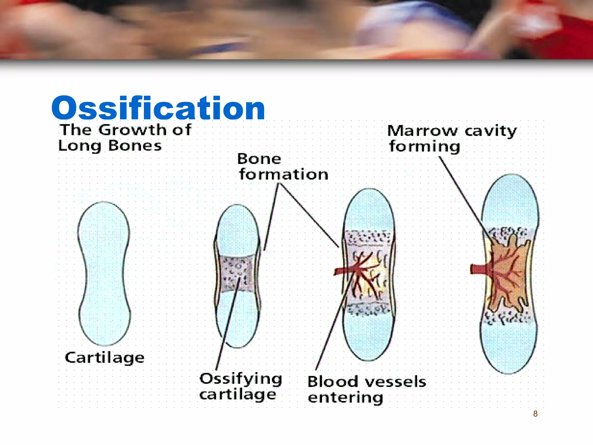
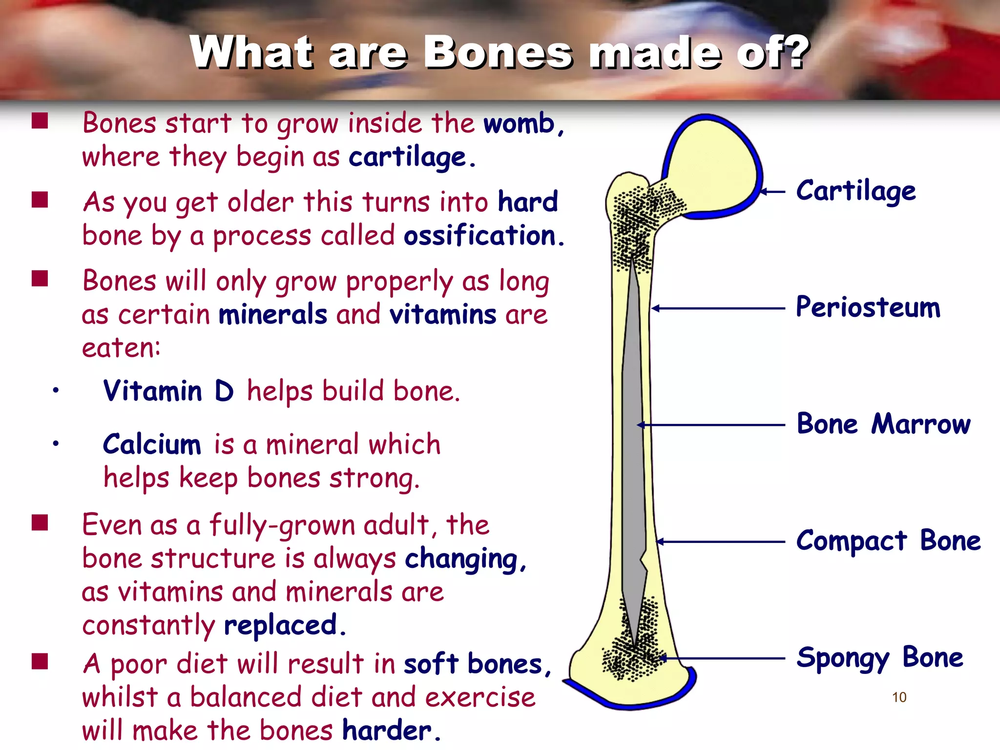
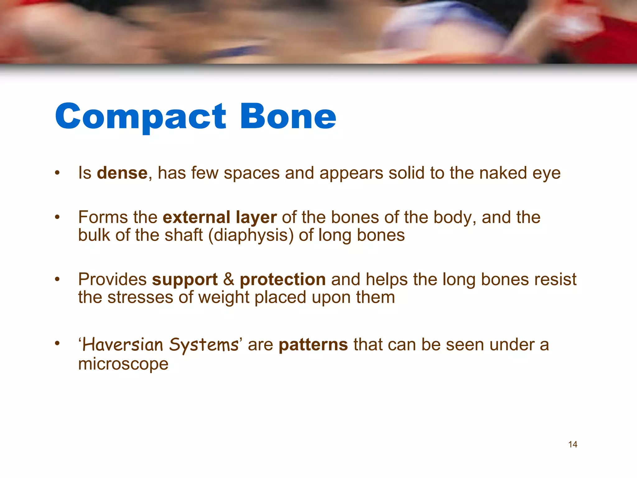
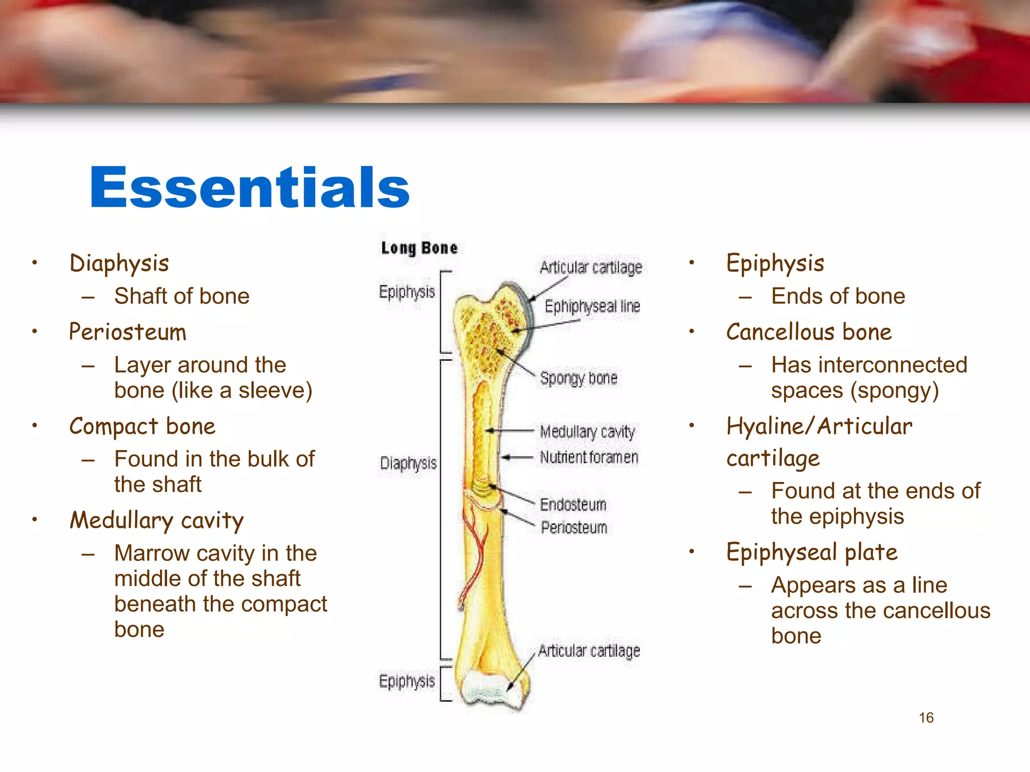
Principles of Anatomy and Physiology in Sport Session 2 – The Skeletal System
Re-Cap Last Session Major bones of the body Axial/Appendicular skeleton Vertebral column - sections
Drag the labels to the correct place on the skeleton... The Skeletal System
Aim  To complete assignment 1 Objectives To be able to describe the functions of the skeletal system To be able to describe the structure of a typical long bone To explain the differences between compact and cancellous bone Aims & Objectives
Activity  With your partner work together to list and briefly describe the 6 functions of the skeleton
Functions of the Skeleton  3. Blood Production Red blood cells are made in the ribs and limb bones. 1.Protection The rib cage protects the delicate heart and lungs. 6. Movement   The bones and joints work with muscles to enable us to walk, run and sprint. 5. Support   The vertebrae support the head. 4. Calcium Storage   Bone is the largest supply of calcium 1. Protection The cranium protects the soft tissue of the brain. 2. Shape   Without the skeleton, the  body would be  flabby and  shapeless.
  Bone Structure Bone is the hardest tissue in the body Compact Bone Found at the  shaft  or diaphysis of long bones External layer  of bone that copes with the stresses of PA Cancellous Bone Referred to as the  spongy  part of a bone Found  under  the external layer of compact bone Bone marrow  is found in this part of the bone
Ossification
Ossification
Bones start to grow inside the  womb,  where they begin as  cartilage. Cartilage Periosteum Bone   Marrow Spongy   Bone Compact   Bone What are Bones made of? Vitamin   D   helps build bone. Calcium   is a mineral which helps keep bones strong. Even as a fully-grown adult, the bone structure is always  changing,  as vitamins and minerals are constantly  replaced. As you get older this turns into  hard  bone by a process called  ossification. Bones will only grow properly as long as certain  minerals  and  vitamins  are eaten: A poor diet will result in  soft   bones,  whilst a balanced diet and exercise will make the bones  harder.
Structure of a Long Bone Consists of a  diaphysis  (shaft) with a bulge called the  epiphysis  at either end Longitudinal  growth occurs between these structures The  medullary  or marrow cavity is situated in middle of the shaft beneath the compact bone; it contains either  red  or  yellow  marrow
Bone Marrow Age is a dependant factor on the colour of the marrow. At birth all bones are filled with red bone marrow which produces both  red & white blood cells With age there is a progressive replacement of the red marrow by that of yellow marrow, which is  fatty  and  non-blood producing
Periosteum Each bone is enclosed in a dense layer of fibrous tissue known as  periosteum There are 2 layers Outer layer Blood vessels & nerves Inner layer Responsible for bone development Bones change in size & shape throughout life and they are  denser  in males
Compact Bone Is  dense , has few spaces and appears solid to the naked eye Forms the  external layer  of the bones of the body, and the bulk of the shaft (diaphysis) of long bones Provides  support  &  protection  and helps the long bones resist the stresses of weight placed upon them ‘ Haversian Systems ’ are  patterns  that can be seen under a microscope
Cancellous Bone Known as  spongy  bone Made up of large  interconnected  spaces, like comic book cheese Is the make up of most flat, short, irregular and the end of long bones It has a  criss cross  structure that gives strength without excess weight, this is known as  trabeculae The spaces between the trabeculae are filled with  red bone marrow
Essentials Diaphysis Shaft of bone Periosteum Layer around the bone (like a sleeve) Compact bone   Found in the bulk of the shaft Medullary cavity Marrow cavity in the middle of the shaft beneath the compact bone Epiphysis Ends of bone Cancellous bone Has interconnected spaces (spongy) Hyaline/Articular cartilage Found at the ends of the epiphysis Epiphyseal plate Appears as a line across the cancellous bone
Summary Functions of skeleton Ossification Cartilage Periosteum Epiphysis Bone Marrow Compact bone Cancellous bone
Session objectives To be able to describe the functions of the skeletal system To be able to describe the structure of a typical long bone To explain the differences between compact and cancellous bone
Next session  Classifications of bone Functions of each classification of bone

Principles of a+p 1112 session 2 - skeletal system (functions of skeletal system and bone structure)

  • 1.
    Principles of Anatomyand Physiology in Sport Session 2 – The Skeletal System
  • 2.
    Re-Cap Last SessionMajor bones of the body Axial/Appendicular skeleton Vertebral column - sections
  • 3.
    Drag the labelsto the correct place on the skeleton... The Skeletal System
  • 4.
    Aim Tocomplete assignment 1 Objectives To be able to describe the functions of the skeletal system To be able to describe the structure of a typical long bone To explain the differences between compact and cancellous bone Aims & Objectives
  • 5.
    Activity Withyour partner work together to list and briefly describe the 6 functions of the skeleton
  • 6.
    Functions of theSkeleton 3. Blood Production Red blood cells are made in the ribs and limb bones. 1.Protection The rib cage protects the delicate heart and lungs. 6. Movement The bones and joints work with muscles to enable us to walk, run and sprint. 5. Support The vertebrae support the head. 4. Calcium Storage Bone is the largest supply of calcium 1. Protection The cranium protects the soft tissue of the brain. 2. Shape Without the skeleton, the body would be flabby and shapeless.
  • 7.
    BoneStructure Bone is the hardest tissue in the body Compact Bone Found at the shaft or diaphysis of long bones External layer of bone that copes with the stresses of PA Cancellous Bone Referred to as the spongy part of a bone Found under the external layer of compact bone Bone marrow is found in this part of the bone
  • 8.
  • 9.
  • 10.
    Bones start togrow inside the womb, where they begin as cartilage. Cartilage Periosteum Bone Marrow Spongy Bone Compact Bone What are Bones made of? Vitamin D helps build bone. Calcium is a mineral which helps keep bones strong. Even as a fully-grown adult, the bone structure is always changing, as vitamins and minerals are constantly replaced. As you get older this turns into hard bone by a process called ossification. Bones will only grow properly as long as certain minerals and vitamins are eaten: A poor diet will result in soft bones, whilst a balanced diet and exercise will make the bones harder.
  • 11.
    Structure of aLong Bone Consists of a diaphysis (shaft) with a bulge called the epiphysis at either end Longitudinal growth occurs between these structures The medullary or marrow cavity is situated in middle of the shaft beneath the compact bone; it contains either red or yellow marrow
  • 12.
    Bone Marrow Ageis a dependant factor on the colour of the marrow. At birth all bones are filled with red bone marrow which produces both red & white blood cells With age there is a progressive replacement of the red marrow by that of yellow marrow, which is fatty and non-blood producing
  • 13.
    Periosteum Each boneis enclosed in a dense layer of fibrous tissue known as periosteum There are 2 layers Outer layer Blood vessels & nerves Inner layer Responsible for bone development Bones change in size & shape throughout life and they are denser in males
  • 14.
    Compact Bone Is dense , has few spaces and appears solid to the naked eye Forms the external layer of the bones of the body, and the bulk of the shaft (diaphysis) of long bones Provides support & protection and helps the long bones resist the stresses of weight placed upon them ‘ Haversian Systems ’ are patterns that can be seen under a microscope
  • 15.
    Cancellous Bone Knownas spongy bone Made up of large interconnected spaces, like comic book cheese Is the make up of most flat, short, irregular and the end of long bones It has a criss cross structure that gives strength without excess weight, this is known as trabeculae The spaces between the trabeculae are filled with red bone marrow
  • 16.
    Essentials Diaphysis Shaftof bone Periosteum Layer around the bone (like a sleeve) Compact bone Found in the bulk of the shaft Medullary cavity Marrow cavity in the middle of the shaft beneath the compact bone Epiphysis Ends of bone Cancellous bone Has interconnected spaces (spongy) Hyaline/Articular cartilage Found at the ends of the epiphysis Epiphyseal plate Appears as a line across the cancellous bone
  • 17.
    Summary Functions ofskeleton Ossification Cartilage Periosteum Epiphysis Bone Marrow Compact bone Cancellous bone
  • 18.
    Session objectives Tobe able to describe the functions of the skeletal system To be able to describe the structure of a typical long bone To explain the differences between compact and cancellous bone
  • 19.
    Next session Classifications of bone Functions of each classification of bone