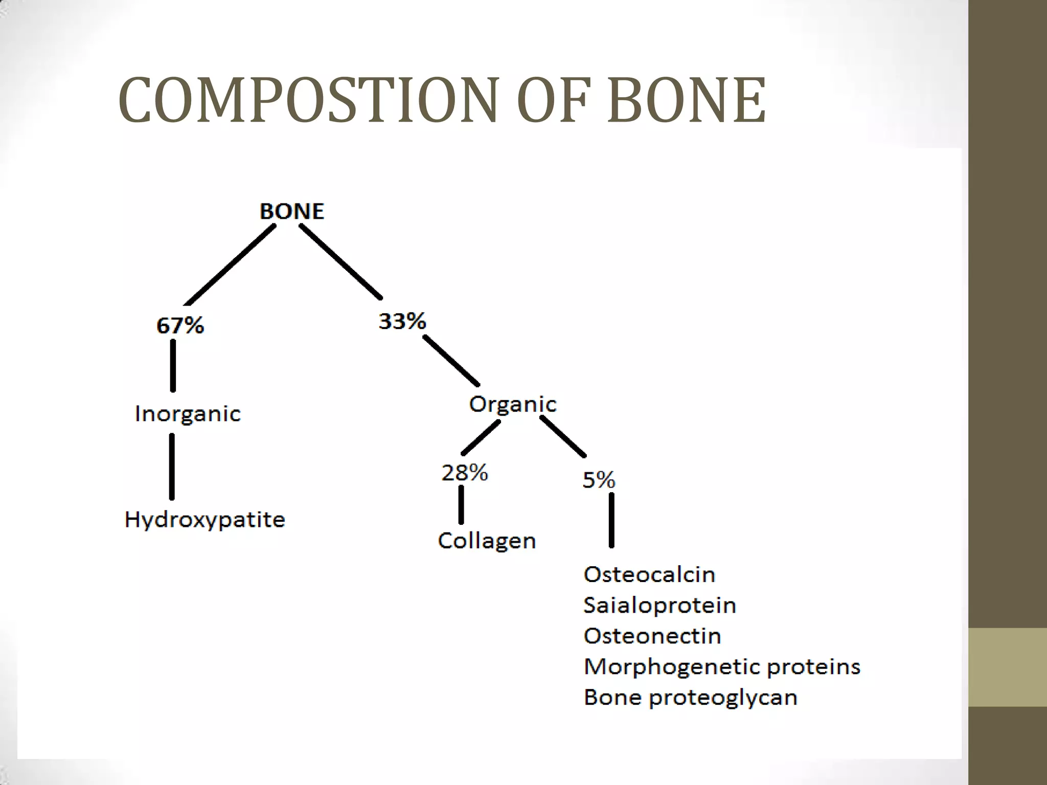
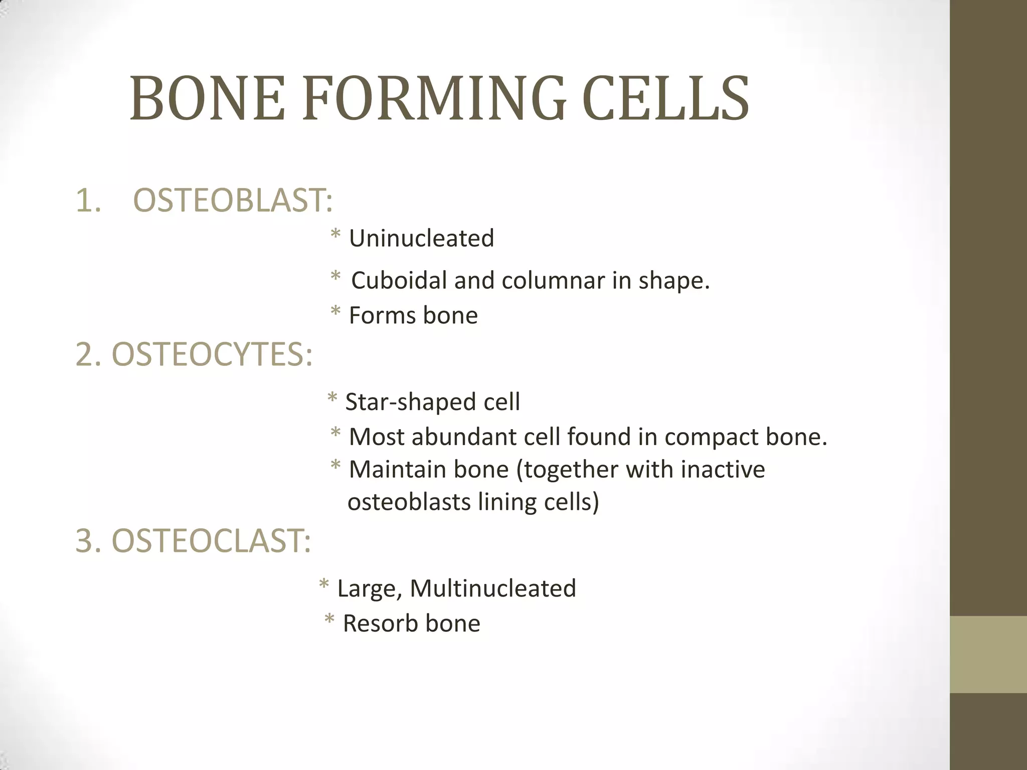
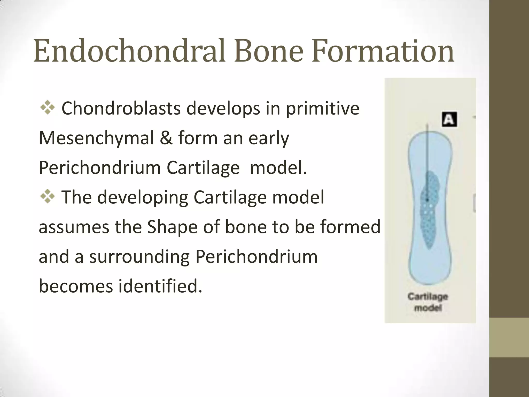
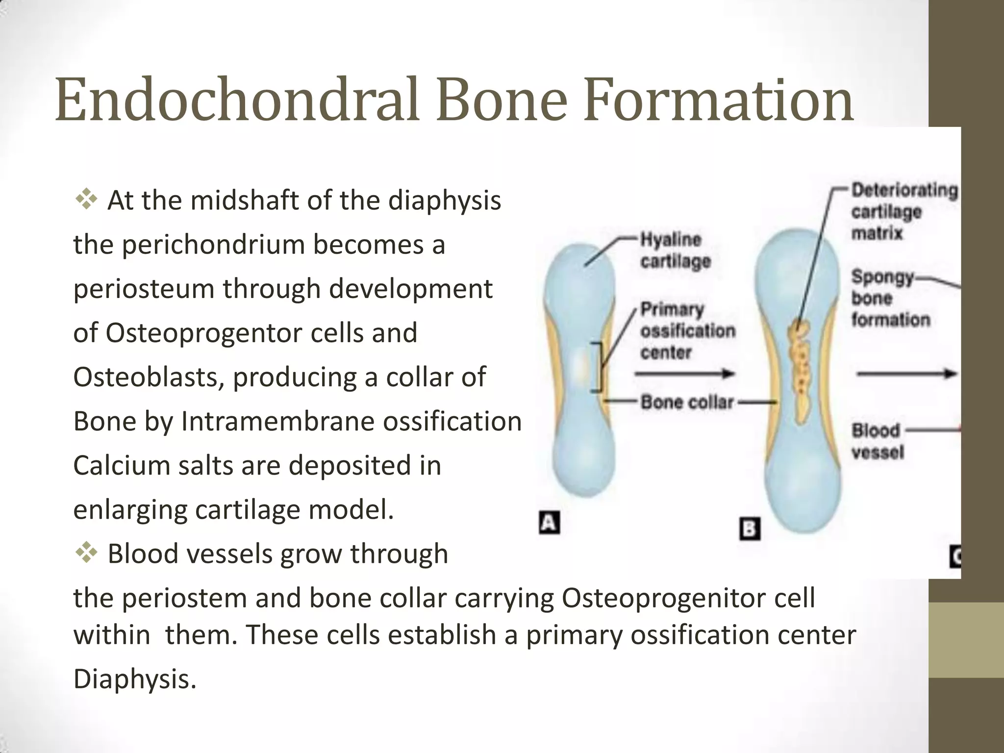
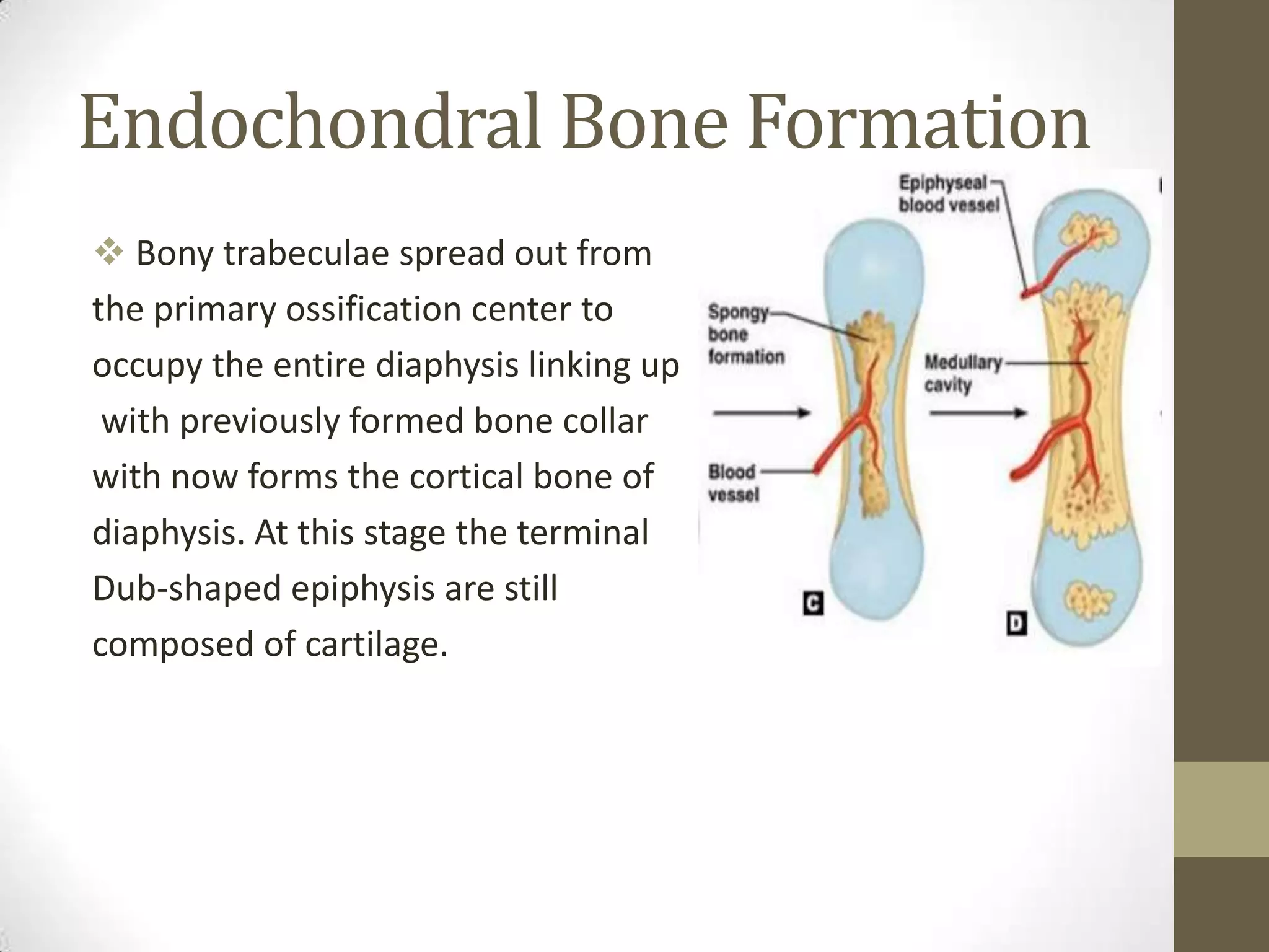
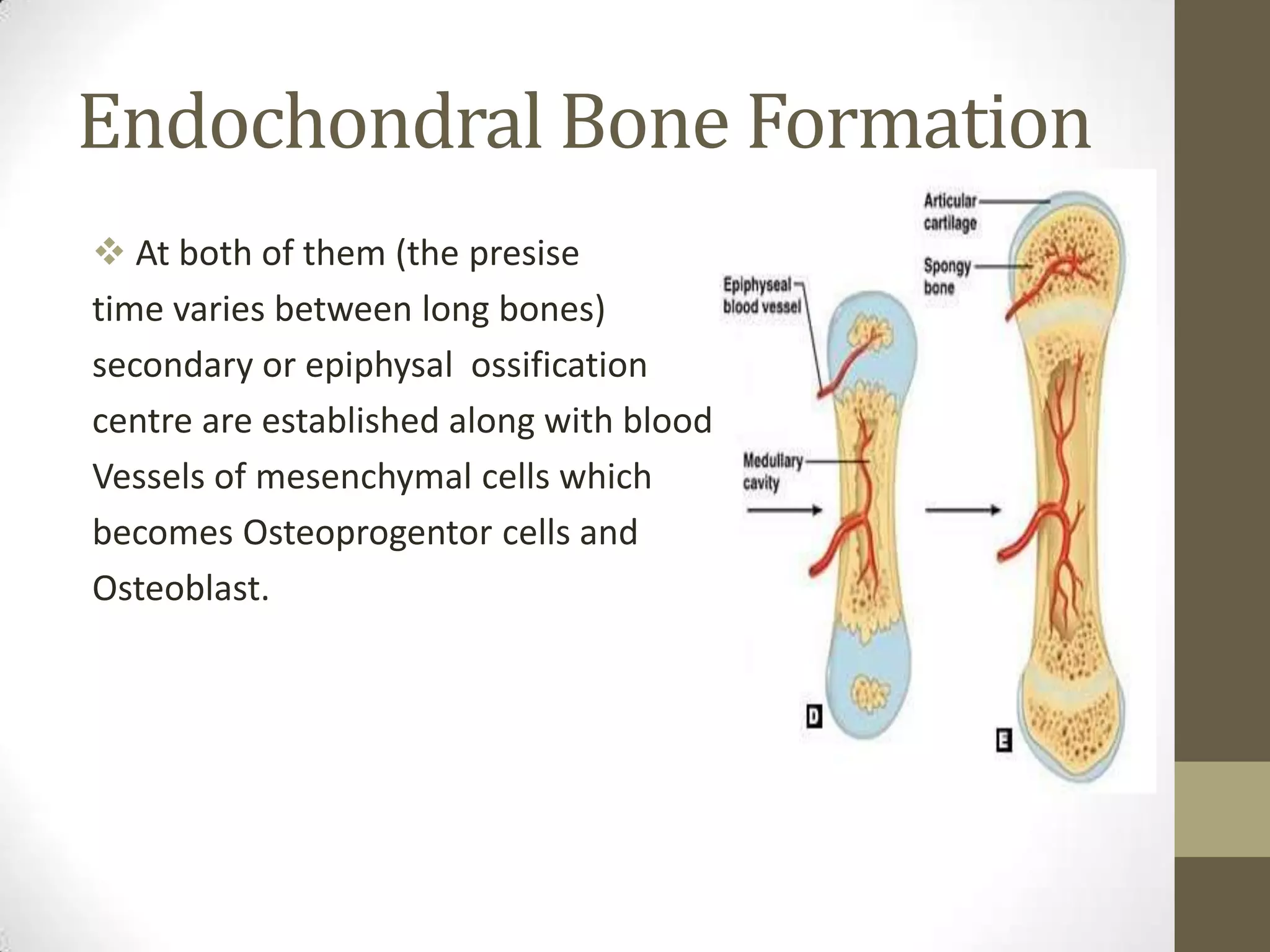
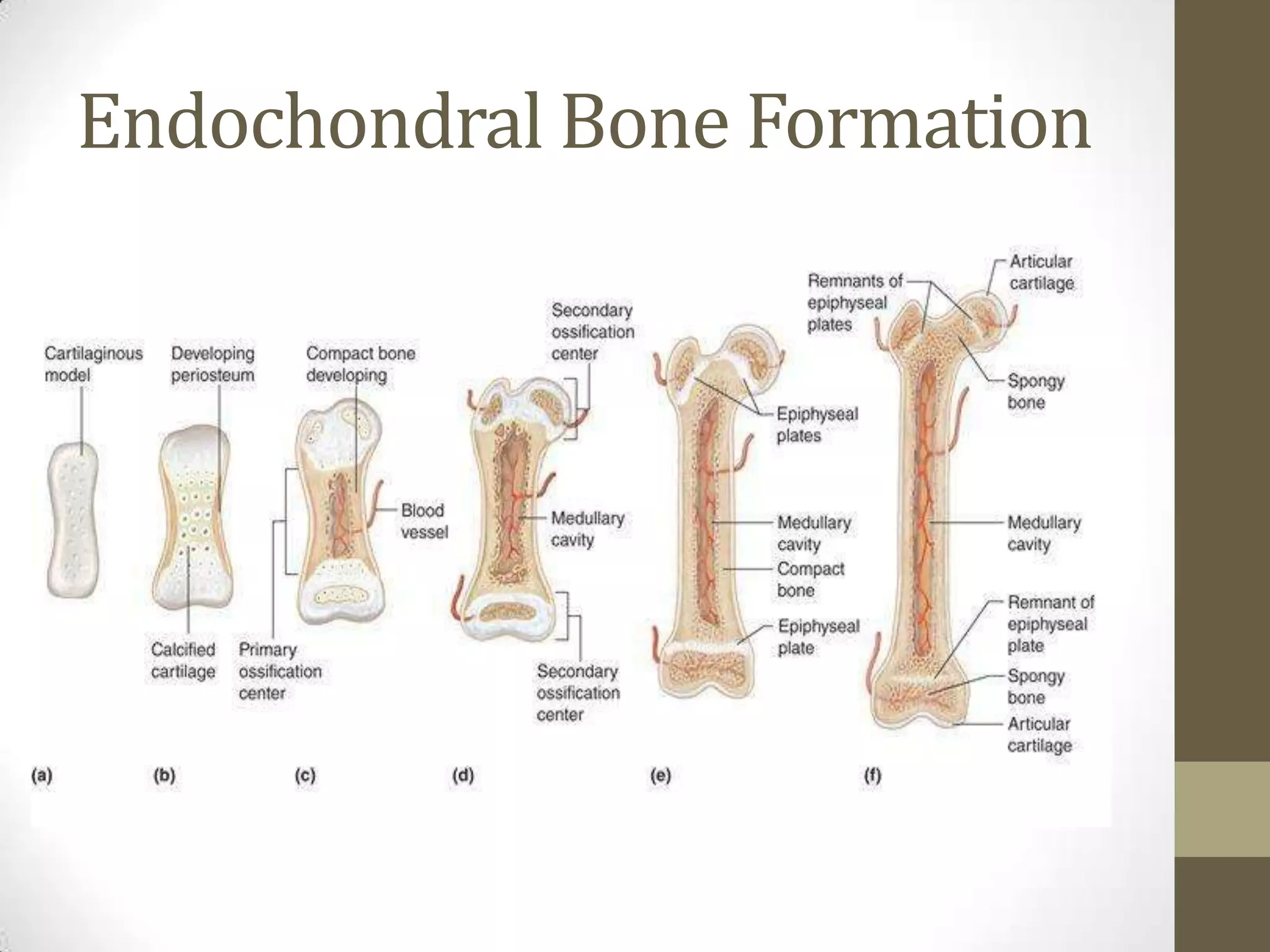
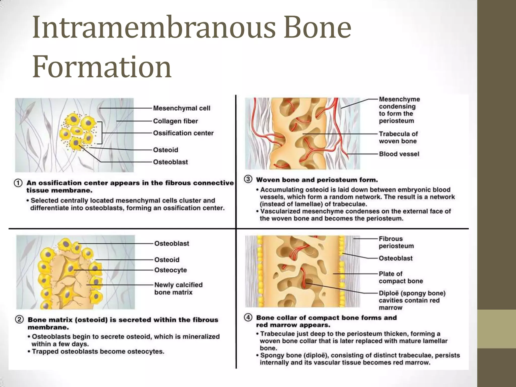
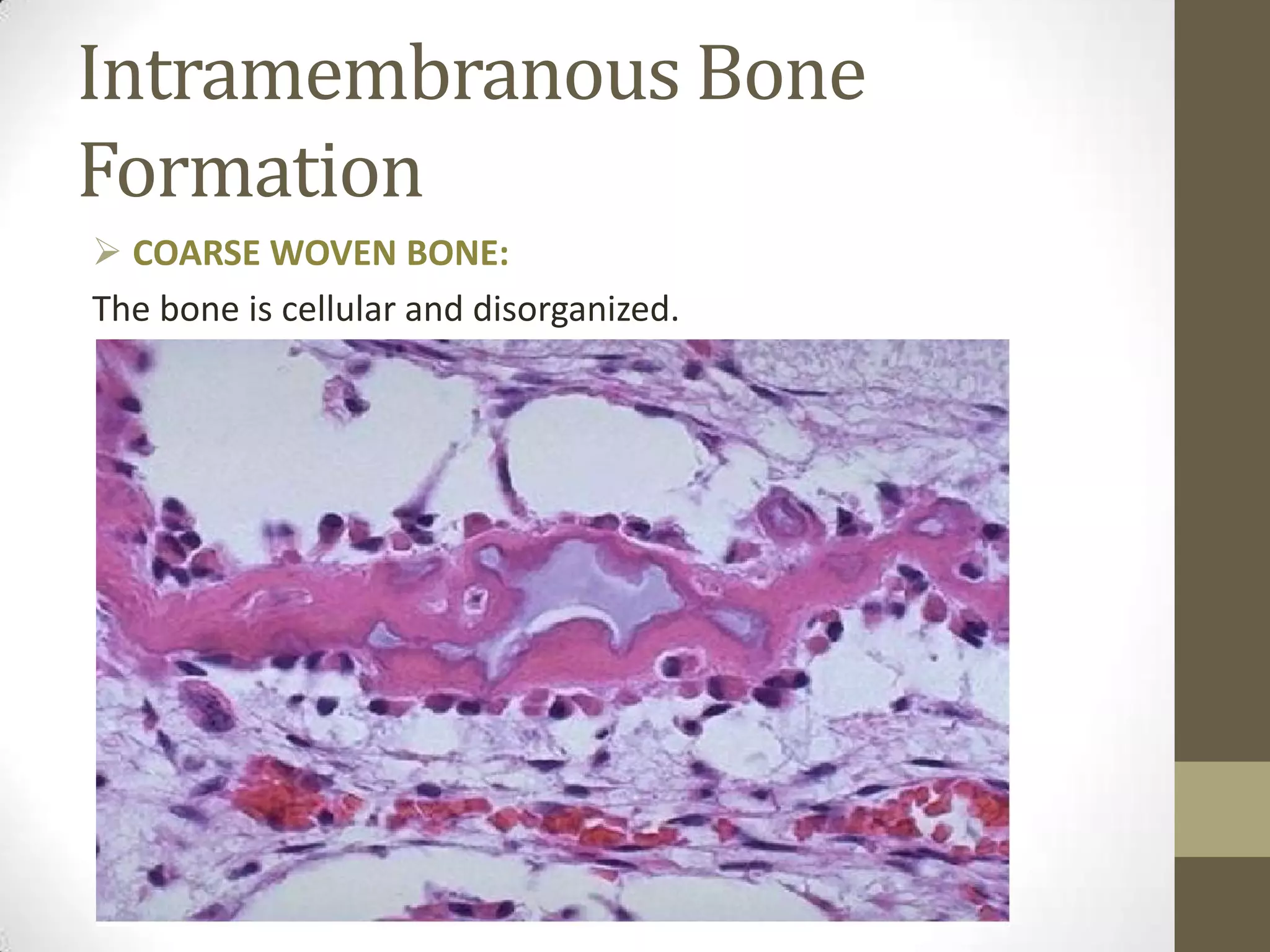
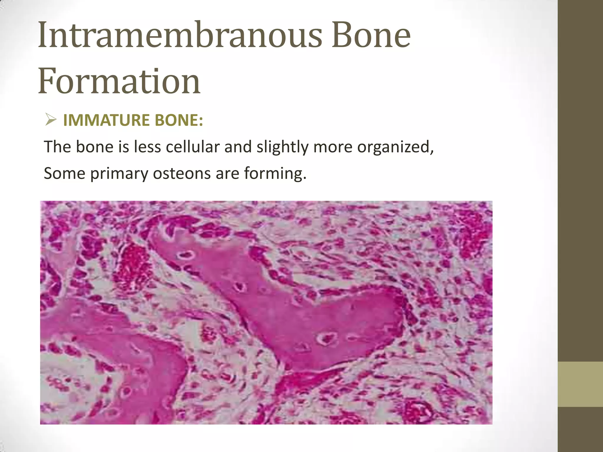
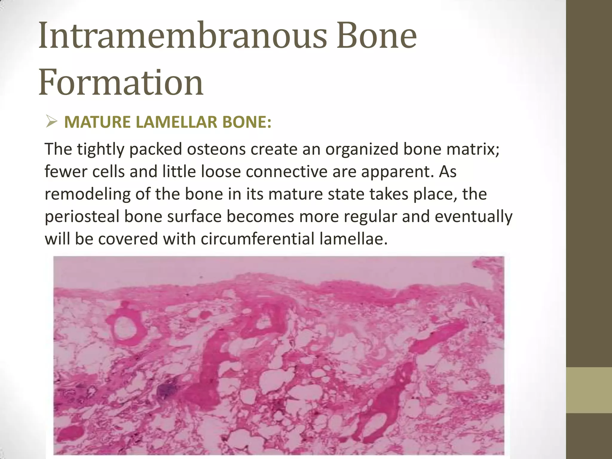
BONE
DEVELOPMENT
By: Madiha Shams
BDS 1st Year,
Dow University Of Health Sciences
BONE
•

Bone is specialized, mineralized connective
tissue consisting by weight of 33% organic
matrix, 28% type l collagen, and 5%
noncollagenous proteins, including
osteonectin, osteocalcin, bone morphogenitic
proteins, bone proteoglycan and bone
sialoprotein.

• This organic matrix is permeated by
hydroxypatite (Ca10 (PO4)6 (0H)2 ), which makes
up the remaining 67% of bone.
COMPOSTION OF BONE
BONE FORMING CELLS
1. OSTEOBLAST:
* Uninucleated

* Cuboidal and columnar in shape.
* Forms bone

2. OSTEOCYTES:
* Star-shaped cell
* Most abundant cell found in compact bone.
* Maintain bone (together with inactive
osteoblasts lining cells)

3. OSTEOCLAST:
* Large, Multinucleated
* Resorb bone
Endochondral Bone Formation
 Chondroblasts develops in primitive
Mesenchymal & form an early
Perichondrium Cartilage model.
 The developing Cartilage model
assumes the Shape of bone to be formed
and a surrounding Perichondrium
becomes identified.
Endochondral Bone Formation
 At the midshaft of the diaphysis
the perichondrium becomes a
periosteum through development
of Osteoprogentor cells and
Osteoblasts, producing a collar of
Bone by Intramembrane ossification
Calcium salts are deposited in
enlarging cartilage model.
 Blood vessels grow through
the periostem and bone collar carrying Osteoprogenitor cell
within them. These cells establish a primary ossification center
Diaphysis.
Endochondral Bone Formation
 Bony trabeculae spread out from
the primary ossification center to
occupy the entire diaphysis linking up
with previously formed bone collar
with now forms the cortical bone of
diaphysis. At this stage the terminal
Dub-shaped epiphysis are still
composed of cartilage.
Endochondral Bone Formation
 At both of them (the presise
time varies between long bones)
secondary or epiphysal ossification
centre are established along with blood
Vessels of mesenchymal cells which
becomes Osteoprogentor cells and
Osteoblast.
Endochondral Bone Formation
Intramembranous Bone
Formation
Intramembranous Bone
Formation
 COARSE WOVEN BONE:
The bone is cellular and disorganized.
Intramembranous Bone
Formation
 IMMATURE BONE:
The bone is less cellular and slightly more organized,
Some primary osteons are forming.
Intramembranous Bone
Formation
 MATURE LAMELLAR BONE:
The tightly packed osteons create an organized bone matrix;
fewer cells and little loose connective are apparent. As
remodeling of the bone in its mature state takes place, the
periosteal bone surface becomes more regular and eventually
will be covered with circumferential lamellae.
Bone  development

Bone development

  • 1.
    BONE DEVELOPMENT By: Madiha Shams BDS1st Year, Dow University Of Health Sciences
  • 2.
    BONE • Bone is specialized,mineralized connective tissue consisting by weight of 33% organic matrix, 28% type l collagen, and 5% noncollagenous proteins, including osteonectin, osteocalcin, bone morphogenitic proteins, bone proteoglycan and bone sialoprotein. • This organic matrix is permeated by hydroxypatite (Ca10 (PO4)6 (0H)2 ), which makes up the remaining 67% of bone.
  • 3.
  • 4.
    BONE FORMING CELLS 1.OSTEOBLAST: * Uninucleated * Cuboidal and columnar in shape. * Forms bone 2. OSTEOCYTES: * Star-shaped cell * Most abundant cell found in compact bone. * Maintain bone (together with inactive osteoblasts lining cells) 3. OSTEOCLAST: * Large, Multinucleated * Resorb bone
  • 5.
    Endochondral Bone Formation Chondroblasts develops in primitive Mesenchymal & form an early Perichondrium Cartilage model.  The developing Cartilage model assumes the Shape of bone to be formed and a surrounding Perichondrium becomes identified.
  • 6.
    Endochondral Bone Formation At the midshaft of the diaphysis the perichondrium becomes a periosteum through development of Osteoprogentor cells and Osteoblasts, producing a collar of Bone by Intramembrane ossification Calcium salts are deposited in enlarging cartilage model.  Blood vessels grow through the periostem and bone collar carrying Osteoprogenitor cell within them. These cells establish a primary ossification center Diaphysis.
  • 7.
    Endochondral Bone Formation Bony trabeculae spread out from the primary ossification center to occupy the entire diaphysis linking up with previously formed bone collar with now forms the cortical bone of diaphysis. At this stage the terminal Dub-shaped epiphysis are still composed of cartilage.
  • 8.
    Endochondral Bone Formation At both of them (the presise time varies between long bones) secondary or epiphysal ossification centre are established along with blood Vessels of mesenchymal cells which becomes Osteoprogentor cells and Osteoblast.
  • 9.
  • 10.
  • 11.
    Intramembranous Bone Formation  COARSEWOVEN BONE: The bone is cellular and disorganized.
  • 12.
    Intramembranous Bone Formation  IMMATUREBONE: The bone is less cellular and slightly more organized, Some primary osteons are forming.
  • 13.
    Intramembranous Bone Formation  MATURELAMELLAR BONE: The tightly packed osteons create an organized bone matrix; fewer cells and little loose connective are apparent. As remodeling of the bone in its mature state takes place, the periosteal bone surface becomes more regular and eventually will be covered with circumferential lamellae.