Computed Tomography (CT), developed by Sir Godfrey Hounsfield in 1970, is a medical imaging technique that creates 3D images from 2D X-ray images using digital geometry processing. The components of a CT scanner include a gantry, X-ray tube, detector, and control console, utilizing various scanning techniques such as axial and volumetric CT. CT imaging provides critical insights into internal body structures, aiding in medical assessments for trauma, tumors, and vascular status.
Overview of the presenter and the topic of Computer Tomography (CT) by Lovnish Thakur from School of Bioscience.
CT is a medical imaging technique that combines X-ray images to create 3D representations of internal body structures.
CT was developed in 1970 by Sir Godfrey Hounsfield, initially focused on neuroradiology.
CT scanners are essential for assessing trauma, locating tumors, and planning surgeries.
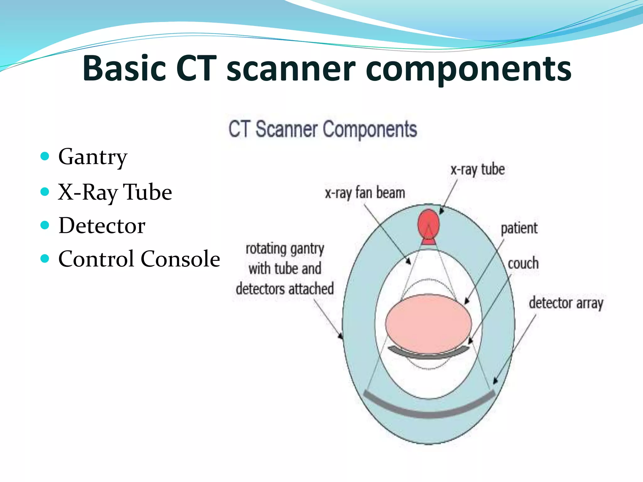
Main components include the gantry, X-ray tube, detector, and control console crucial for CT functions.
Discusses digital and conventional CT methods, including digital projection, axial CT, and helical CT. Explains how X-ray attenuation differs among structures, influencing image formation and brightness.
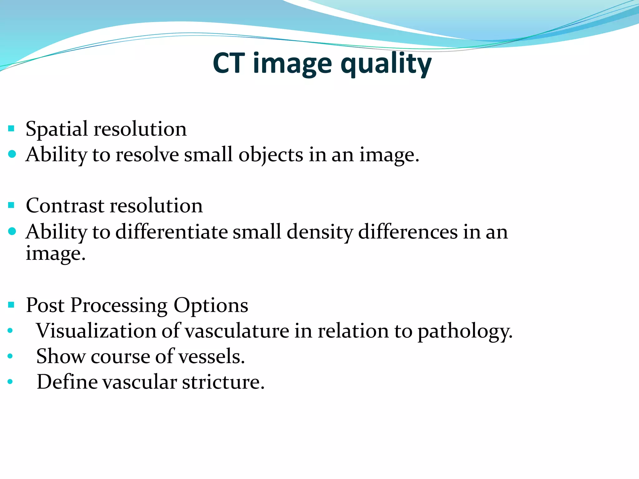
Describes image quality factors such as spatial resolution, contrast resolution, and post-processing for enhanced visualization.
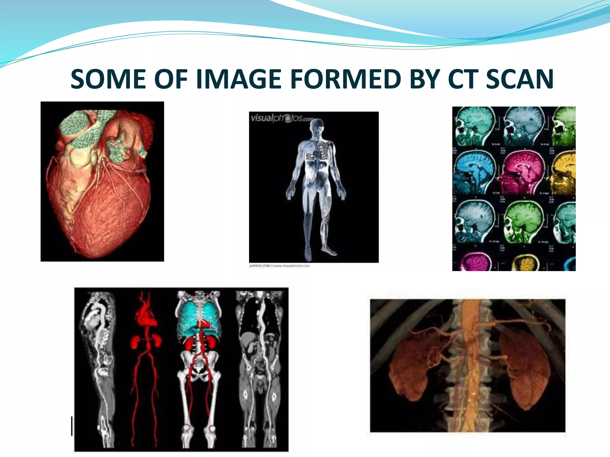
Displays various images generated from CT scans, showcasing practical applications of the technology.
Closing remarks and thank you note from the presenter Lovnish Thakur.
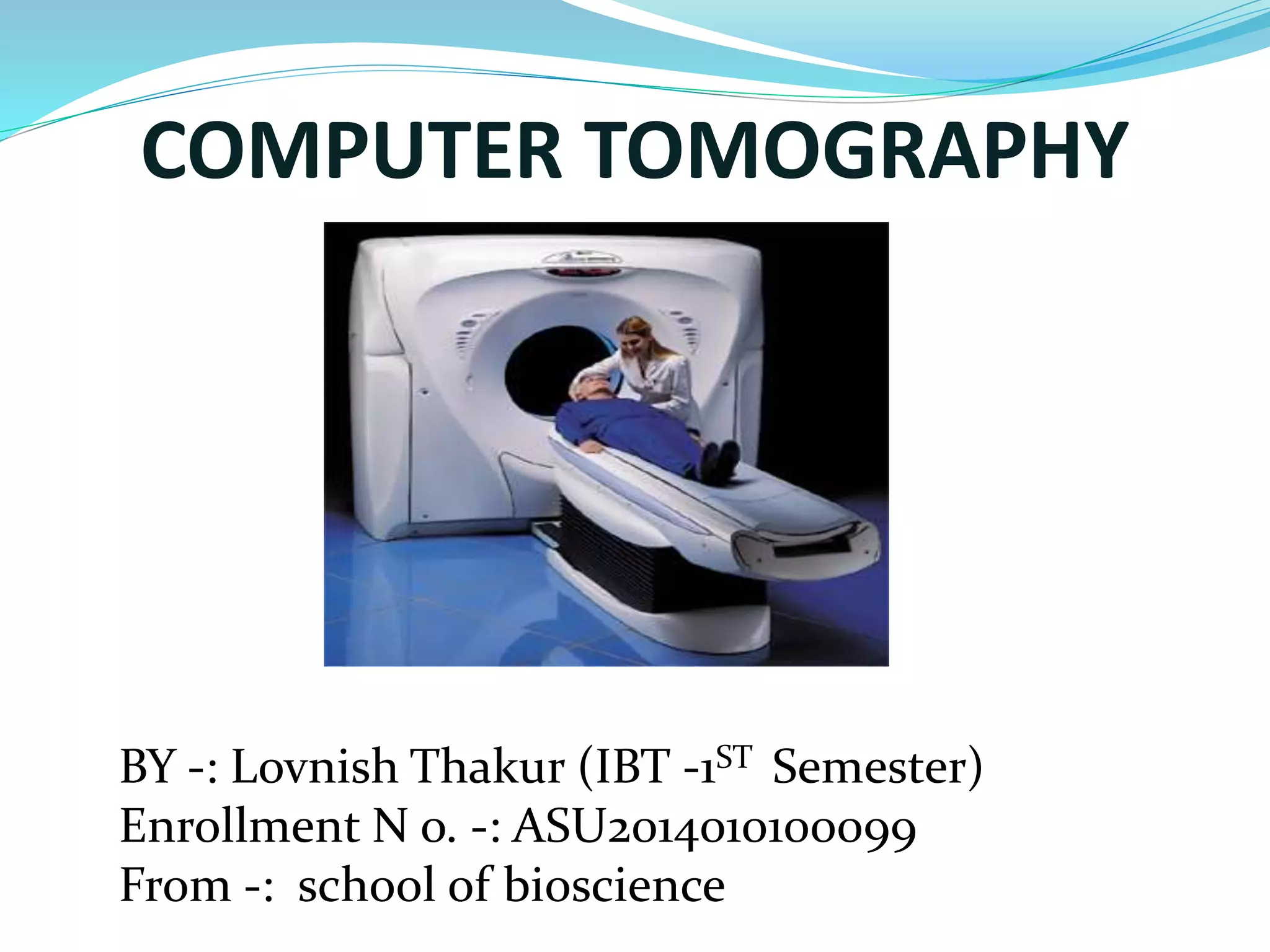
COMPUTER TOMOGRAPHY
BY-: Lovnish Thakur (IBT -1ST Semester)
Enrollment N o. -: ASU2014010100099
From -: school of bioscience
2.
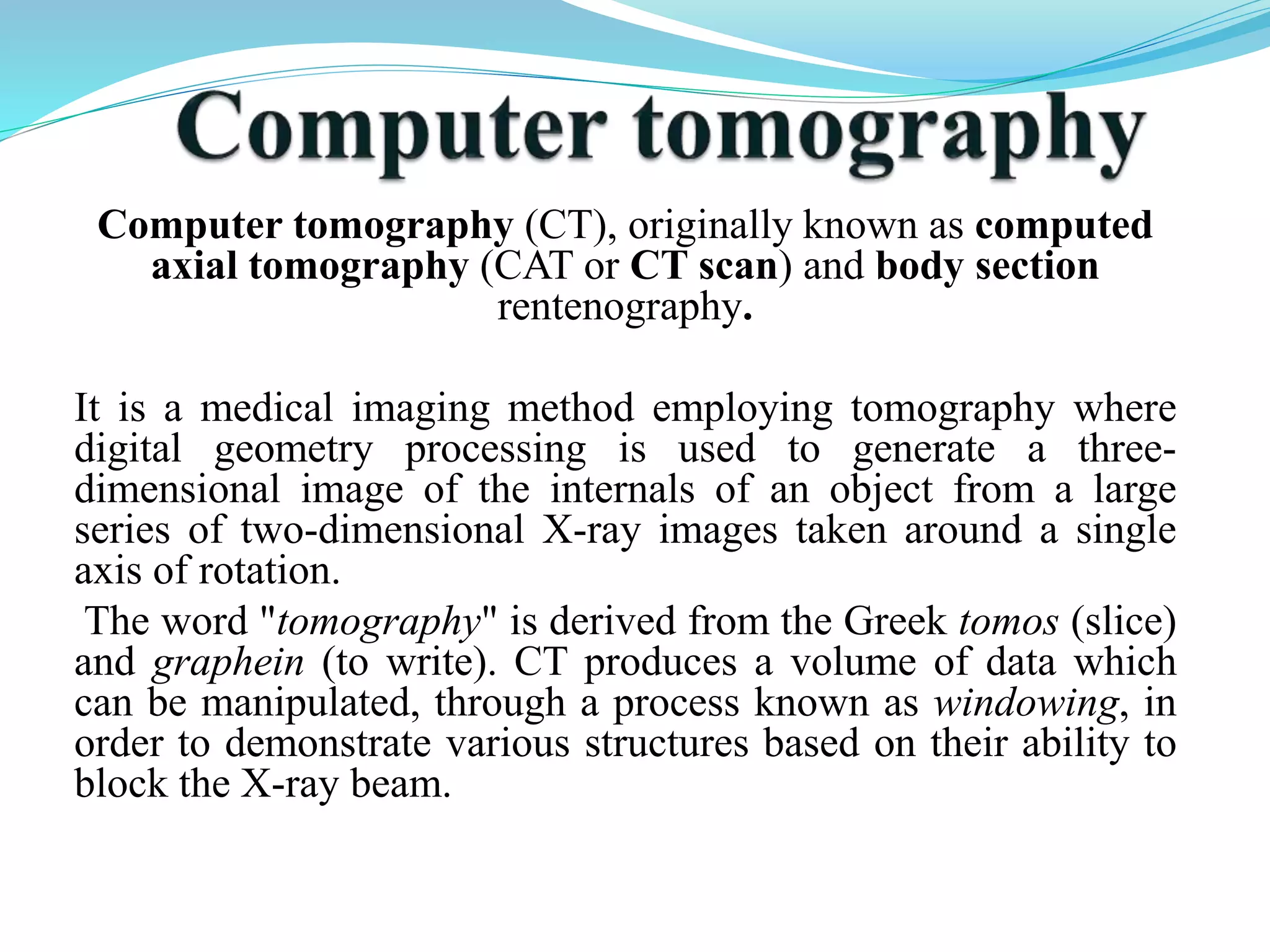
Computer tomography (CT),originally known as computed
axial tomography (CAT or CT scan) and body section
rentenography.
It is a medical imaging method employing tomography where
digital geometry processing is used to generate a three-dimensional
image of the internals of an object from a large
series of two-dimensional X-ray images taken around a single
axis of rotation.
The word "tomography" is derived from the Greek tomos (slice)
and graphein (to write). CT produces a volume of data which
can be manipulated, through a process known as windowing, in
order to demonstrate various structures based on their ability to
block the X-ray beam.
3.
CT: The beginning
CT founded in 1970 by Sir Godfrey Hounsfield
first applications were in neuroradiology.
4.
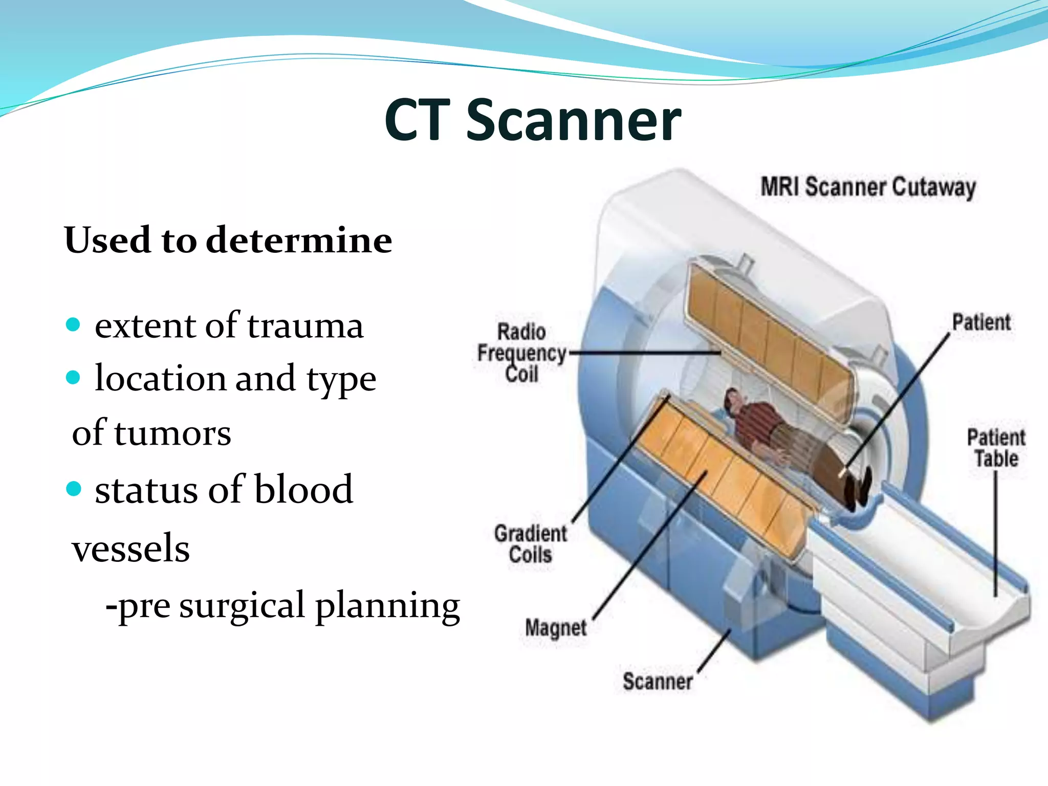
CT Scanner
Usedto determine
extent of trauma
location and type
of tumors
status of blood
vessels
-pre surgical planning
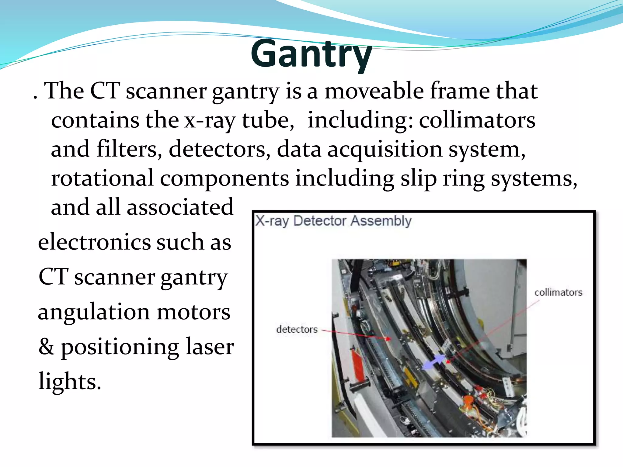
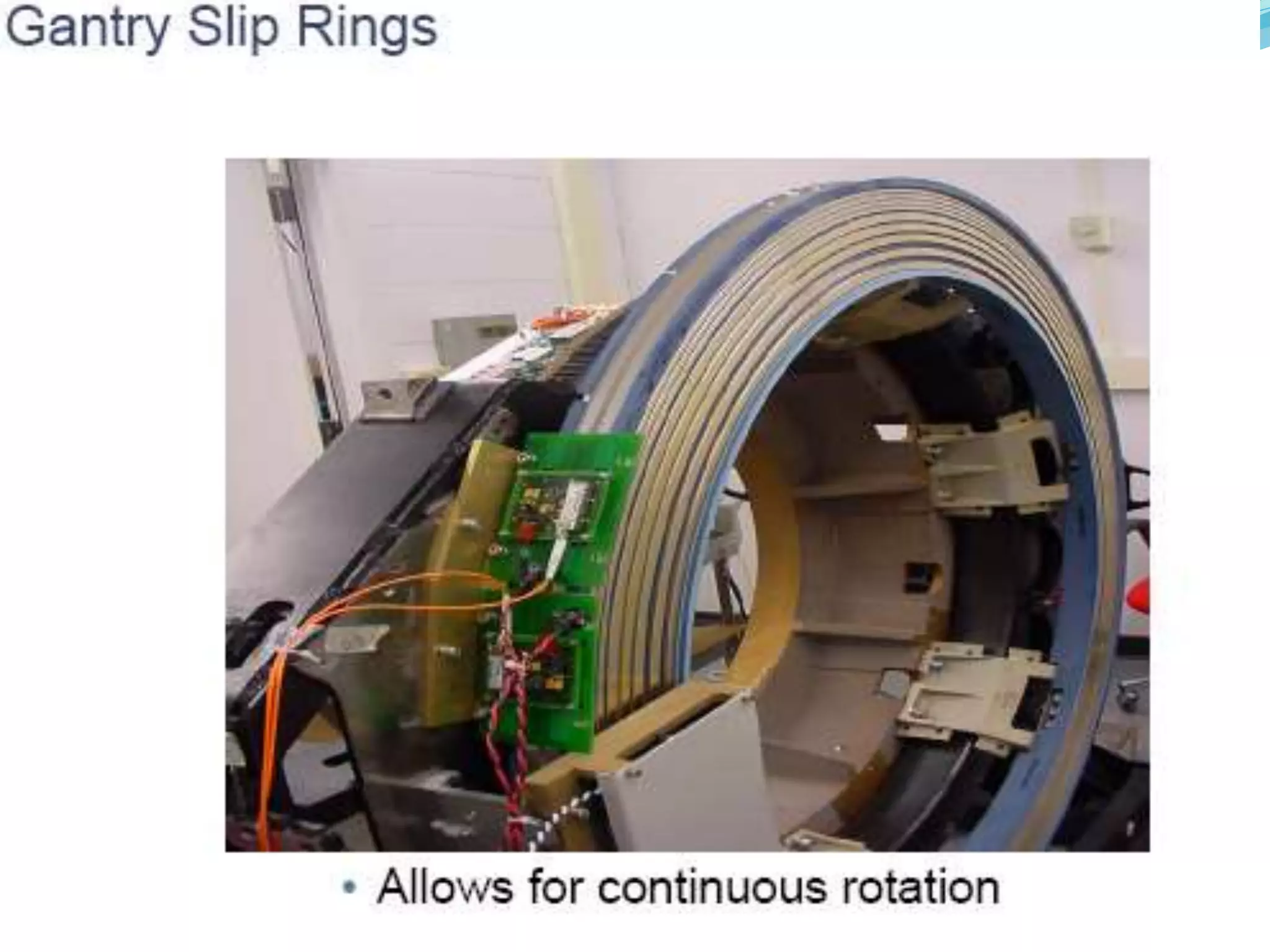
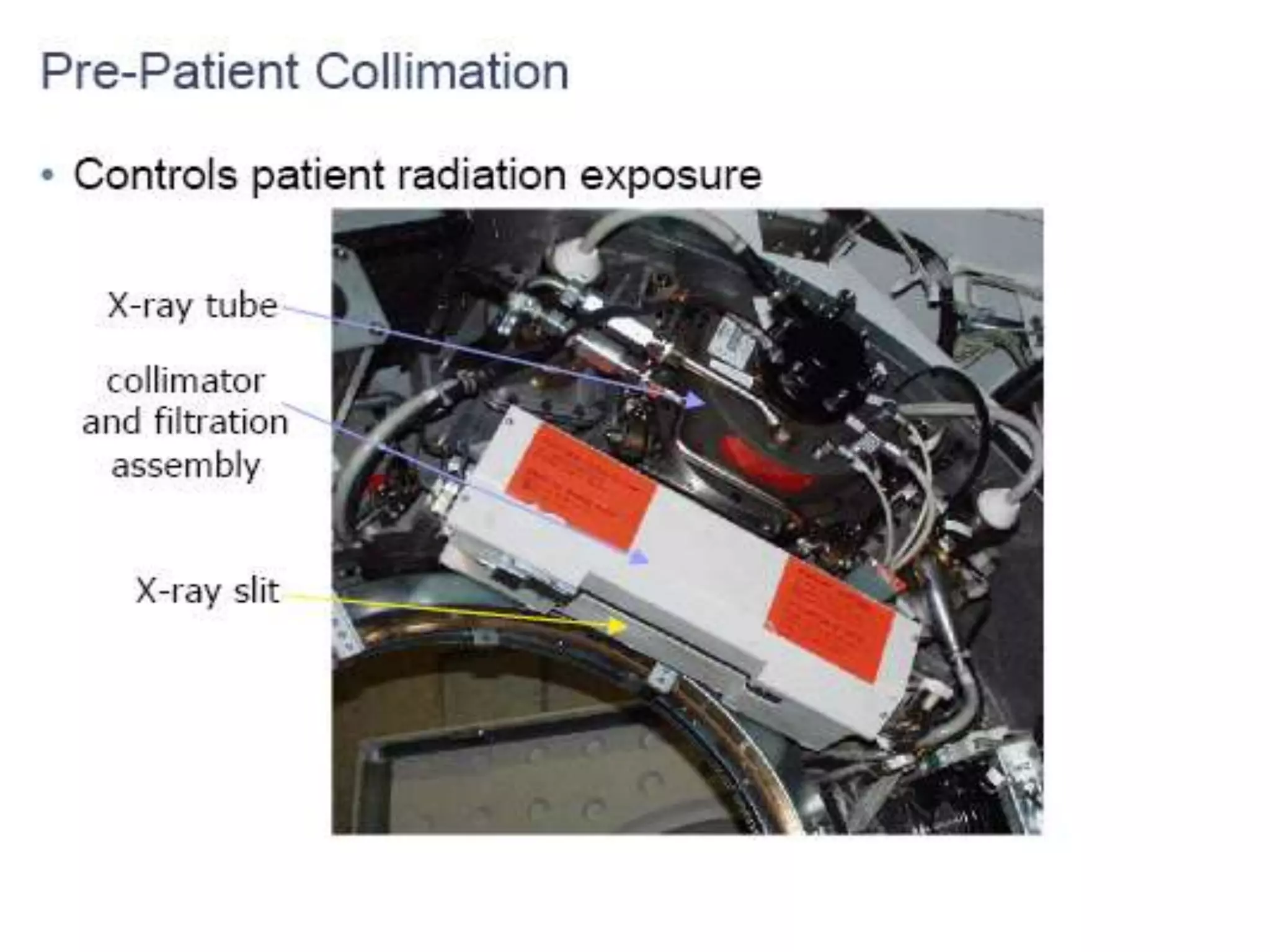
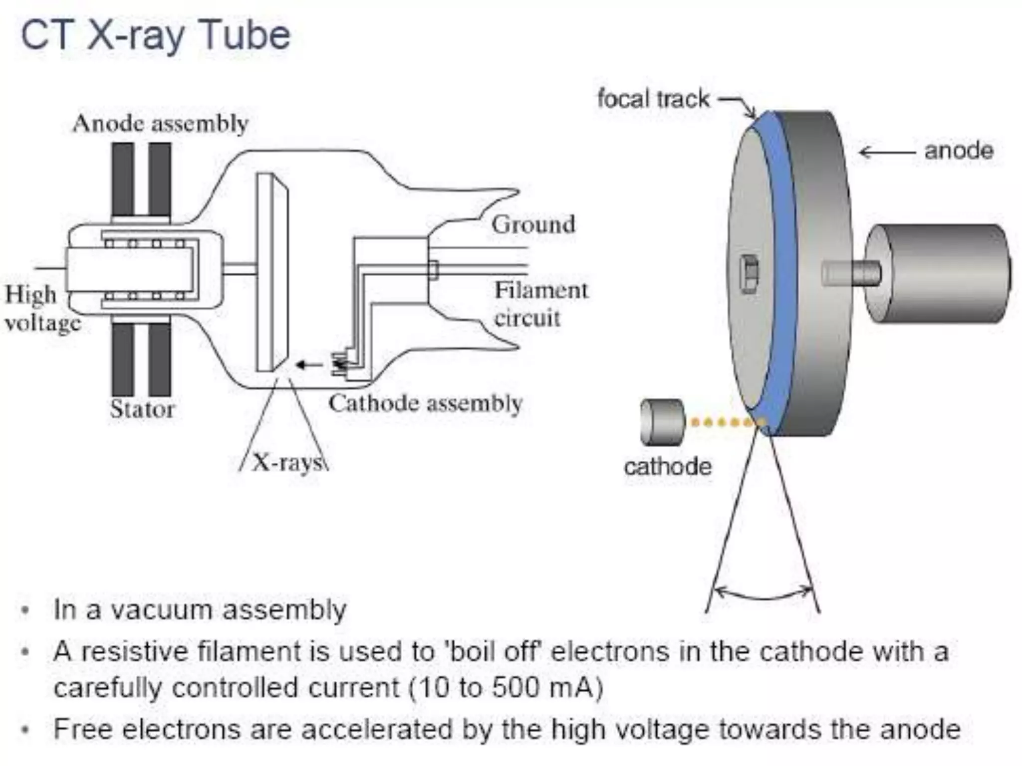
Gantry
. TheCT scanner gantry is a moveable frame that
contains the x-ray tube, including: collimators
and filters, detectors, data acquisition system,
rotational components including slip ring systems,
and all associated
electronics such as
CT scanner gantry
angulation motors
& positioning laser
lights.
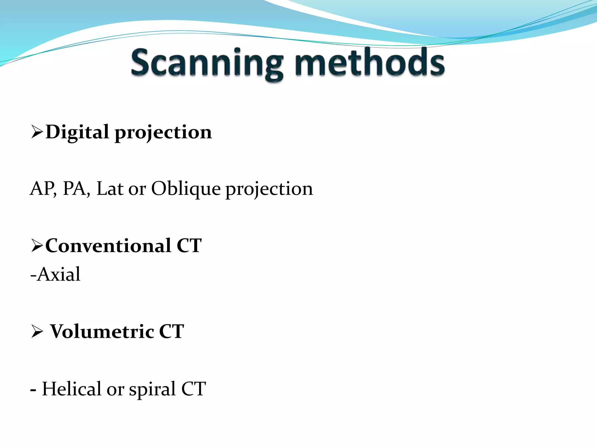
Digital projection
AP,PA, Lat or Oblique projection
Conventional CT
-Axial
Volumetric CT
- Helical or spiral CT
13.
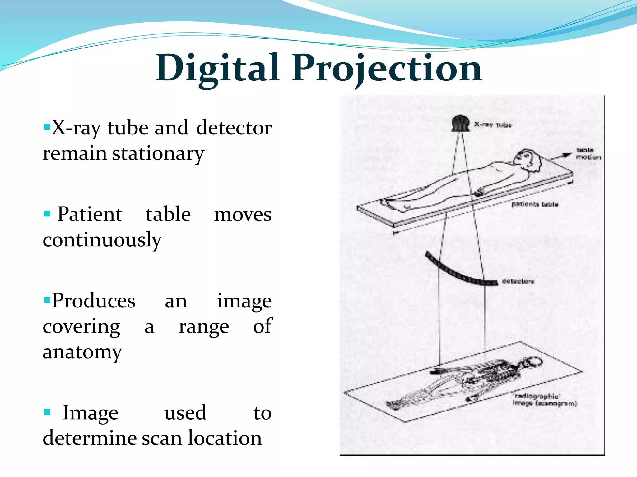
Digital Projection
X-raytube and detector
remain stationary
Patient table moves
continuously
Produces an image
covering a range of
anatomy
Image used to
determine scan location
14.
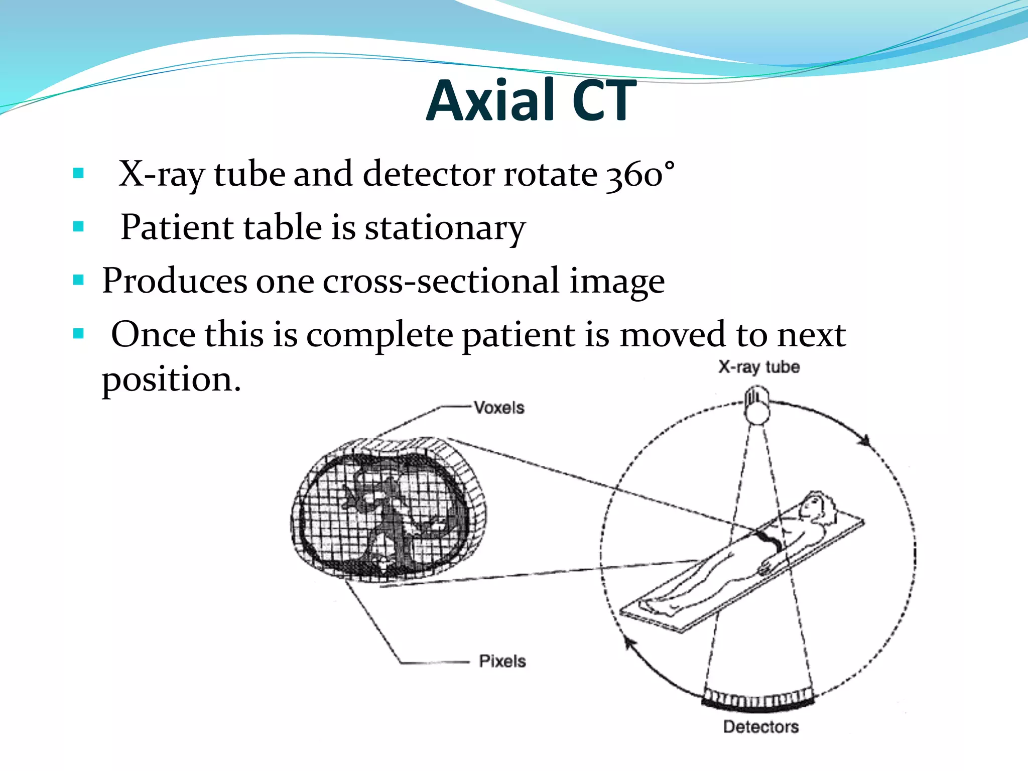
Axial CT
X-ray tube and detector rotate 360°
Patient table is stationary
Produces one cross-sectional image
Once this is complete patient is moved to next
position.
15.
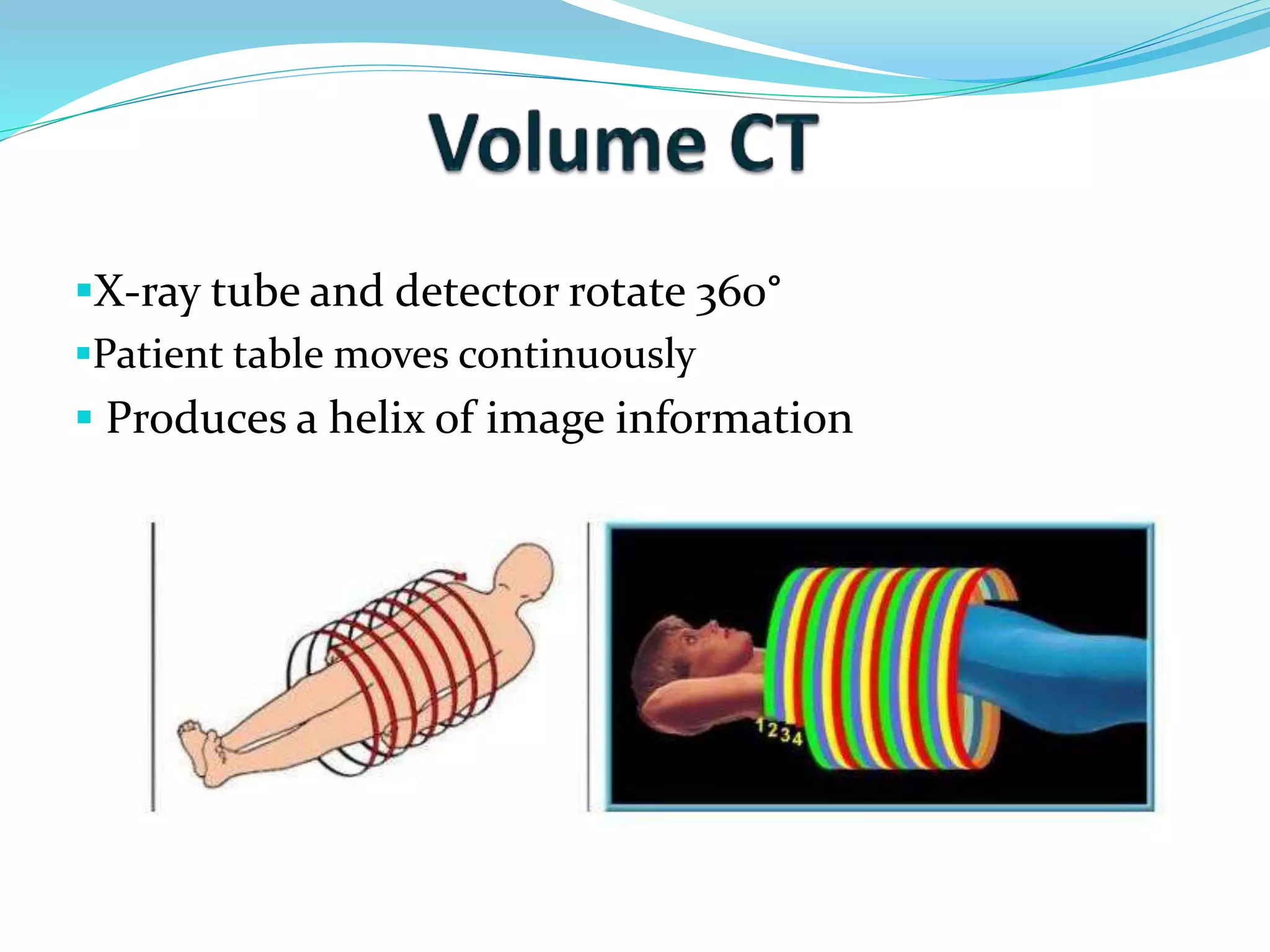
X-ray tube anddetector rotate 360°
Patient table moves continuously
Produces a helix of image information
16.
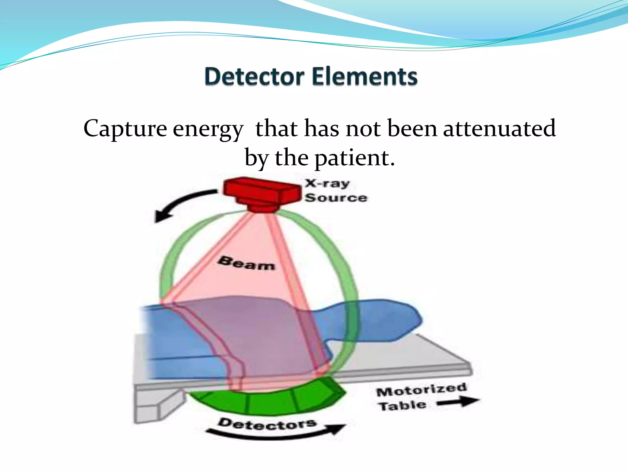
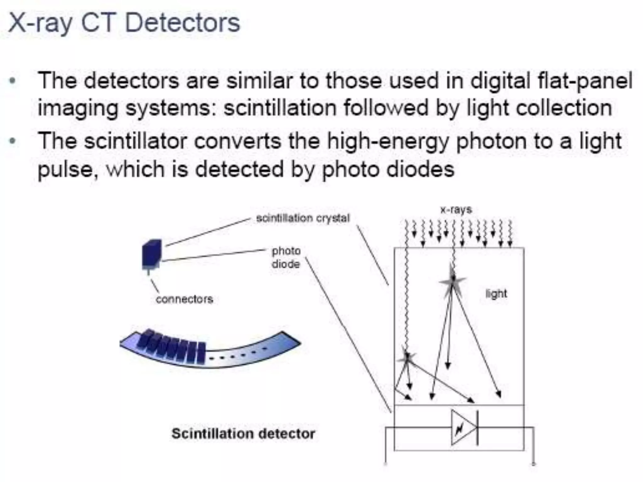
Attenuation
X-raybeam passes through patient
Each structure attenuates X-ray beam differently
According to individual densities
Radiation received by detector varies according to
these densities
17.
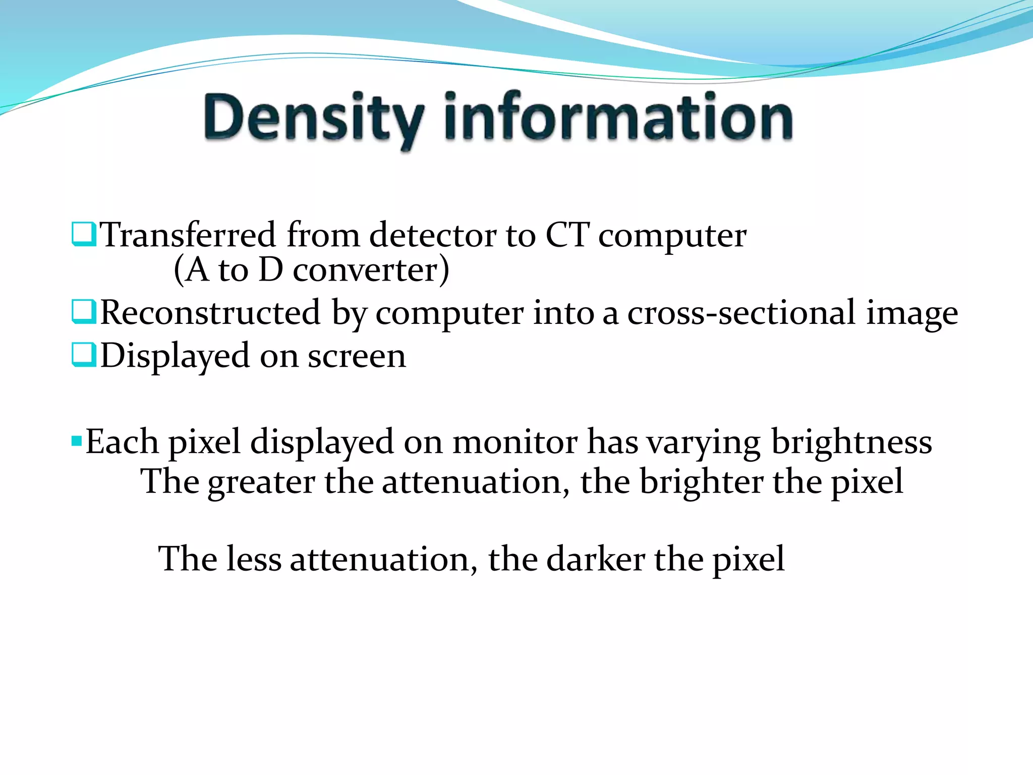
Transferred from detectorto CT computer
(A to D converter)
Reconstructed by computer into a cross-sectional image
Displayed on screen
Each pixel displayed on monitor has varying brightness
The greater the attenuation, the brighter the pixel
The less attenuation, the darker the pixel
18.
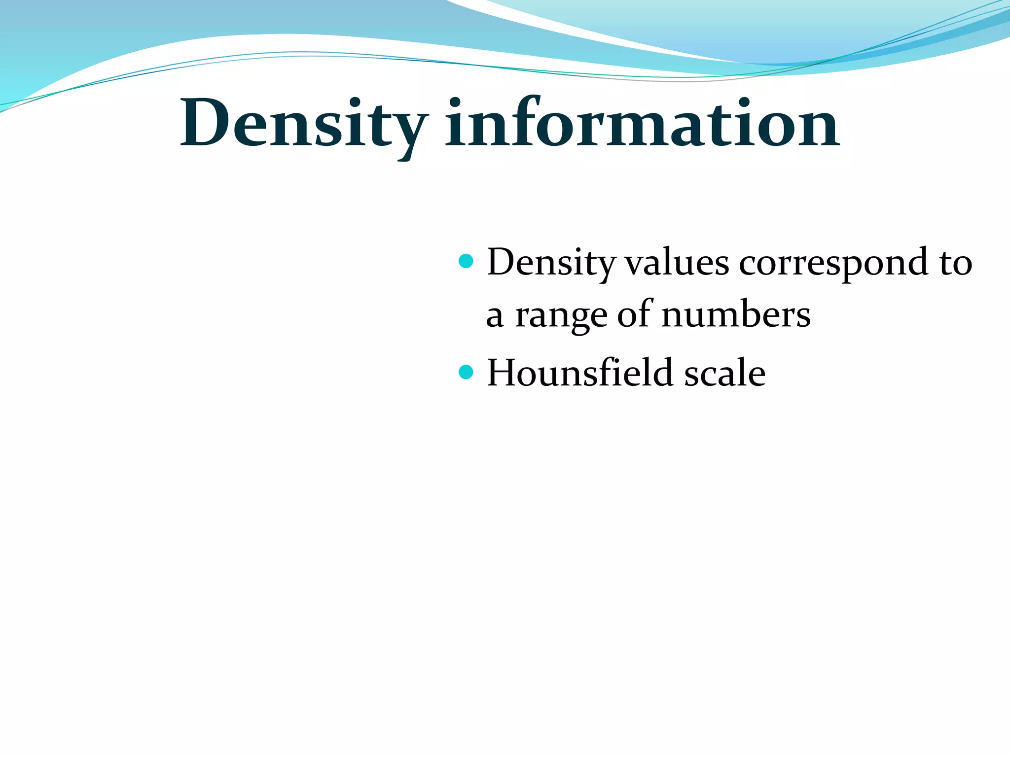
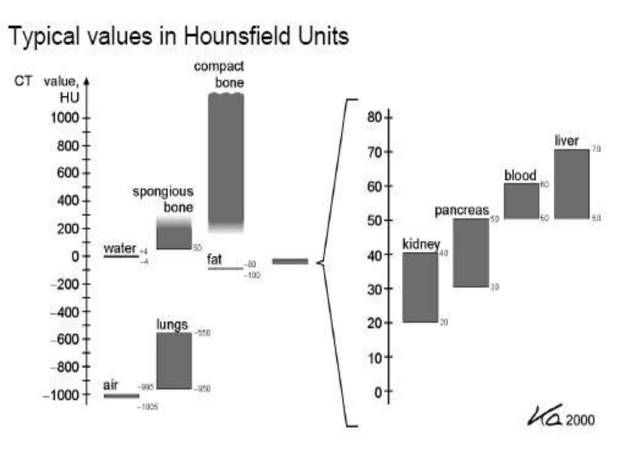
Density information
Density values correspond to
a range of numbers
Hounsfield scale
20.
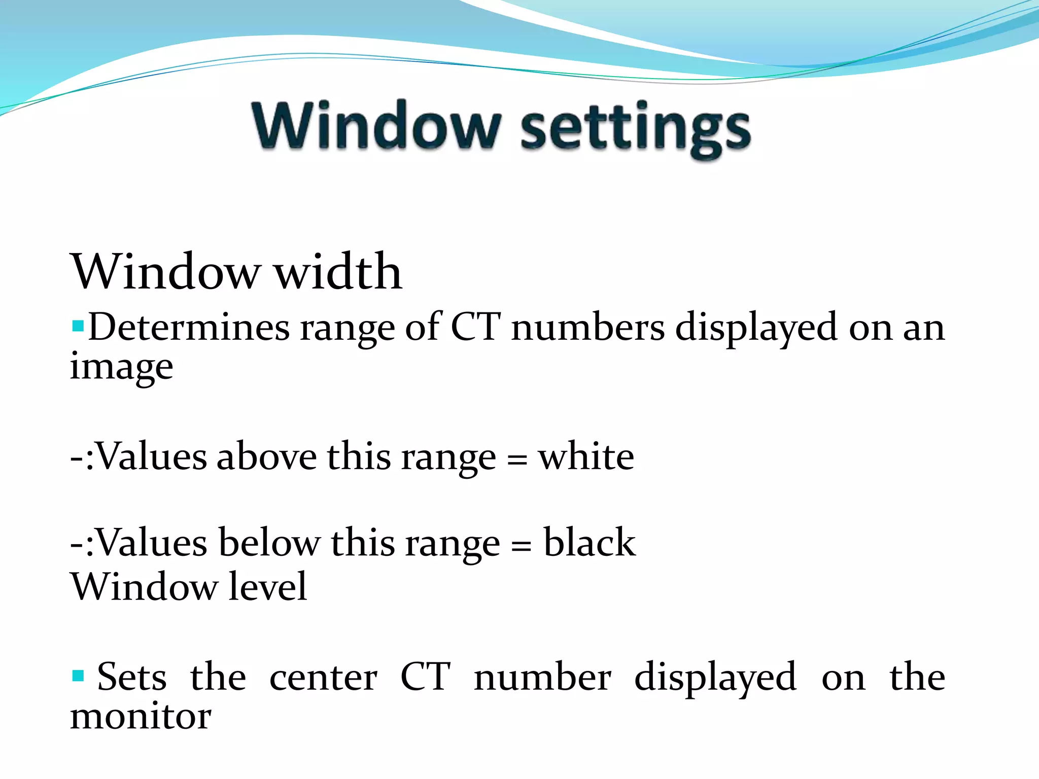
Window width
Determinesrange of CT numbers displayed on an
image
-:Values above this range = white
-:Values below this range = black
Window level
Sets the center CT number displayed on the
monitor
21.
CT image quality
Spatial resolution
Ability to resolve small objects in an image.
Contrast resolution
Ability to differentiate small density differences in an
image.
Post Processing Options
• Visualization of vasculature in relation to pathology.
• Show course of vessels.
• Define vascular stricture.