

Recommended
More Related Content
What's hot
What's hot (20)
Viewers also liked
Viewers also liked (9)
Similar to Endoüroloji 2011 ankara
Similar to Endoüroloji 2011 ankara (20)
More from Mahmut Gündoğan
More from Mahmut Gündoğan (13)
Recently uploaded
Recently uploaded (20)
Endoüroloji 2011 ankara
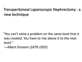
- 1. Transperitoneal Laparoscopic Nephrectomy : a new technique "You can't solve a problem on the same level that it was created. You have to rise above it to the next level." —Albert Einstein (1879-1955)
- 2. Standard technique (transperitoneal laparoscopic nephrectomy) 1. port placement, 2. mobilization of colon, 3. dissection of the ureter, 4. dissection of lower renal pole, 5. dissection of renal hilum, 6. occlusion and division of the renal artery and vein, 7. completion of nephrectomy.
- 3. Mobilization of the Colon ( right laparoscopic nephrectomy) Standard New • Colon is mobilized by incising the • The part of the peritoneum ipsilateral line of Toldt. immeditially under the liver is • Peritoneal incision extends from incised from triangular ligament the right common iliac artery, to the vena cava coursing lateral to the cecum and • At the vena cava, the insicion of ascending colon and around the the peritoneum is extended hepatic flexure. inferiorly to the lower pole of • At its cephalad end, the incision is the kidney, carried medially in a horizontal • Ascending colon and hepatic manner between the liver and flexure are rolled medially. transverse colon • During this procedure you can • Ascending colon and hepatic see the second part of the flexure are rolled medially. duodenum and it is easily • Duodenum is dissected medially retracted medially. until the anterior surface of the inferior vena cava is clearly seen (endpoint of dissection).
- 4. • In the standard technique the peritoneal incision starts inferior and goes superior . • Our technique the incision starts superior and goes inferior.
- 5. Finding the Ureter (Standard technique ) • The ureter is identified in the retroperitoneal fat just medial to the psoas muscle. • In general, the ureter is located more medially than expected. • If the ureter is hard to find: 1. Identify the gonadal vessels that course anterior and parallel to the midureter 2. Gently stroke the retroperitoneal fat in a horizontal manner (at right angles to the longitudinal axis of the ureter) with an atraumatic grasper, and look for ureteral peristalsis; 3. Look for the ureter where it crosses the common iliac vessels.
- 6. Dissection and retraction of the Ureter (Standard technique) • The ureter is mobilized and retracted laterally. Atraumatic forceps Percutaneously placed suture to loop the ureter. Alternatively, the ureter can be clipped and divided at this point. • Lateral traction of the ureter, whether the ureter is intact or divided, helps to expose the renal hilum for disection. Glenns urologic surgery,
- 7. • In laparoscopic surgery : Disecting and retracting the ureter take time Require another port Retracting the ureter actually makes the disection of the renal hilum more difficult.
- 8. Renal Polar Dissection and Retraction(Standard technique) • Gerotas fascia is entered and the lower renal pole is identified and mobilized circumferentially. • The upper renal pole is detached from the adrenal gland and mobilized. • Upper and lower renal poles are retracted laterally by an atraumatic grasper.
- 11. Modification of the standard transperitoneal laparoscopic nephrectomy technique seems to significantly facilitate the procedure • February 2004 –April 2010 • Patients underwent laparoscopic surgery at Gazi University Department of Urology and other University and training hospitals. • Indications for surgery RCC 96 patients TCC 10 patients Nonfunctional kidney 77 patients • Conventional technique 85 patients • New Technique 98 patients • Mean operation time(after port placement until the specimen is placed in the endobag)
- 12. RESULTS n age Op. Time Blood loss Hospital stay (Minute) (ml) (days) Convansionel 85 51 85.9±3.9 150 3 New tecnique 98 53 30.1±7.5 30 2 • Operation time was significantly shorter in new technique group (p<0.001) • Mean intraoperative blood loss was lower in new technique group (p<0.05).
- 13. Conclusions: • We recommend initially exposing the upper kidney pole followed by dissection of the renal hilum rather than dissection of the ureter and dissection of the lower renal pole during L-RN, L-NU and L-SN • This change makes these procedures easier and quicker.
- 14. In laparoscopic nephrectomy which procedure is safe for renal pedicle control? • Vascular stapler • Polymer clip • Titanium clip • Suture • Ligasure
- 16. En Bloc Ligation A- V Fistula? • The first case of fistula formation after en bloc ligation of the renal pedicle was reported by Hollingsworth (1934) in a patient with tuberculosis renal disease. • Approximately 60 cases of fistula formation have been reported. • In all these cases en bloc ligation was performed
- 17. White WM, Klein FA, Gash J, Waters WB. Prospective radiographic followup after en bloc ligation of the renal hilum. J Urol. 2007; 178: 1888-91 • Prospective study • All patients underwent en bloc ligation of the renal hilum during nephrectomy for malignant diseases • AVF signs: Hypertension, abdominal bruit, new onset congestive heart failure. • Follow up time: 12 months • CT- arteriography: to assess arteriovenous fistula
- 18. White WM, Klein FA, Gash J, Waters WB. Prospective radiographic followup after en bloc ligation of the renal hilum. J Urol. 2007; 178: 1888-91 • In the 40 patients who underwent computerized tomographic arteriography no fistulas were noted. • Conclusions: after en bloc ligation of the renal hilum with a titanium endovascular stapler, risk of arteriovenous fistula formation is very low.
- 20. In laparoscopic nephrectomy, which is safe for use on the renal pedicle? • Two clinical studies, total patients = 248 En bloc vascular stapler n = 158 Single titanium stapler n = 90 • Postoperatively, no arteriovenous fistulas or other complications were seen in either group • Conclusion: vascular stapler is safe for controlling the renal pedicle, and en bloc control does not increase the risk of arteriovenous fistula
- 21. Conclusions • Based on clinical followup and prospective radiographic evaluation there appears to no risk of arteriovenous fistula formation after en bloc ligation of the renal hilum using a titanium endovascular stapler.
- 25. Conclusion • What is now proved was once only imagined. – William Blake • Whatever technique you are considering, if your “gut” says yes, then you probably should use it. -- Lutfi Tunc