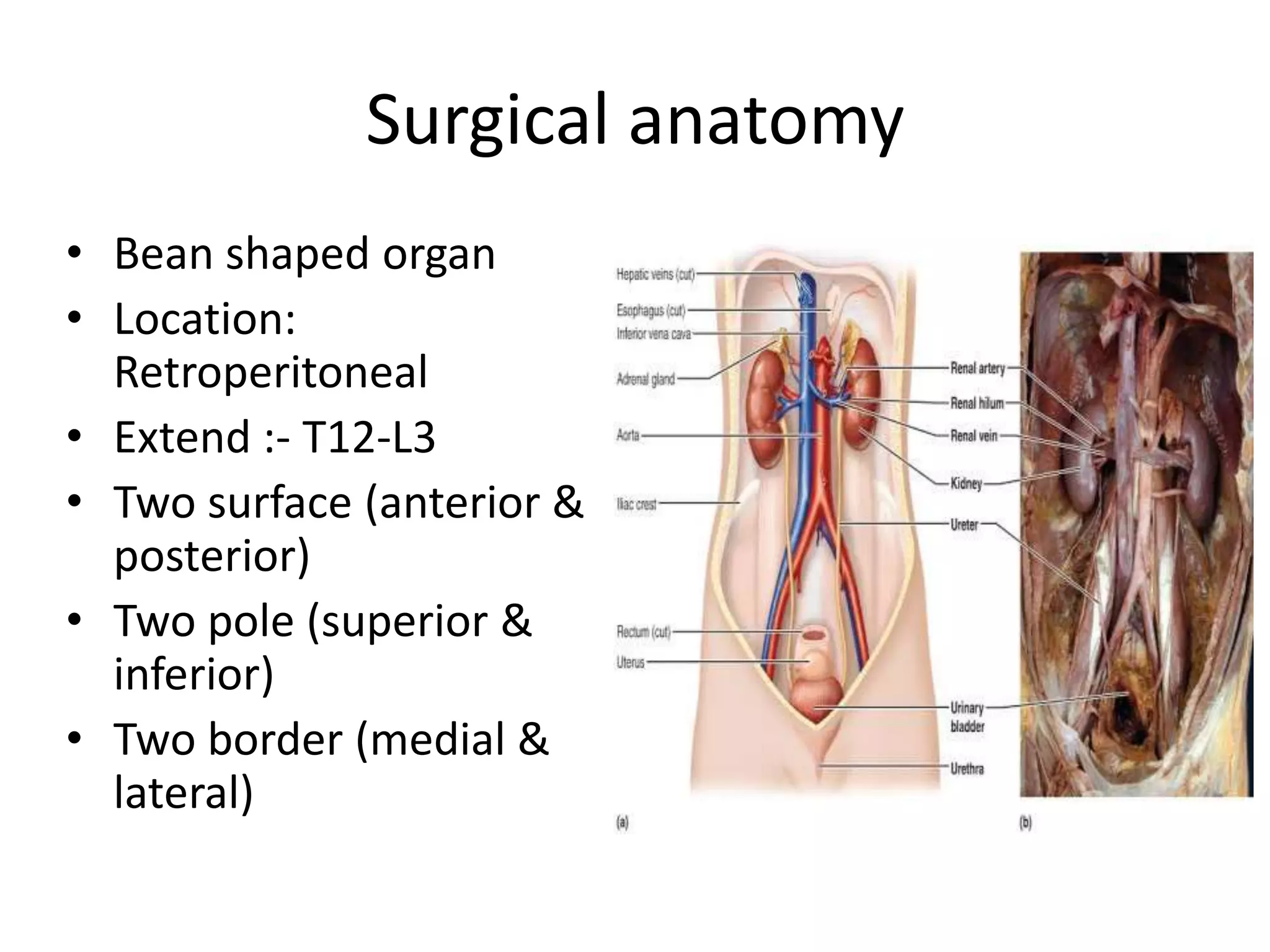
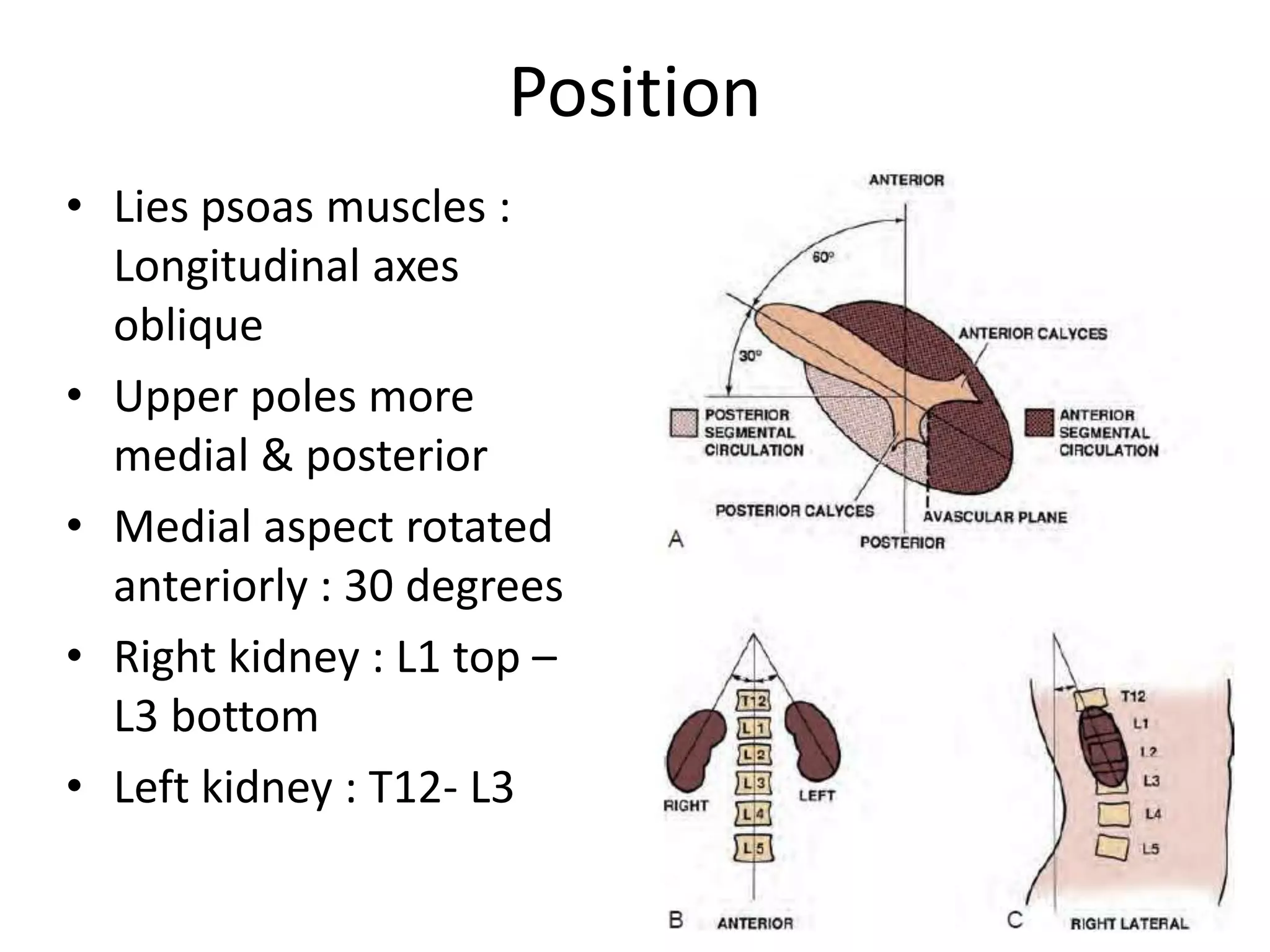
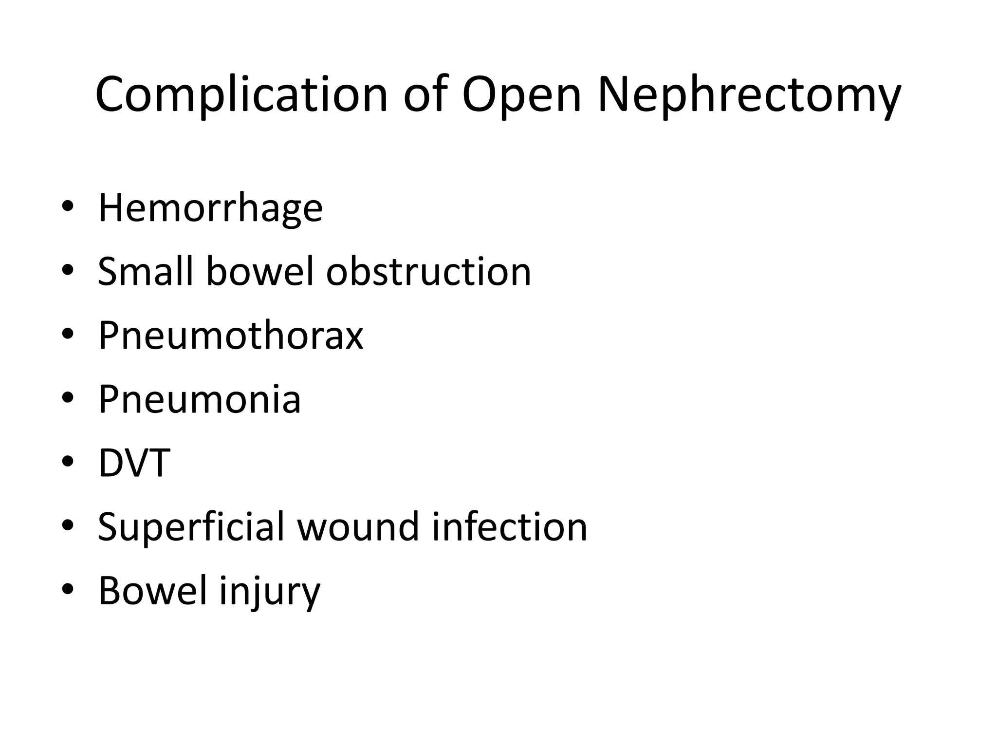
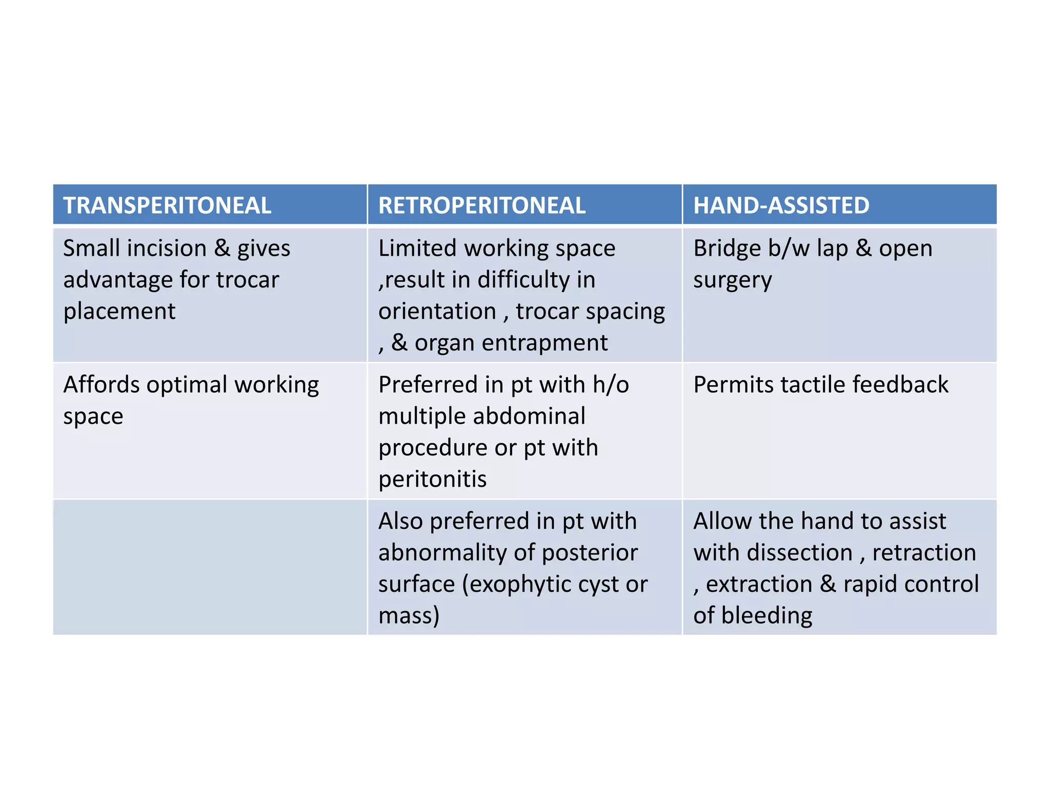
Operative techniques: Nephrectomy
Presenter: Dr Sangamesh S K
Moderator: Col S Tripathy
Overview of presentation
• Surgical anatomy
• Indication of Nephrectomy
• Types of Nephrectomy
– Simple, Partial & Radical nephrectomy
– Donor Nephrectomy
• Different technique of Nephrectomy
• Complications
Surgical anatomy
• Bean shaped organ
• Location:
Retroperitoneal
• Extend :- T12-L3
• Two surface (anterior &
posterior)
• Two pole (superior &
inferior)
• Two border (medial &
lateral)
Position
• Lies psoas muscles :
Longitudinal axes
oblique
• Upper poles more
medial & posterior
• Medial aspect rotated
anteriorly : 30 degrees
• Right kidney : L1 top –
L3 bottom
• Left kidney : T12- L3
Anterior relation of kidney
Posterior relation of kidney
Axial section imaging
Renal vasculature
Segmental blood supply
Arterial supply
Indication of Nephrectomy
• Severe trauma
• Renal infection
– Xanthogranulomatous & Emphysematous Pyelonephritis
• Malignancy
• Non functioning kidney
– Stones & obstruction
• Renal fistula
• Renal vascular Hypertension
– All medical & surgical therapy have failed
• Transplantation
Types of Nephrectomy
• Simple Nephrectomy
– Removal of kidney within Gerota fascia
• Partial Nephrectomy
– Small-sized renal cancer
– Benign diseases- excised renal capsule (renorrhaphy)
• Radical Nephrectomy
– Complete removal of kidney outside Gerota fascia
– Ipsilateral adrenal gland
– Complete regional lymphadenectomy (crus of
diaphragm - aortic bifurcation)
Different surgical approach
1. Open approach of Nephrectomy
2. Laparoscopic
• Transperitoneal
• Retroperitoneal
• Hand assisted
3. Robotic assisted
Pre- op evaluation
• Global assessment of pt’s renal function is done:
– Urine analysis & culture
– Serum Creatinine
– GFR evaluation
– Cardiac & pulmonary status: Positioning & bleeding
– Cross sectional imaging (CT/MRI) :- Surgical planning
– Locally advanced metastatic lesion : Screen hepatic
status (stauffer syndrome)
– Renal Artery Embolisation (RAE) : Large tumor
Instruments
• General set
• Abdominal drainage tube
• Self-retaining retractors : omni tract, balfour
• Long genitourinary surgical instruments
• Bulldog &/or Satinsky vascular pedicle clamps
• Mixters right angle forceps
• Bulldog arterial clamp
• Retractor- doyen’s and deiver’s
• Kidney pedicle clamp
Positioning
• Induction & ET tube
placement
• Catheterization done
• Pt : Lateral decubitus
• Table flexed b/w iliac crest &
costal margin
• Head supported: Avoid
excessive flexion
• Pt back supported by blanket
• Dependent leg flexed & top
leg straight
• All pressure point : padded
surface
• Pt secured to table : tape
Surgical approach to kidney
Anterior approach of kidney
INCISION INDICATION BENEFIT LIMITATION
Midline
Transperitoneal
Trauma, IVC
Thrombus , B/L
Renal or Ureteral
disease , Horseshoe
Rapid ,Early
vascular control ,
Access both kidney
Limited exposure to
kidney & bowel
Subcostal Radical
nephrectomy ,
UPJO
Incision can extend
to chevron , early
vascular control
Bowel complication
Hilum access poor
Chevron B/L renal tumor ,
IVC thrombus
Excellent b/l
exposure ,
Injury to Liver ,
Spleen , Transection
of large muscle
Transverse
abdominal
Wilms tumor Easy access to
pedicle &
retroperitoneal
node
Modified
Thoracoabdominal
Radical
nephrectomy ,
lymphadenectomy
versatile Bowel
complication,
Transection of large
muscle
Flank incision
INCISION INDICATION BENEFIT LIMITATION
11th or 12th rib
supracostal
Partial
nephrectomy , &
Simple
nephrectomy
Good renal &
retroperitoneal
exposure
Pleural injury
11th rib Transcostal Partial and Simple
nephrectomy
Good renal &
retroperitoneal
exposure
Pleural injury &
Noticeable flank
defect
Thoracoabdominal Large renal mass ,
IVC thrombus ,
involvement of
surrounding
structure
Excellent exposure ,
Can approach
completely
extraperitoneally
Pleural injury ,
Transection of large
muscles,
Steps of flank approach
• After Gerota fascia incised & kidney is
dissected free from surrounding perinephric
fat.
• Renal artery identified & ligated before vein
• Renal vein ligated
Simple nephrectomy
Indications
• Poor functionaing kidney
– Obstruction, infection, trauma, stones,
nephrosclerosis, vesicoureteral reflux, polycystic
kidney, or congenital dysplasia
• Functional kidney
– Relieve intractable symptoms : bleeding, pain,
hypertension, or persistent infection
Simple nephrectomy
• Flank incision : retroperitoneal access
• Renal fascia incised , perirenal fat is separated
from kidney
• Aberrant vessels : near the poles
• Large hydronephrotic : puncture and aspirate
• Adrenal dissectd in upper pole
• Lower pole mobilised and ureter isolated
• Division of ureter : access to hilar structures
Right kidney approach
Left kidney approach
Radical nephrectomy : Right
• Additional mobilization of liver, avascular Rt triangular
ligament is incised
• White line of Toldt : pelvis to hepatic flexure
• 2nd part of duodenum : Kocher maneuver
• Dissection anterior to IVC : Identification of renal vein
& gonadal vein
• Ligature of ureter below : kidney lifted , artery more
exposed
• Difficult hilar dissections : Dissect in the interaortocaval
region
• Lumbar veins close approximation
Radical nephrectomy : left
• White line of Toldt : splenic flexure to descending
colon is reflected medially.
• Renocolic ligament is divided and extreme care is
taken to avoid injury to the tail of the pancreas
• Left renal vein is identified using the anterior
surface of the aorta as a guide
• Left renal artery is usually located cranial and
posterior to the left renal vein
• Further mobilization of the lower pole of the
kidney, the left ureter and the left gonadal vein
PARTIAL NEPHRECTOMY:- INDICATION
1) Absolute:- i) Single kidney
ii) Bilateral renal tumor
iii) Severe renal failure
2) Relative:- i) Abnormal contralateral
ii) Metabolic disease associated with
renal failure
iii) Genetic syndrome with tumor
multifocality (e.g VHL syndrome)
3) Elective:- i) Tumor < 4cm in young & healthy pt
ii) Peripheral tumor
Partial Nephrectomy : Relative
Contraindication
Technical issue
– Cold ischemia time >45min (consider extracorporeal
approach)
– Less than 20% of global nephron mass retained
Cancer related issue
– Diffuse encasement of renal pedicle by tumor
– Diffuse invasion of central collecting system
– Tumor thrombus involving major renal veins
– Adjacent organ invasion (stage cT4)
– Regional lymphadenopathy (stage cTxN1)
Partial nephrectomy
• Avascular plane : Segmental artery clamping
– 5ml Indigo carmine : clamped artey
– Cooled down to 20o
• Hyperfiltration injury :
– Over decades, FSGS : proteinuria and progressive
renal failure (nephron mass reduced by 80%)
• Vascular clamping: Ischemia & hypothermia
Partial Nephrectomy :- Wedge
Resection
Partial Nephrectomy: Segmental Polar
Resection
Complication of partial nephrectomy
• Urinary fistula
• Post operative bleeding
• Renal insufficiency
Wound closure
• Hemostasis and evaluate adjacent organs for any signs
of injury
• Pleural injury,
– retroperitoneum is filled to level of the flank incision with
saline.
– anesthesiologist then inflates lungs with high inspiratory
volumes
– Bubbling of saline irrigation in the retroperitoneum with
deep inspiration would suggest a pneumothorax
• Fascial layers approximated : two layers
– Transversus abdominis & internal oblique fasciae are
approximated together
– External oblique fascia is approximated as a separate layer
Donor Nephrectomy
• Kidney is mobilized, only remaining attachments are
the ureter, renal vein, and renal artery
• 12.5 g of mannitol and 20 mg of furosemide are rapidly
infused intravenously
• Immediately after dividing renal vessels, placed on
workbench : Pan of ice slush covered with a towel.
• Flushed intra-arterially by gravity flow with renal
preservation solution at 6°C
• Flushing should continue until it is cooled & renal
effluent is clear (~500 to 1000 mL)
• Kept in ice slush basin during procedure : Hypothermia
Donor nephrectomy
• Renal artery & vein are flushed independently
with preservation solution : Bleeding
• Retrograde flushing of ureter : Collecting system
leaks
• Transplanted into either lower quadrant,
transferred to iliac fossa
• Renal vein anastomosed : External iliac vein.
• Renal artery anastomosis
– End-to-end anastomosis to hypogastric artery
– End-to-side anastomosis with external iliac artery
Donor nephrectomy
• During anastomosis, vessels should be irrigated :
Heparin solution (10,000 U Heparin in 100 mL NS)
• Surgeon should consider injecting
– 10 mg Verapamil into renal artery following
anastomosis : Vasodilation.
• Ureter is implanted into dome of bladder with a
tension-free anastomosis
• Prior to completion ureteral anastomosis:
Ureteral stent is placed
Complication of Open Nephrectomy
• Hemorrhage
• Small bowel obstruction
• Pneumothorax
• Pneumonia
• DVT
• Superficial wound infection
• Bowel injury
LAPROSCOPIC NEPHRECTOMY
SURGICAL APPROACHES:-
1) Transperitoneal
2) Retroperitoneal
3) Hand-Assisted
TRANSPERITONEAL RETROPERITONEAL HAND-ASSISTED
Small incision & gives
advantage for trocar
placement
Limited working space
,result in difficulty in
orientation , trocar spacing
, & organ entrapment
Bridge b/w lap & open
surgery
Affords optimal working
space
Preferred in pt with h/o
multiple abdominal
procedure or pt with
peritonitis
Permits tactile feedback
Also preferred in pt with
abnormality of posterior
surface (exophytic cyst or
mass)
Allow the hand to assist
with dissection , retraction
, extraction & rapid control
of bleeding
PATIENT EVALUATION & PREPARATION
• Prior abdominal surgery
– Transperitoneal & retroperitoneal
– Pt positioning ,
– Placement of trocars
• Pre-op abdominal CT :- useful in surgical planning
• Rest of evaluation similar to open nephrectomy
Patient positioning & trocar sites
OPERATING ROOM
Procedure of transperitoneal approach
• Reflection of colon
• Dissection of ureter
• Identification of renal hilum
• Securing of renal blood vessel
• Isolation of upper pole
• Organ entrapment
Retroperitoneal Approach
• Retroperitoneal approach used for posterior or
lower pole lesion
• Pt in full flank position ,10mm trocar is placed in
posterior axillary line , halfway b/w iliac crest &
12th rib
• Pneumo-peritonium is established & anterior wall
is identified with gentle dissection of
retroperitoneal fat
Retroperitoneal approach
Retroperitoneal Approach
• Second 12mm port is placed under direct
vision in anterior axillary line
• A third port 5mm is placed superior to 2nd port
below rib cage
• Using the perinephric fat to elevate kidney ,
surgeon readily sees the lower pole &
posterior surface
Complication of laparoscopic renal
surgery
1. Access related problems:-
i. Solid organ injury
ii. Bowel injury
iii. Abdominal wall hematoma
iv. Epigastric vessel injury
2. Hemorrhage
3. Pneumonia
4. Pulmonary embolus
5. Unrecognized bowel injury
6. Incisional hernia :- after intact specimen removal
Nephrectomy: Robotic Port Placement
Robotic Nephrectomy : Operating room
Advantage of robotic over laparoscopic
• Minimal invasive approach
• 3D visual & depth perception
Thank you

Nephrectomy : Operative Technique

  • 1.
    Operative techniques: Nephrectomy Presenter:Dr Sangamesh S K Moderator: Col S Tripathy
  • 2.
    Overview of presentation •Surgical anatomy • Indication of Nephrectomy • Types of Nephrectomy – Simple, Partial & Radical nephrectomy – Donor Nephrectomy • Different technique of Nephrectomy • Complications
  • 3.
    Surgical anatomy • Beanshaped organ • Location: Retroperitoneal • Extend :- T12-L3 • Two surface (anterior & posterior) • Two pole (superior & inferior) • Two border (medial & lateral)
  • 4.
    Position • Lies psoasmuscles : Longitudinal axes oblique • Upper poles more medial & posterior • Medial aspect rotated anteriorly : 30 degrees • Right kidney : L1 top – L3 bottom • Left kidney : T12- L3
  • 5.
  • 6.
  • 7.
  • 8.
  • 9.
  • 10.
  • 11.
    Indication of Nephrectomy •Severe trauma • Renal infection – Xanthogranulomatous & Emphysematous Pyelonephritis • Malignancy • Non functioning kidney – Stones & obstruction • Renal fistula • Renal vascular Hypertension – All medical & surgical therapy have failed • Transplantation
  • 12.
    Types of Nephrectomy •Simple Nephrectomy – Removal of kidney within Gerota fascia • Partial Nephrectomy – Small-sized renal cancer – Benign diseases- excised renal capsule (renorrhaphy) • Radical Nephrectomy – Complete removal of kidney outside Gerota fascia – Ipsilateral adrenal gland – Complete regional lymphadenectomy (crus of diaphragm - aortic bifurcation)
  • 13.
    Different surgical approach 1.Open approach of Nephrectomy 2. Laparoscopic • Transperitoneal • Retroperitoneal • Hand assisted 3. Robotic assisted
  • 14.
    Pre- op evaluation •Global assessment of pt’s renal function is done: – Urine analysis & culture – Serum Creatinine – GFR evaluation – Cardiac & pulmonary status: Positioning & bleeding – Cross sectional imaging (CT/MRI) :- Surgical planning – Locally advanced metastatic lesion : Screen hepatic status (stauffer syndrome) – Renal Artery Embolisation (RAE) : Large tumor
  • 15.
    Instruments • General set •Abdominal drainage tube • Self-retaining retractors : omni tract, balfour • Long genitourinary surgical instruments • Bulldog &/or Satinsky vascular pedicle clamps • Mixters right angle forceps • Bulldog arterial clamp • Retractor- doyen’s and deiver’s • Kidney pedicle clamp
  • 16.
    Positioning • Induction &ET tube placement • Catheterization done • Pt : Lateral decubitus • Table flexed b/w iliac crest & costal margin • Head supported: Avoid excessive flexion • Pt back supported by blanket • Dependent leg flexed & top leg straight • All pressure point : padded surface • Pt secured to table : tape
  • 17.
  • 18.
    Anterior approach ofkidney INCISION INDICATION BENEFIT LIMITATION Midline Transperitoneal Trauma, IVC Thrombus , B/L Renal or Ureteral disease , Horseshoe Rapid ,Early vascular control , Access both kidney Limited exposure to kidney & bowel Subcostal Radical nephrectomy , UPJO Incision can extend to chevron , early vascular control Bowel complication Hilum access poor Chevron B/L renal tumor , IVC thrombus Excellent b/l exposure , Injury to Liver , Spleen , Transection of large muscle Transverse abdominal Wilms tumor Easy access to pedicle & retroperitoneal node Modified Thoracoabdominal Radical nephrectomy , lymphadenectomy versatile Bowel complication, Transection of large muscle
  • 19.
    Flank incision INCISION INDICATIONBENEFIT LIMITATION 11th or 12th rib supracostal Partial nephrectomy , & Simple nephrectomy Good renal & retroperitoneal exposure Pleural injury 11th rib Transcostal Partial and Simple nephrectomy Good renal & retroperitoneal exposure Pleural injury & Noticeable flank defect Thoracoabdominal Large renal mass , IVC thrombus , involvement of surrounding structure Excellent exposure , Can approach completely extraperitoneally Pleural injury , Transection of large muscles,
  • 20.
  • 21.
    • After Gerotafascia incised & kidney is dissected free from surrounding perinephric fat. • Renal artery identified & ligated before vein • Renal vein ligated
  • 22.
    Simple nephrectomy Indications • Poorfunctionaing kidney – Obstruction, infection, trauma, stones, nephrosclerosis, vesicoureteral reflux, polycystic kidney, or congenital dysplasia • Functional kidney – Relieve intractable symptoms : bleeding, pain, hypertension, or persistent infection
  • 23.
    Simple nephrectomy • Flankincision : retroperitoneal access • Renal fascia incised , perirenal fat is separated from kidney • Aberrant vessels : near the poles • Large hydronephrotic : puncture and aspirate • Adrenal dissectd in upper pole • Lower pole mobilised and ureter isolated • Division of ureter : access to hilar structures
  • 24.
  • 25.
  • 26.
    Radical nephrectomy :Right • Additional mobilization of liver, avascular Rt triangular ligament is incised • White line of Toldt : pelvis to hepatic flexure • 2nd part of duodenum : Kocher maneuver • Dissection anterior to IVC : Identification of renal vein & gonadal vein • Ligature of ureter below : kidney lifted , artery more exposed • Difficult hilar dissections : Dissect in the interaortocaval region • Lumbar veins close approximation
  • 27.
    Radical nephrectomy :left • White line of Toldt : splenic flexure to descending colon is reflected medially. • Renocolic ligament is divided and extreme care is taken to avoid injury to the tail of the pancreas • Left renal vein is identified using the anterior surface of the aorta as a guide • Left renal artery is usually located cranial and posterior to the left renal vein • Further mobilization of the lower pole of the kidney, the left ureter and the left gonadal vein
  • 28.
    PARTIAL NEPHRECTOMY:- INDICATION 1)Absolute:- i) Single kidney ii) Bilateral renal tumor iii) Severe renal failure 2) Relative:- i) Abnormal contralateral ii) Metabolic disease associated with renal failure iii) Genetic syndrome with tumor multifocality (e.g VHL syndrome) 3) Elective:- i) Tumor < 4cm in young & healthy pt ii) Peripheral tumor
  • 29.
    Partial Nephrectomy :Relative Contraindication Technical issue – Cold ischemia time >45min (consider extracorporeal approach) – Less than 20% of global nephron mass retained Cancer related issue – Diffuse encasement of renal pedicle by tumor – Diffuse invasion of central collecting system – Tumor thrombus involving major renal veins – Adjacent organ invasion (stage cT4) – Regional lymphadenopathy (stage cTxN1)
  • 30.
    Partial nephrectomy • Avascularplane : Segmental artery clamping – 5ml Indigo carmine : clamped artey – Cooled down to 20o • Hyperfiltration injury : – Over decades, FSGS : proteinuria and progressive renal failure (nephron mass reduced by 80%) • Vascular clamping: Ischemia & hypothermia
  • 31.
    Partial Nephrectomy :-Wedge Resection
  • 32.
  • 33.
    Complication of partialnephrectomy • Urinary fistula • Post operative bleeding • Renal insufficiency
  • 34.
    Wound closure • Hemostasisand evaluate adjacent organs for any signs of injury • Pleural injury, – retroperitoneum is filled to level of the flank incision with saline. – anesthesiologist then inflates lungs with high inspiratory volumes – Bubbling of saline irrigation in the retroperitoneum with deep inspiration would suggest a pneumothorax • Fascial layers approximated : two layers – Transversus abdominis & internal oblique fasciae are approximated together – External oblique fascia is approximated as a separate layer
  • 35.
    Donor Nephrectomy • Kidneyis mobilized, only remaining attachments are the ureter, renal vein, and renal artery • 12.5 g of mannitol and 20 mg of furosemide are rapidly infused intravenously • Immediately after dividing renal vessels, placed on workbench : Pan of ice slush covered with a towel. • Flushed intra-arterially by gravity flow with renal preservation solution at 6°C • Flushing should continue until it is cooled & renal effluent is clear (~500 to 1000 mL) • Kept in ice slush basin during procedure : Hypothermia
  • 36.
    Donor nephrectomy • Renalartery & vein are flushed independently with preservation solution : Bleeding • Retrograde flushing of ureter : Collecting system leaks • Transplanted into either lower quadrant, transferred to iliac fossa • Renal vein anastomosed : External iliac vein. • Renal artery anastomosis – End-to-end anastomosis to hypogastric artery – End-to-side anastomosis with external iliac artery
  • 37.
    Donor nephrectomy • Duringanastomosis, vessels should be irrigated : Heparin solution (10,000 U Heparin in 100 mL NS) • Surgeon should consider injecting – 10 mg Verapamil into renal artery following anastomosis : Vasodilation. • Ureter is implanted into dome of bladder with a tension-free anastomosis • Prior to completion ureteral anastomosis: Ureteral stent is placed
  • 38.
    Complication of OpenNephrectomy • Hemorrhage • Small bowel obstruction • Pneumothorax • Pneumonia • DVT • Superficial wound infection • Bowel injury
  • 39.
    LAPROSCOPIC NEPHRECTOMY SURGICAL APPROACHES:- 1)Transperitoneal 2) Retroperitoneal 3) Hand-Assisted
  • 40.
    TRANSPERITONEAL RETROPERITONEAL HAND-ASSISTED Smallincision & gives advantage for trocar placement Limited working space ,result in difficulty in orientation , trocar spacing , & organ entrapment Bridge b/w lap & open surgery Affords optimal working space Preferred in pt with h/o multiple abdominal procedure or pt with peritonitis Permits tactile feedback Also preferred in pt with abnormality of posterior surface (exophytic cyst or mass) Allow the hand to assist with dissection , retraction , extraction & rapid control of bleeding
  • 41.
    PATIENT EVALUATION &PREPARATION • Prior abdominal surgery – Transperitoneal & retroperitoneal – Pt positioning , – Placement of trocars • Pre-op abdominal CT :- useful in surgical planning • Rest of evaluation similar to open nephrectomy
  • 42.
  • 43.
  • 44.
    Procedure of transperitonealapproach • Reflection of colon • Dissection of ureter • Identification of renal hilum • Securing of renal blood vessel • Isolation of upper pole • Organ entrapment
  • 46.
    Retroperitoneal Approach • Retroperitonealapproach used for posterior or lower pole lesion • Pt in full flank position ,10mm trocar is placed in posterior axillary line , halfway b/w iliac crest & 12th rib • Pneumo-peritonium is established & anterior wall is identified with gentle dissection of retroperitoneal fat
  • 47.
  • 48.
    Retroperitoneal Approach • Second12mm port is placed under direct vision in anterior axillary line • A third port 5mm is placed superior to 2nd port below rib cage • Using the perinephric fat to elevate kidney , surgeon readily sees the lower pole & posterior surface
  • 49.
    Complication of laparoscopicrenal surgery 1. Access related problems:- i. Solid organ injury ii. Bowel injury iii. Abdominal wall hematoma iv. Epigastric vessel injury 2. Hemorrhage 3. Pneumonia 4. Pulmonary embolus 5. Unrecognized bowel injury 6. Incisional hernia :- after intact specimen removal
  • 50.
  • 51.
    Robotic Nephrectomy :Operating room
  • 52.
    Advantage of roboticover laparoscopic • Minimal invasive approach • 3D visual & depth perception
  • 53.

Editor's Notes

  • #9 Level of IV disk between LV1 and LV2. Longer right renal artery passes posterior to IVC. Rt vein: 2-4cm, Left vein : 6-10cm
  • #10 A small Apical segmental branch : post br, but MC : Ant div. Posterior segmental artery : the posterior division passes posterior to the renal pelvis, while others pass anterior to the renal pelvis. Accessory renal arteries are seen in 25% to 28%
  • #34 renal artery is clamped with a vascular bulldo