Downloaded 620 times


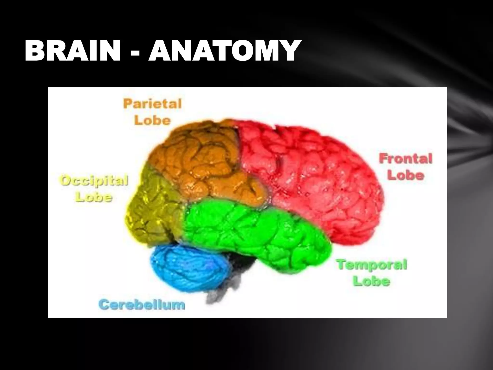
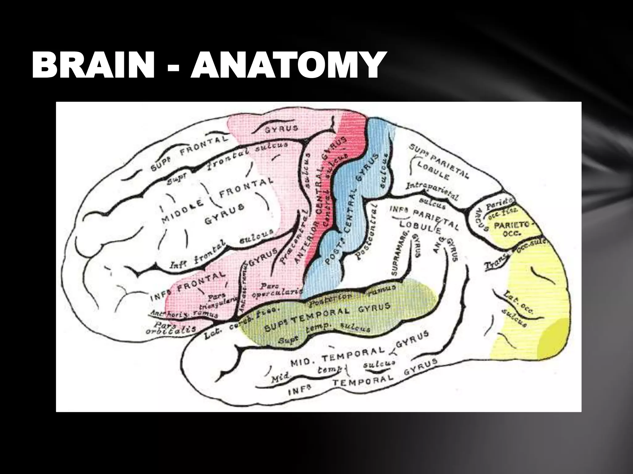


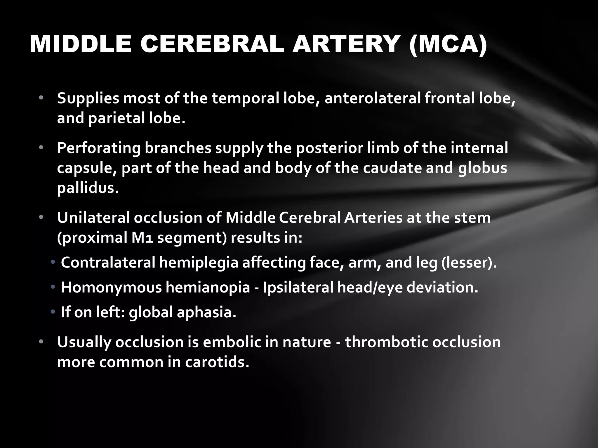
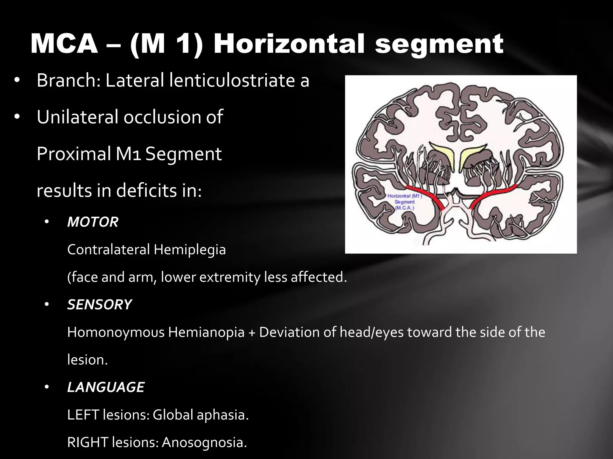


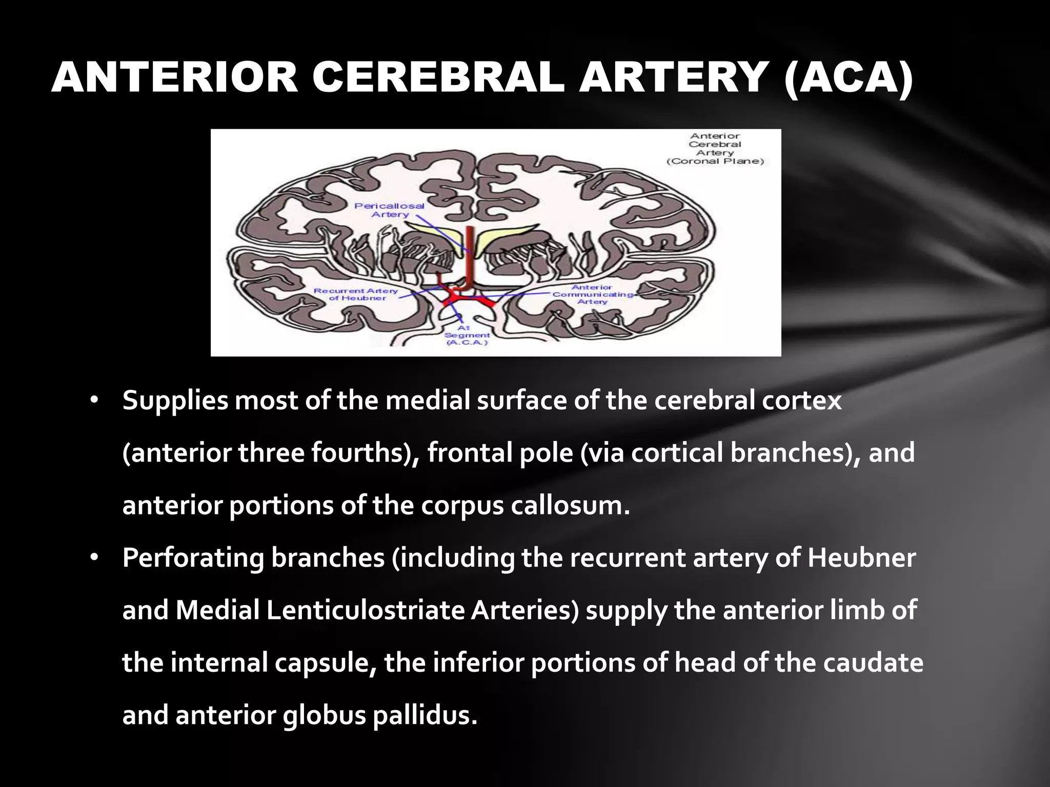

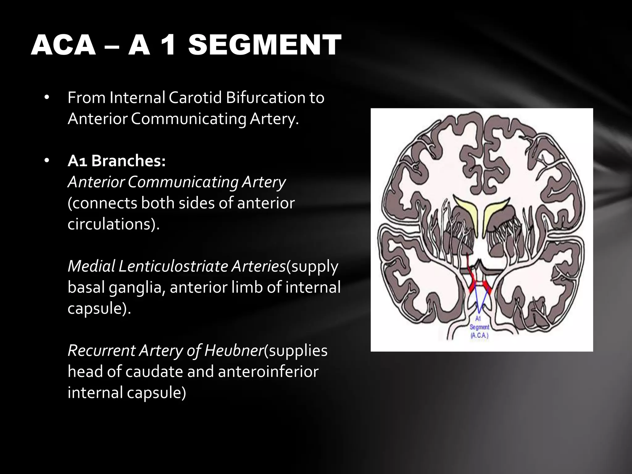


The document discusses the anterior cerebral circulation, including the internal carotid artery, anterior cerebral artery, and middle cerebral artery. It describes the typical vascular territories and clinical deficits that can result from occlusions or infarctions in different segments of these arteries. Key points include that unilateral middle cerebral artery occlusion can cause contralateral hemiplegia and homonymous hemianopia, while bilateral anterior cerebral artery occlusion can lead to paraplegia and urinary incontinence.


