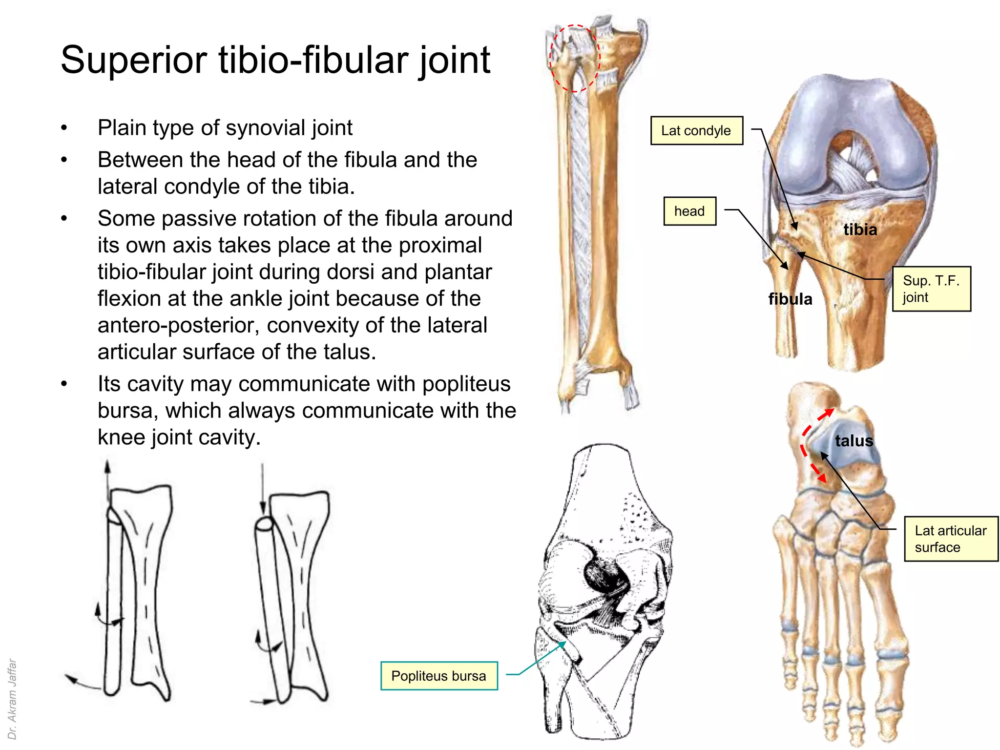
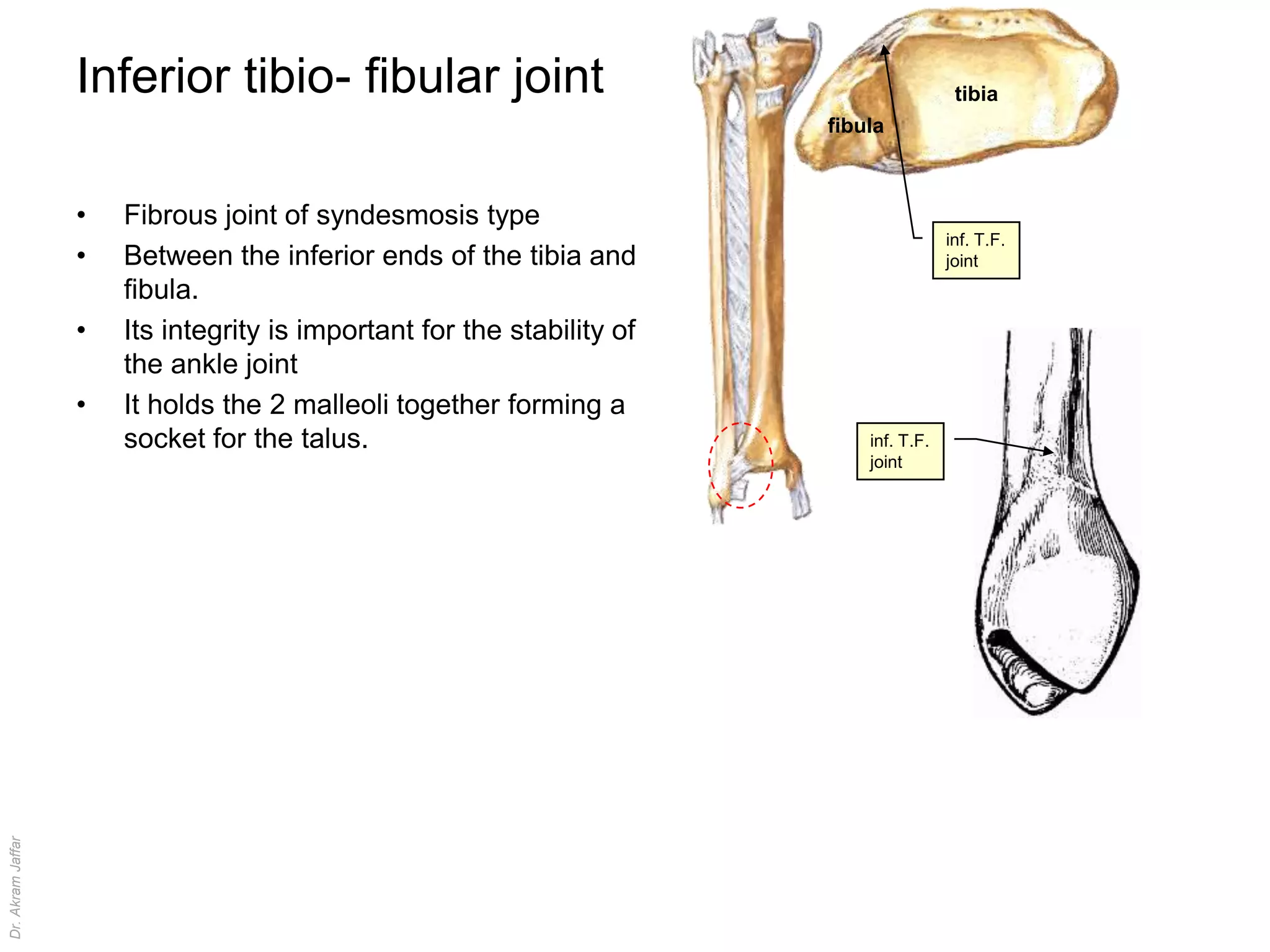
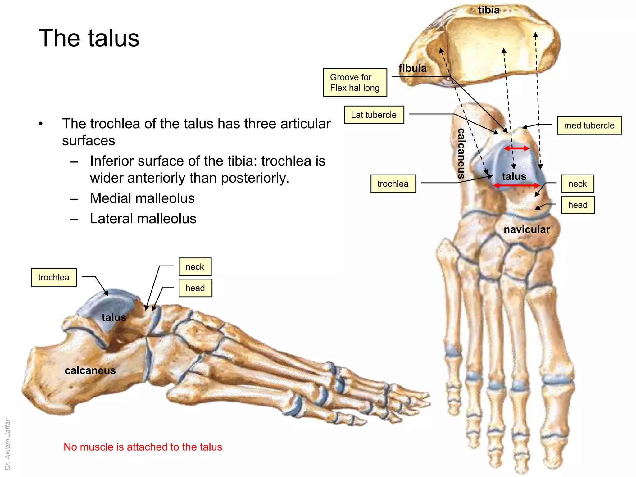
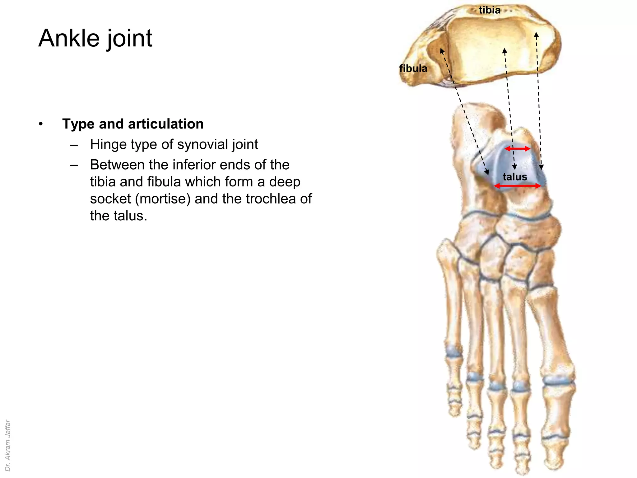
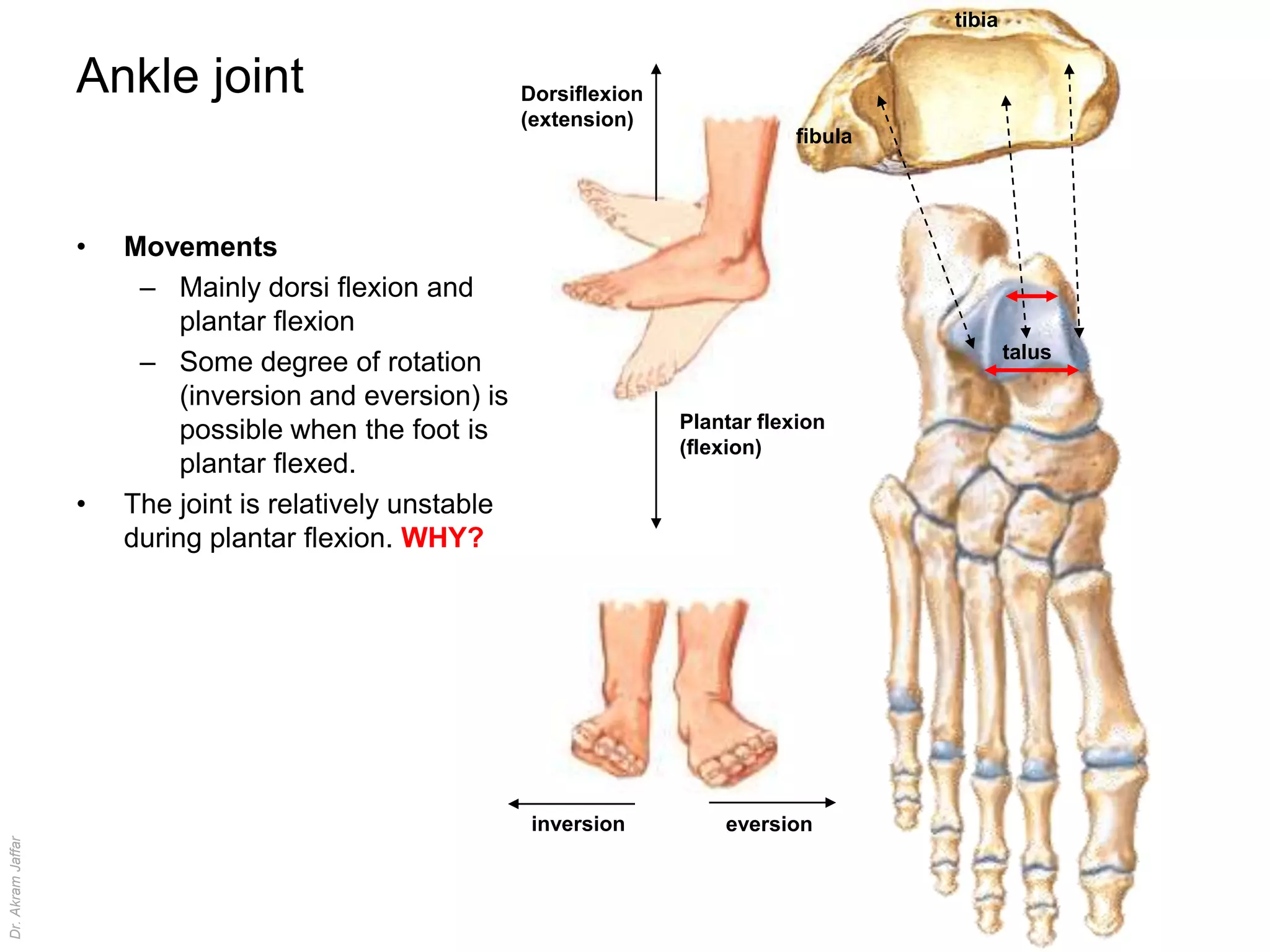
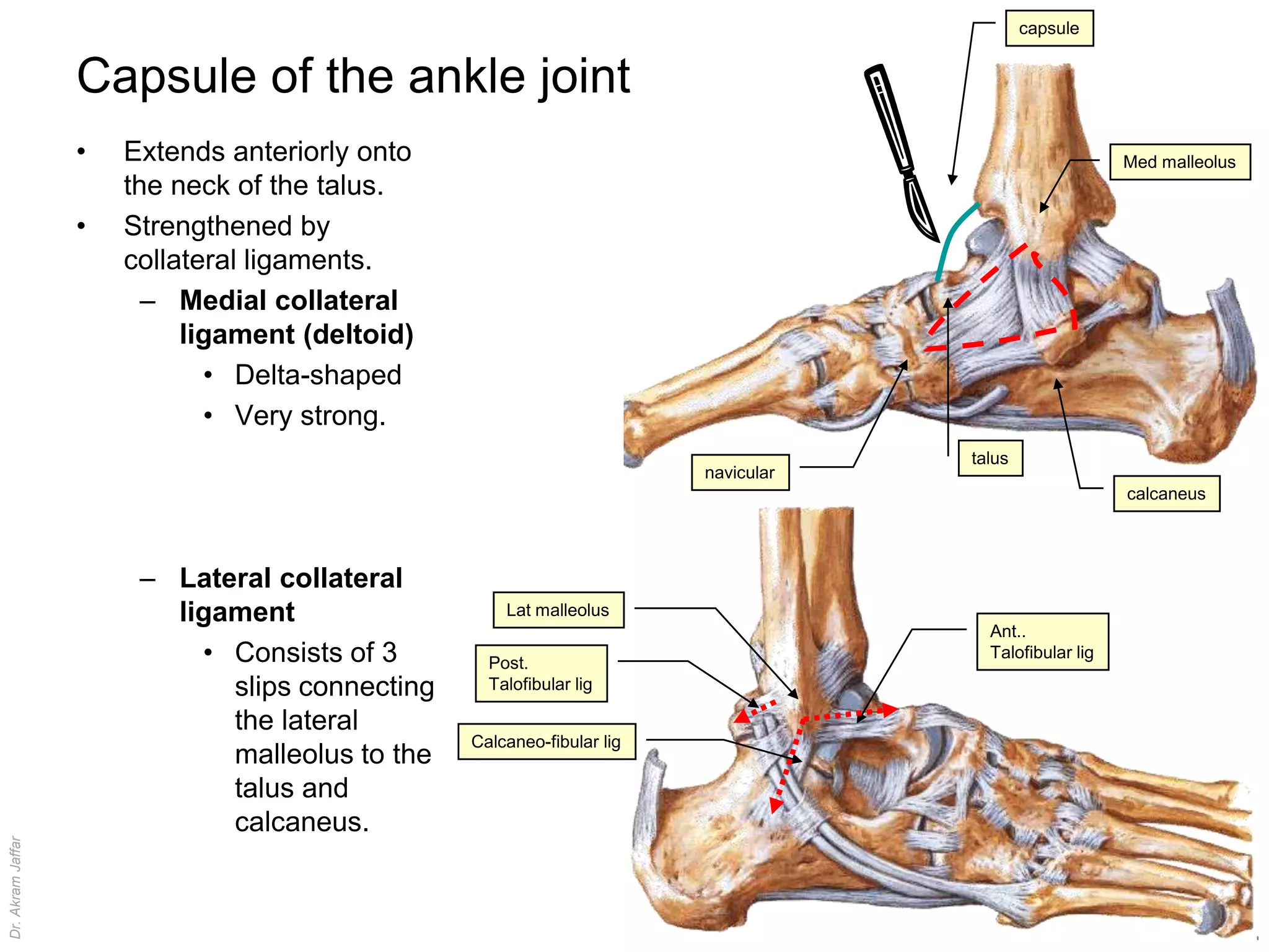
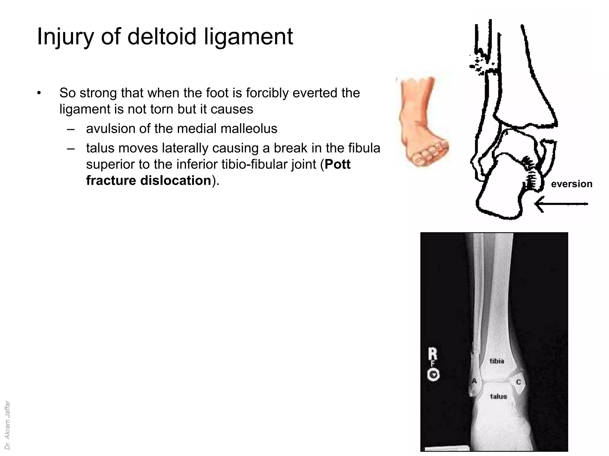
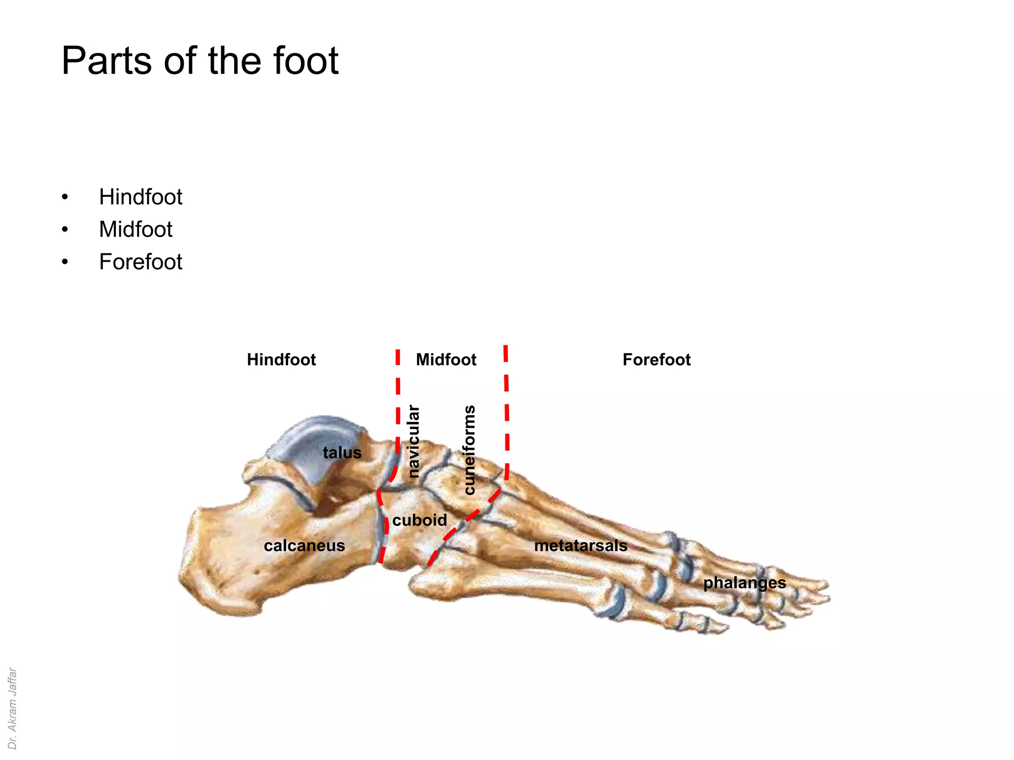
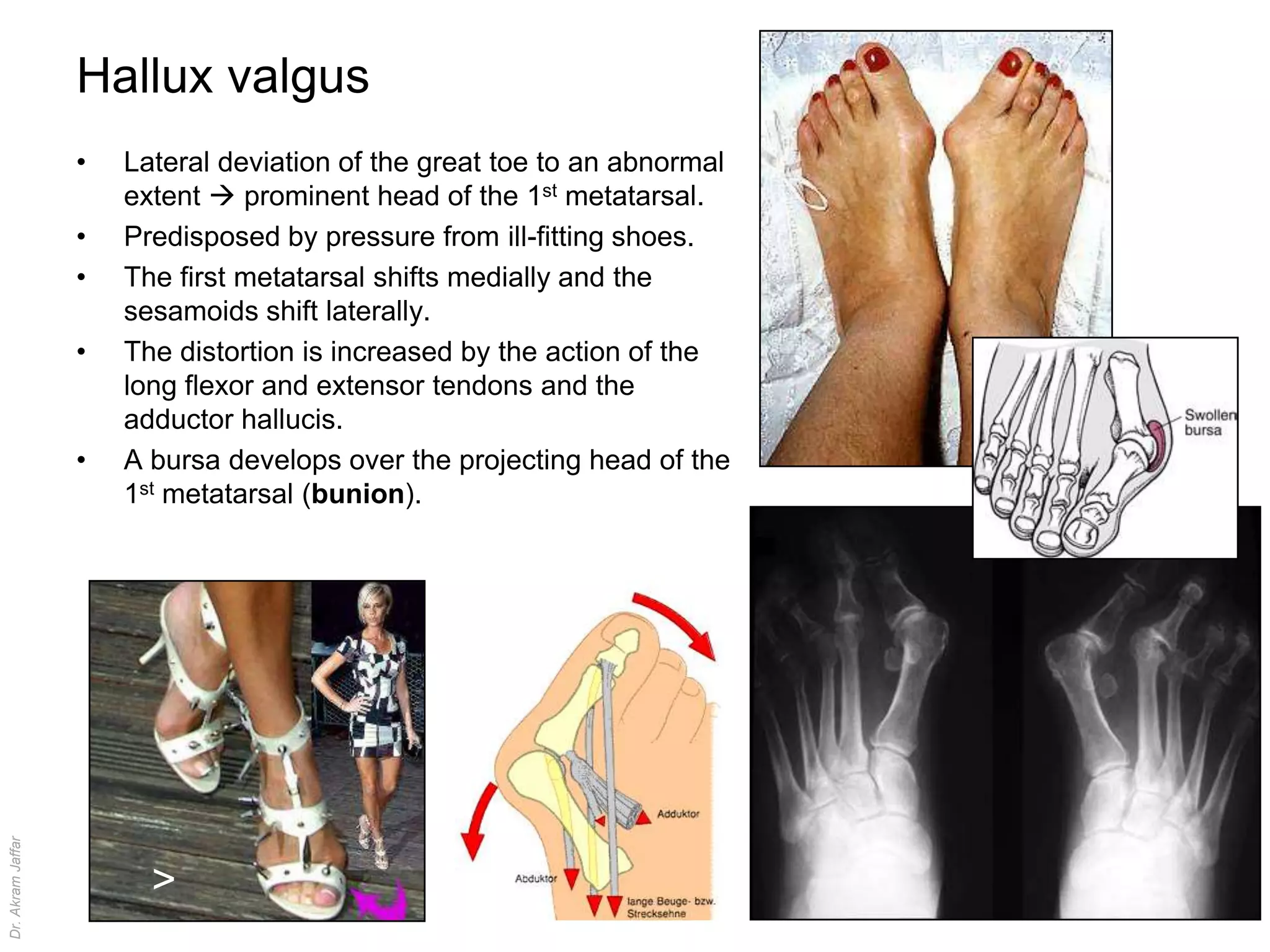
The Ankle & Joints of the Foot

                            Dr. Akram Jaffar
                                  Ph.D.
Dr. Akram Jaffar


                                                Dr. Akram Jaffar
References and suggested reading


                   •   Moore KL & Dalley AF (2006): Clinically oriented anatomy. 5th ed. Lippincott Williams
                       & Wilkins. Baltimore
                   •   Snell RS (2006): Clinical anatomy by systems. Lippincott Williams & Wilkins.
                       Baltimore
Dr. Akram Jaffar


                                                                                                         Dr. Akram Jaffar
Superior tibio-fibular joint
                   •   Plain type of synovial joint                    Lat condyle

                   •   Between the head of the fibula and the
                       lateral condyle of the tibia.
                   •   Some passive rotation of the fibula around        head
                                                                                               tibia
                       its own axis takes place at the proximal
                       tibio-fibular joint during dorsi and plantar                                    Sup. T.F.
                       flexion at the ankle joint because of the                     fibula            joint

                       antero-posterior, convexity of the lateral
                       articular surface of the talus.
                   •   Its cavity may communicate with popliteus
                       bursa, which always communicate with the
                       knee joint cavity.                                                     talus


                                                                                                        Lat articular
                                                                                                        surface
Dr. Akram Jaffar


                                                     Popliteus bursa


                                                                                                       Dr. Akram Jaffar
Inferior tibio- fibular joint                                          tibia
                                                                         fibula


                   •   Fibrous joint of syndesmosis type
                                                                                         inf. T.F.
                   •   Between the inferior ends of the tibia and                        joint
                       fibula.
                   •   Its integrity is important for the stability of
                       the ankle joint
                   •   It holds the 2 malleoli together forming a
                       socket for the talus.                                 inf. T.F.
                                                                             joint
Dr. Akram Jaffar


                                                                                                     Dr. Akram Jaffar
tibia


                   The talus
                                                                                         fibula
                                                                     Groove for
                                                                     Flex hal long


                                                                          Lat tubercle
                   •     The trochlea of the talus has three articular                                                            med tubercle


                                                                                                  calcaneus
                         surfaces
                          – Inferior surface of the tibia: trochlea is
                              wider anteriorly than posteriorly.                                                      talus
                                                                                 trochlea                                          neck
                          – Medial malleolus                                                                                      head
                          – Lateral malleolus
                                                                                                                      navicular


                                                   neck
                   trochlea
                                                   head


                                 talus


                         calcaneus
Dr. Akram Jaffar


                         No muscle is attached to the talus

                                                                                                                                     Dr. Akram Jaffar
tibia


                   Ankle joint
                                                                 fibula


                   •   Type and articulation
                        – Hinge type of synovial joint
                        – Between the inferior ends of the
                                                                                  talus
                          tibia and fibula which form a deep
                          socket (mortise) and the trochlea of
                          the talus.
Dr. Akram Jaffar


                                                                                          Dr. Akram Jaffar
tibia


                   Ankle joint                            Dorsiflexion
                                                          (extension)
                                                                                    fibula


                   •   Movements
                        – Mainly dorsi flexion and
                            plantar flexion
                                                                                                     talus
                        – Some degree of rotation
                            (inversion and eversion) is
                            possible when the foot is                    Plantar flexion
                                                                         (flexion)
                            plantar flexed.
                   •   The joint is relatively unstable
                       during plantar flexion. WHY?


                                                           inversion         eversion
Dr. Akram Jaffar


                                                                                                             Dr. Akram Jaffar
capsule


                   Capsule of the ankle joint
                   •   Extends anteriorly onto                                                          Med malleolus
                       the neck of the talus.
                   •   Strengthened by
                       collateral ligaments.
                         – Medial collateral
                            ligament (deltoid)
                              • Delta-shaped
                              • Very strong.
                                                                                    talus
                                                                        navicular
                                                                                                        calcaneus


                        – Lateral collateral
                          ligament                   Lat malleolus
                                                                                      Ant..
                            • Consists of 3        Post.
                                                                                      Talofibular lig

                              slips connecting     Talofibular lig

                              the lateral
                                                 Calcaneo-fibular lig
                              malleolus to the
                              talus and
                              calcaneus.
Dr. Akram Jaffar


                                                                                                           Dr. Akram Jaffar
Ankle sprain
Dr. Akram Jaffar


                                  Dr. Akram Jaffar
Injury of lateral collateral ligament

                   •   When the foot is forcibly inverted as when the
                       weight-bearing foot trips on an uneven
                       surface.
                   •   The anterior talofibular ligament is the
                       most vulnerable and most commonly torn.
                   •   In severe cases, the calcaneofibular ligament
                       is torn and the lateral malleolus is fractured.


                         Inf tibiofibular joint


                             Lat malleolus
                             fracture
Dr. Akram Jaffar


                                                                         Dr. Akram Jaffar
Injury of deltoid ligament

                   •   So strong that when the foot is forcibly everted the
                       ligament is not torn but it causes
                         – avulsion of the medial malleolus
                         – talus moves laterally causing a break in the fibula
                            superior to the inferior tibio-fibular joint (Pott
                            fracture dislocation).                               eversion
Dr. Akram Jaffar


                                                                                 Dr. Akram Jaffar
Posterior tibio-fibular ligament

                    •      Between malleolar fossa of the fibula to the
                           posterior edge of the tibia.
                                                                                            Post. Tibio-fibular lig.
                    •      Prevents the leg to slide foreword on the
                           talus under the influence of gravity when the
                           foot is plantar flexed at the take off stage in
                                                                                               Post. Talo-fibular lig.
                           walking.


                                                                             inf. T.F.
                                                                             joint

                   Post. Tibio-fibular lig.


                                                                             Malleolar
                                                                             facet
Dr. Akram Jaffar


                                                                                Malleolar
                                                                                fossa
                                                                                                                         Dr. Akram Jaffar
tibia


                   Stability of the ankle joint
                   •   Bone: mortise                                        fibula

                   •   Ligaments: collateral & tibiofibular
                   •   Muscle: surrounding tendons.

                   •   Forward sliding of the leg on the talus is prevented by:
                        – The mortise is deepened at the back                                 talus
                             • Posterior lip of the tibia
                             • Posterior tibio-fibular ligament
                        – The superior articular surface of the talus is wider in
                           front than behind.
                             • The joint is unstable with the foot plantar flexed
                                (walking downhill)  ankles are more commonly
                                sprained whilst walking downstairs rather than when
                                going upstairs.
Dr. Akram Jaffar


                                                                                                      Dr. Akram Jaffar
Parts of the foot


                   •   Hindfoot
                   •   Midfoot
                   •   Forefoot


                                  Hindfoot                 Midfoot                       Forefoot


                                                                  cuneiforms
                                                      navicular

                                             talus


                                                     cuboid
                                   calcaneus                                   metatarsals

                                                                                                    phalanges
Dr. Akram Jaffar


                                                                                                                Dr. Akram Jaffar
Joints of inversion and eversion:       anatomical classification

                   •   Subtalar joint                                                        Subtalar j.
                        – Synovial joint between the inferior         talus
                           surface of the body of the talus
                           and the superior surface of the                       cuboid
                           calcaneus.                           calcaneus
                   •   talocalcaneonavicular joint
                        – Synovial joint between the head of                             Calcaneo-cuboid j.
                           the talus on one side and the
                           posterior surface of the
                           navicular, superior surface of the
                           spring ligament, and the
                                                                                                                       talus


                                                                                                           navicular
                           sustentaculum tali of the
                           calcaneus on the other side.
                   •   calcaneocuboid joint                                                                              calcaneus
                        – Synovial joint between the anterior
                           surface of the calcaneus and the
                           posterior surface of the cuboid.
                                                                Talo-calcaneo-cavicular j.
Dr. Akram Jaffar


                                                                                                                               Dr. Akram Jaffar
Spring ligament

                   •   Officially, plantar calcaneonavicular
                       ligament
                   •   Between the sustentaculum tali and
                       the navicular bone.
                   •   Forms part of the socket for the head
                       of the talus.
                   •   Important in maintaining the medial
                       longitudinal arch of the foot.
                                                               Spring lig.
                                                                                                         Sustentaculum tali


                                                                                                         talus


                                                                                             navicular
                                                                                                           calcaneus


                                                                        Sustentaculum tali
Dr. Akram Jaffar


                                   Sustentaculum tali


                                                                                                                 Dr. Akram Jaffar
Joints of inversion and eversion:               functional classification

                                          talus

                   Subtalar j.


                                                                                                                     calcaneus
                                     calcaneus


                                                                                                     Midtarsal j.                  talus

                                                                      talus                                         cuboid       navicular

                                                                                       Subtalar j.
                                                                        calcaneus


                         •       Mid-tarsal joint (transverse tarsal joint): Articulation of the head of
                                 the talus with the navicular lies in line with the calcaneocuboid
                                 joint.
                         •       Subtalar joint: Articulation of the head of the talus with the spring
                                 ligament and the sustentaculum tali + anatomical subtalar joint
Dr. Akram Jaffar


                                                                                                                                     Dr. Akram Jaffar
Foot amputation


                   •   Transection across the transverse
                       tarsal joint is a standard method for
                       surgical amputation of the foot.
Dr. Akram Jaffar


                       Diabetic foot
                                                               Dr. Akram Jaffar
Avulsion fracture of the 5th metatarsal


                   •   Forcible inversion of the foot  Avulsion of the
                       tuberosity of the 5th metatarsal bone by the
                       attached tendon on peroneus brevis
Dr. Akram Jaffar


                                                                          Dr. Akram Jaffar
“March” fracture of the 2nd metatarsal bone
                   •   The base of the 2nd metatarsal is firmly
                       fixed between the anterior ends of the
                       medial and lateral cuneiforms.
                   •   The 2nd metatarsal and toe form the axis
                       of the foot.
                   •   The immobility of the 2nd metatarsal and
                       the slenderness of its shaft contribute to
                       its „spontaneous‟ fracture following mild
                       repetitive trauma (stress fracture).
Dr. Akram Jaffar


                                                                    Dr. Akram Jaffar
Hallux valgus
                   •   Lateral deviation of the great toe to an abnormal
                       extent  prominent head of the 1st metatarsal.
                   •   Predisposed by pressure from ill-fitting shoes.
                   •   The first metatarsal shifts medially and the
                       sesamoids shift laterally.
                   •   The distortion is increased by the action of the
                       long flexor and extensor tendons and the
                       adductor hallucis.
                   •   A bursa develops over the projecting head of the
                       1st metatarsal (bunion).
Dr. Akram Jaffar


                         >
                                                                           Dr. Akram Jaffar
Long and short plantar ligaments

                   •   Long plantar ligament
                        – Passes from the plantar
                          surface of the
                          calcaneus to the cuboid
                          bone
                        – Bridges the groove on
                          the cuboid and converts
                          it into a tunnel for the
                          passage of the tendon
                          of peroneus longus
                          muscle.                                     cuboid
                                                     Groove for
                   •   Short plantar ligament        peroneus long.               Peroneus
                                                                                  longus
                        – Deep to the long plantar
                          ligament
                        – From the plantar                                     long plantar lig.
                          surface of the
                          calcaneus to the cuboid                                   Short plantar lig.
                          bone.
Dr. Akram Jaffar


                                                                                                         Dr. Akram Jaffar
Formation of the arches of the foot
                                                                                    lateral long. arch


                                               cuboid
                             calcaneus                  2 metatarsals                                             cuneiforms


                                                                                                         cuboid
                                                                                                                               Transverse arch
                       Medial long. arch                   cuneiforms
                                                                                        talus
                                                                        navicular

                                           3 metatarsals
                                                                                           calcaneus


                   •   Medial longitudinal arch
                        – formed by the calcaneus, talus, navicular, 3 cuneiform bones, and the medial
                           three metatarsals
                   •   Lateral longitudinal arch
                        – formed by the calcaneus, cuboid, and the lateral two metatarsals
Dr. Akram Jaffar


                   •   Transverse arch
                        – formed by the cuneiforms, cuboid, and the bases of the metatarsals.
                                                                                                                                     Dr. Akram Jaffar
Function of the arches of the foot
                   •   Support and divide the body weight about equally
                       between the calcaneus and the heads of the metatarsal         80 Kg
                       bones.
                   •   Propel the body in walking or running:
                        – Allow the long flexors and the muscles of the foot to
                           act on the bones of the fore part of the foot and toes
                           (take-off part) and greatly assist the propulsive force
                           of gastricnemius and soleus muscles.
                   •   Shock absorption.
                   •   Adapt to changes when walking on uneven surfaces.
Dr. Akram Jaffar


                                                                                             Dr. Akram Jaffar
Mechanism of arch (bridge) support


                                                                                     Tie beam
                             Key stone
                                                                   staples


                   •   Shape of the stones: Wedge-shaped stones with
                       the thin edge of the wedge lying inferiorly. This is
                       especially true for the stone at the center of the
                       arch “key stone”.                                      suspension
                   •   Staples: Tie the inferior edges of the stones.
                   •   Tie beams: Connect the pillars and prevent their
                       separation.
                   •   Suspension by slings
Dr. Akram Jaffar


                                                                                                Dr. Akram Jaffar
Mechanism of foot arch support


                         •     Shape of bones: e.g. head of the talus “key stone”.
                         •     Staples: e.g. the long and short plantar ligaments.
                         •     Tie beam: e.g. tendon of flexor hallucis longus.
                         •     Suspension: e.g. tendons of tibialis anterior, tibialis
                               posterior, and the peroneii
                                                                                              Short plantar lig.

                                                                                               staples
                                                                                              Long plantar lig.


                                                      Tibialis ant.
                               suspension
                   Tibialis post.


                                                                                         cuneiform

                                                                                                                   talus
Dr. Akram Jaffar


                                                                                               Key stones
                                                    Tie beam
                                    Flex hal long
                                                                                                                           Dr. Akram Jaffar
Factors maintaining the arches of the foot


                   •   Bones, ligaments, and muscles.
                   •   Passive support: Ligaments are sufficient to
                       support the arches when standing still
                   •   Active support: muscles are brought into action to
                       support the arches during running or walking


                                                                            **
                                                                               ligaments
Dr. Akram Jaffar


                                    muscles                                  bones

                                                                                     Dr. Akram Jaffar
Flat feet (pes planus)
                   •   The arch of the foot collapses, with the entire sole of the
                       foot coming into contact with the ground.
                   •   Normal before age of 3 years.
                   •   Affects the medial longitudinal arch.
                   •   Causes: failure of factors maintining the arches
                         – Bone: deformity  rigid flat foot
                         – Ligaments: loose or degenerated  flexible flat foot
                            (only when weight bearing)
                         – Muscle: trauma, degeneration, or denervation 
                            acquired flat foot.
Dr. Akram Jaffar


                                                                                     Dr. Akram Jaffar
head

                                                 sesamoid

                                                                               head
                                                           base
                                      Metatarsal 1                                         neck


                                                                           navicular
                                                                                                    tibia


                                                                                       talus                           fibula
                   phalanx
                                    Metatarsal 5
                                                                  cuboid

                                                                                                                trochlea

                                    tuberosity                                                              condyle

                                                                                        calcaneus

                                    Calcaneocuboid joint
                                                                                                              tuberosity
Dr. Akram Jaffar


                                                                                                                           Dr. Akram Jaffar

Anatomy of the ankle and joints of foot

  • 1.
    The Ankle &Joints of the Foot Dr. Akram Jaffar Ph.D. Dr. Akram Jaffar Dr. Akram Jaffar
  • 2.
    References and suggestedreading • Moore KL & Dalley AF (2006): Clinically oriented anatomy. 5th ed. Lippincott Williams & Wilkins. Baltimore • Snell RS (2006): Clinical anatomy by systems. Lippincott Williams & Wilkins. Baltimore Dr. Akram Jaffar Dr. Akram Jaffar
  • 3.
    Superior tibio-fibular joint • Plain type of synovial joint Lat condyle • Between the head of the fibula and the lateral condyle of the tibia. • Some passive rotation of the fibula around head tibia its own axis takes place at the proximal tibio-fibular joint during dorsi and plantar Sup. T.F. flexion at the ankle joint because of the fibula joint antero-posterior, convexity of the lateral articular surface of the talus. • Its cavity may communicate with popliteus bursa, which always communicate with the knee joint cavity. talus Lat articular surface Dr. Akram Jaffar Popliteus bursa Dr. Akram Jaffar
  • 4.
    Inferior tibio- fibularjoint tibia fibula • Fibrous joint of syndesmosis type inf. T.F. • Between the inferior ends of the tibia and joint fibula. • Its integrity is important for the stability of the ankle joint • It holds the 2 malleoli together forming a socket for the talus. inf. T.F. joint Dr. Akram Jaffar Dr. Akram Jaffar
  • 5.
    tibia The talus fibula Groove for Flex hal long Lat tubercle • The trochlea of the talus has three articular med tubercle calcaneus surfaces – Inferior surface of the tibia: trochlea is wider anteriorly than posteriorly. talus trochlea neck – Medial malleolus head – Lateral malleolus navicular neck trochlea head talus calcaneus Dr. Akram Jaffar No muscle is attached to the talus Dr. Akram Jaffar
  • 6.
    tibia Ankle joint fibula • Type and articulation – Hinge type of synovial joint – Between the inferior ends of the talus tibia and fibula which form a deep socket (mortise) and the trochlea of the talus. Dr. Akram Jaffar Dr. Akram Jaffar
  • 7.
    tibia Ankle joint Dorsiflexion (extension) fibula • Movements – Mainly dorsi flexion and plantar flexion talus – Some degree of rotation (inversion and eversion) is possible when the foot is Plantar flexion (flexion) plantar flexed. • The joint is relatively unstable during plantar flexion. WHY? inversion eversion Dr. Akram Jaffar Dr. Akram Jaffar
  • 8.
    capsule Capsule of the ankle joint • Extends anteriorly onto Med malleolus the neck of the talus. • Strengthened by collateral ligaments. – Medial collateral ligament (deltoid) • Delta-shaped • Very strong. talus navicular calcaneus – Lateral collateral ligament Lat malleolus Ant.. • Consists of 3 Post. Talofibular lig slips connecting Talofibular lig the lateral Calcaneo-fibular lig malleolus to the talus and calcaneus. Dr. Akram Jaffar Dr. Akram Jaffar
  • 9.
    Ankle sprain Dr. AkramJaffar Dr. Akram Jaffar
  • 10.
    Injury of lateralcollateral ligament • When the foot is forcibly inverted as when the weight-bearing foot trips on an uneven surface. • The anterior talofibular ligament is the most vulnerable and most commonly torn. • In severe cases, the calcaneofibular ligament is torn and the lateral malleolus is fractured. Inf tibiofibular joint Lat malleolus fracture Dr. Akram Jaffar Dr. Akram Jaffar
  • 11.
    Injury of deltoidligament • So strong that when the foot is forcibly everted the ligament is not torn but it causes – avulsion of the medial malleolus – talus moves laterally causing a break in the fibula superior to the inferior tibio-fibular joint (Pott fracture dislocation). eversion Dr. Akram Jaffar Dr. Akram Jaffar
  • 12.
    Posterior tibio-fibular ligament • Between malleolar fossa of the fibula to the posterior edge of the tibia. Post. Tibio-fibular lig. • Prevents the leg to slide foreword on the talus under the influence of gravity when the foot is plantar flexed at the take off stage in Post. Talo-fibular lig. walking. inf. T.F. joint Post. Tibio-fibular lig. Malleolar facet Dr. Akram Jaffar Malleolar fossa Dr. Akram Jaffar
  • 13.
    tibia Stability of the ankle joint • Bone: mortise fibula • Ligaments: collateral & tibiofibular • Muscle: surrounding tendons. • Forward sliding of the leg on the talus is prevented by: – The mortise is deepened at the back talus • Posterior lip of the tibia • Posterior tibio-fibular ligament – The superior articular surface of the talus is wider in front than behind. • The joint is unstable with the foot plantar flexed (walking downhill)  ankles are more commonly sprained whilst walking downstairs rather than when going upstairs. Dr. Akram Jaffar Dr. Akram Jaffar
  • 14.
    Parts of thefoot • Hindfoot • Midfoot • Forefoot Hindfoot Midfoot Forefoot cuneiforms navicular talus cuboid calcaneus metatarsals phalanges Dr. Akram Jaffar Dr. Akram Jaffar
  • 15.
    Joints of inversionand eversion: anatomical classification • Subtalar joint Subtalar j. – Synovial joint between the inferior talus surface of the body of the talus and the superior surface of the cuboid calcaneus. calcaneus • talocalcaneonavicular joint – Synovial joint between the head of Calcaneo-cuboid j. the talus on one side and the posterior surface of the navicular, superior surface of the spring ligament, and the talus navicular sustentaculum tali of the calcaneus on the other side. • calcaneocuboid joint calcaneus – Synovial joint between the anterior surface of the calcaneus and the posterior surface of the cuboid. Talo-calcaneo-cavicular j. Dr. Akram Jaffar Dr. Akram Jaffar
  • 16.
    Spring ligament • Officially, plantar calcaneonavicular ligament • Between the sustentaculum tali and the navicular bone. • Forms part of the socket for the head of the talus. • Important in maintaining the medial longitudinal arch of the foot. Spring lig. Sustentaculum tali talus navicular calcaneus Sustentaculum tali Dr. Akram Jaffar Sustentaculum tali Dr. Akram Jaffar
  • 17.
    Joints of inversionand eversion: functional classification talus Subtalar j. calcaneus calcaneus Midtarsal j. talus talus cuboid navicular Subtalar j. calcaneus • Mid-tarsal joint (transverse tarsal joint): Articulation of the head of the talus with the navicular lies in line with the calcaneocuboid joint. • Subtalar joint: Articulation of the head of the talus with the spring ligament and the sustentaculum tali + anatomical subtalar joint Dr. Akram Jaffar Dr. Akram Jaffar
  • 18.
    Foot amputation • Transection across the transverse tarsal joint is a standard method for surgical amputation of the foot. Dr. Akram Jaffar Diabetic foot Dr. Akram Jaffar
  • 19.
    Avulsion fracture ofthe 5th metatarsal • Forcible inversion of the foot  Avulsion of the tuberosity of the 5th metatarsal bone by the attached tendon on peroneus brevis Dr. Akram Jaffar Dr. Akram Jaffar
  • 20.
    “March” fracture ofthe 2nd metatarsal bone • The base of the 2nd metatarsal is firmly fixed between the anterior ends of the medial and lateral cuneiforms. • The 2nd metatarsal and toe form the axis of the foot. • The immobility of the 2nd metatarsal and the slenderness of its shaft contribute to its „spontaneous‟ fracture following mild repetitive trauma (stress fracture). Dr. Akram Jaffar Dr. Akram Jaffar
  • 21.
    Hallux valgus • Lateral deviation of the great toe to an abnormal extent  prominent head of the 1st metatarsal. • Predisposed by pressure from ill-fitting shoes. • The first metatarsal shifts medially and the sesamoids shift laterally. • The distortion is increased by the action of the long flexor and extensor tendons and the adductor hallucis. • A bursa develops over the projecting head of the 1st metatarsal (bunion). Dr. Akram Jaffar > Dr. Akram Jaffar
  • 22.
    Long and shortplantar ligaments • Long plantar ligament – Passes from the plantar surface of the calcaneus to the cuboid bone – Bridges the groove on the cuboid and converts it into a tunnel for the passage of the tendon of peroneus longus muscle. cuboid Groove for • Short plantar ligament peroneus long. Peroneus longus – Deep to the long plantar ligament – From the plantar long plantar lig. surface of the calcaneus to the cuboid Short plantar lig. bone. Dr. Akram Jaffar Dr. Akram Jaffar
  • 23.
    Formation of thearches of the foot lateral long. arch cuboid calcaneus 2 metatarsals cuneiforms cuboid Transverse arch Medial long. arch cuneiforms talus navicular 3 metatarsals calcaneus • Medial longitudinal arch – formed by the calcaneus, talus, navicular, 3 cuneiform bones, and the medial three metatarsals • Lateral longitudinal arch – formed by the calcaneus, cuboid, and the lateral two metatarsals Dr. Akram Jaffar • Transverse arch – formed by the cuneiforms, cuboid, and the bases of the metatarsals. Dr. Akram Jaffar
  • 24.
    Function of thearches of the foot • Support and divide the body weight about equally between the calcaneus and the heads of the metatarsal 80 Kg bones. • Propel the body in walking or running: – Allow the long flexors and the muscles of the foot to act on the bones of the fore part of the foot and toes (take-off part) and greatly assist the propulsive force of gastricnemius and soleus muscles. • Shock absorption. • Adapt to changes when walking on uneven surfaces. Dr. Akram Jaffar Dr. Akram Jaffar
  • 25.
    Mechanism of arch(bridge) support Tie beam Key stone staples • Shape of the stones: Wedge-shaped stones with the thin edge of the wedge lying inferiorly. This is especially true for the stone at the center of the arch “key stone”. suspension • Staples: Tie the inferior edges of the stones. • Tie beams: Connect the pillars and prevent their separation. • Suspension by slings Dr. Akram Jaffar Dr. Akram Jaffar
  • 26.
    Mechanism of footarch support • Shape of bones: e.g. head of the talus “key stone”. • Staples: e.g. the long and short plantar ligaments. • Tie beam: e.g. tendon of flexor hallucis longus. • Suspension: e.g. tendons of tibialis anterior, tibialis posterior, and the peroneii Short plantar lig. staples Long plantar lig. Tibialis ant. suspension Tibialis post. cuneiform talus Dr. Akram Jaffar Key stones Tie beam Flex hal long Dr. Akram Jaffar
  • 27.
    Factors maintaining thearches of the foot • Bones, ligaments, and muscles. • Passive support: Ligaments are sufficient to support the arches when standing still • Active support: muscles are brought into action to support the arches during running or walking ** ligaments Dr. Akram Jaffar muscles bones Dr. Akram Jaffar
  • 28.
    Flat feet (pesplanus) • The arch of the foot collapses, with the entire sole of the foot coming into contact with the ground. • Normal before age of 3 years. • Affects the medial longitudinal arch. • Causes: failure of factors maintining the arches – Bone: deformity  rigid flat foot – Ligaments: loose or degenerated  flexible flat foot (only when weight bearing) – Muscle: trauma, degeneration, or denervation  acquired flat foot. Dr. Akram Jaffar Dr. Akram Jaffar
  • 29.
    head sesamoid head base Metatarsal 1 neck navicular tibia talus fibula phalanx Metatarsal 5 cuboid trochlea tuberosity condyle calcaneus Calcaneocuboid joint tuberosity Dr. Akram Jaffar Dr. Akram Jaffar