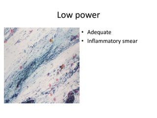
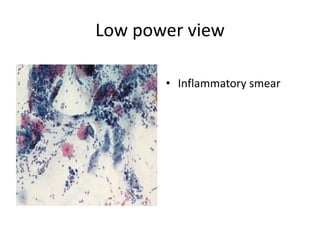
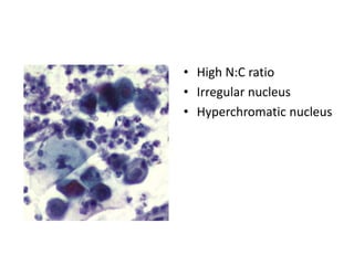
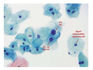
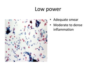
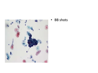
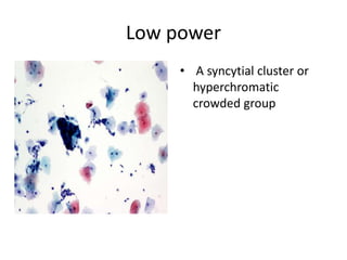
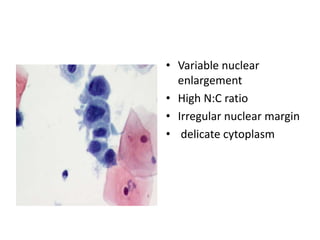
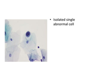
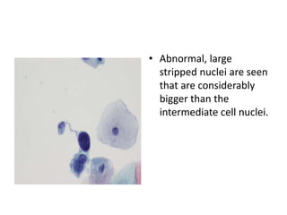
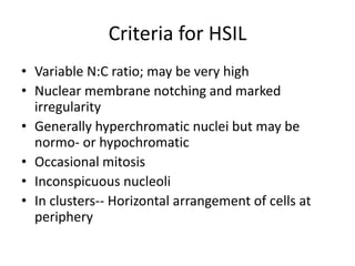
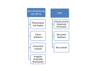
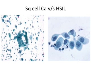
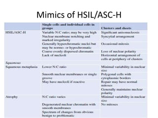
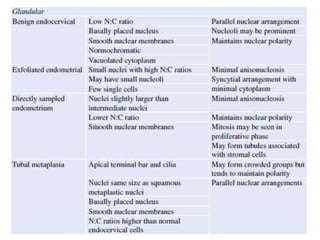
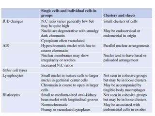
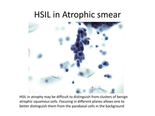
Patient details
• Pt is a 39 yr female, a housewife
• HIV positive since 2001
• Is on Regular ART
H/O present illness
• Pt visited gynecology OPD in December 2014
for a routine examination.
• No h/o dysmenrrhoea, white discharge p/v.
• No h/o irregular menstruation.
• No h/o chronic disease
• No h/o blood transfusion or surgery
Obstetric history:
P1L1, FT ND, male child, 19 yrs back
Menstrual history:
Regular cycle of menstruation
Menarche at 13 years
On Examination
P/A– Soft, non tender, no organomegaly
P/S– Cervix, Vagina healthy
P/V– uterus normal size, AV, FF, NFT
RS/CVS/CNS– NAD
PAP smear was done
Criteria for ASC-H
• Cells are the size of a metaplastic cells with
nuclei 1.5 to 2 times the size of normal.
• Variable N:C ratio; may be very high
• Prominent nuclear irregularity
• Generally hyperchromatic nuclei but may be
normo- or hypochromatic.
• Lack of nucleoli
ASCUS
• Nuclei are 2.5 to 3
times the nucleus of a
normal intermediate Sq
cell or twice the size
• of a squamous
metaplastic cell nucleus
• Slightly increased N:C
ratio
• Minimal nuclear
hyperchromasia
LSIL
Mature squamous cells
with enlarged nuclei
Koilocytosis or perinuclear
cavitation
Binucleation/multinucleati
on is common
Variable size nuclei
• Revisited the OPD on 30th May 2015.
• No specific gynecological complaints.
On examination:
P/A : soft, non-tender
P/S : Cx, vagina healthy
P/V : uterus normal size, FF, NFT.
• USG: Normal study, No obvious mass/focal
lesion in Cervix.
• PAP smear
Criteria for HSIL
• Variable N:C ratio; may be very high
• Nuclear membrane notching and marked
irregularity
• Generally hyperchromatic nuclei but may be
normo- or hypochromatic
• Occasional mitosis
• Inconspicuous nucleoli
• In clusters-- Horizontal arrangement of cells at
periphery
Non keratinizing
Sq cell Ca
Pleomorphic
cell shapes
Tumor
diathesis
Prominent
nucleoli
Irregular
chromatin
distribution
HSIL
Coarse evenly
dispersed
chromatin
No tumor
diathesis
No nucleoli
HSIL in Atrophic smear
HSIL in atrophy may be difficult to distinguish from clusters of benign
atrophic squamous cells. Focusing in different planes allows one to
better distinguish them from the parabasal cells in the background
Kindly Leave a message if
you like the presentation
and follow for further
updates.