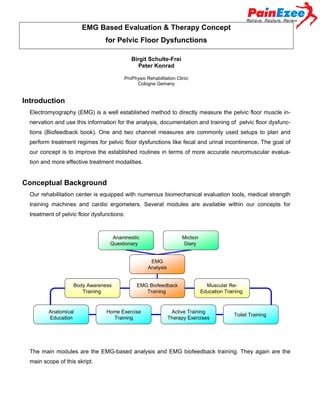
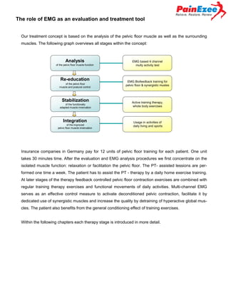
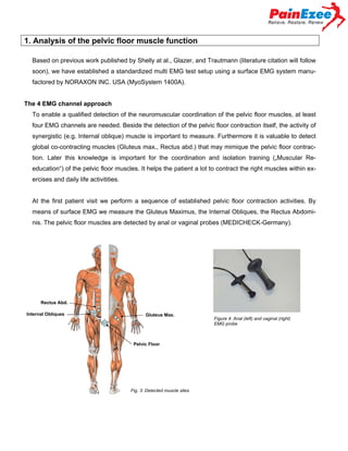
(1) EMG is used to directly measure pelvic floor muscle function and innervation for evaluating and treating dysfunctions like incontinence. (2) A 4-channel EMG system measures the pelvic floor and surrounding muscles during standardized tests to analyze coordination and identify issues. (3) Biofeedback training then focuses on re-educating the pelvic floor muscle through isolated activation exercises and integrating it into whole body movements and daily activities. (4) Retests assess changes in muscle activation, endurance, and coordination from the training.
Non-uniform electromyographic activity during fatigue and recovery of the vas...Nosrat hedayatpour
The aim of the study was to investigate EMG signal features
during fatigue and recovery at three locations of the vastus
medialis and lateralis muscles.
Review of Software to Analyse the Physical Conditions of the Athletes using sEMGijtsrd
Electromyography measures muscle responses of a nerve's simulation of the muscle. EMG is generally measured or recorded through surface, needle or wired electrodes. The surface electromyography is a commonly used technique for measuring the muscle exhilaration. The purpose of this project is to evaluate the use of sEMG in the practical context and to translate the given context to the appropriate analysis. The sEMG are used on the athletes while they are running and respective results are being noted. By using this technique our project wishes to implement an android iOS application to calculate the corresponding values which are being noted by the particular device which we have been made. The signals which are being given by the device is converted into the appropriate percentage values or graphs which can be determined into giving a complete overview about the person whom he is checking and can suggest the diets and exercises to make that person fit to the expectations. This software is mainly look forward for the development of the future athletes which can win the prizes. This platform provides immense forms of diets which are based on the values or results which have been depicted. Performance analysis in sports is considered to be an integral component of understanding the requirements of the optimal performance. Several measurement techniques have been used to inspect the performance of the best athletes today. it is mostly commonly done in laboratory where physiology and bio mechanics can be analyzed. in this system first, the coaches conduct a study about the agility, strength and nutrition of the excellent players of the country. Then the coaches of the respective clubs or the schools check each and every student's physical condition and compare with the stored data in order to train them. The project has got direct advantage to the aspiring future athletes of the country and also to the health conscious society by providing them a device to calculate on their body metrics and work around to improve on it. Tenwin James K | Varun Vincent | Lino Louis | Vishnuraj T | Aneesh Chandran "Review of Software to Analyse the Physical Conditions of the Athletes using sEMG" Published in International Journal of Trend in Scientific Research and Development (ijtsrd), ISSN: 2456-6470, Volume-4 | Issue-4 , June 2020, URL: https://www.ijtsrd.com/papers/ijtsrd31546.pdf Paper Url :https://www.ijtsrd.com/computer-science/embedded-system/31546/review-of-software-to-analyse-the-physical-conditions-of-the-athletes-using-semg/tenwin-james-k
Presented by Brad Aiken
Doctor and science-fiction writer Brad Aiken presents on new and upcoming technologies in neurological rehabilitation. Topics include breakthrough advances that can help people recover from stroke, brain injury, and spinal cord injury. Current, cutting-edge technologies will be discussed, as well as likely upcoming advances in this field.
As expected when the ActivMotion Bar was compared to medicine balls and standard bars in the same movements there was consistently much higher muscle activation all over the body!
PhD Dissertation Defense - Melissa Mazzo - Estimates of neural drive and the ...Melissa Mazzo
A brief background on motor unit physiology and some of the findings from my PhD research projects involving quantification of the neural command to the calf muscles and adjustments in neural drive in response to stretching.
Reduced Short- and Long-Latency Afferent Inhibition Following Acute Muscle Pa...Antonio Martinez
Corticomotor output is reduced in re-
sponse to acute muscle pain, yet the mechanisms
that underpin this effect remain unclear. Here the au-
thors investigate the effect of acute muscle pain on
short-latency afferent inhibition, long-latency afferent
inhibition, and long-interval intra-cortical inhibition to
determine whether these mechanisms could plausibly
contribute to reduced motor output in pain.
Biofeedback machines commonly used in the physiotherapy practice are described along with available evidences for clinical use.
Most of the feedback modalities are described along with its use and the mechanism behind it.
22- Force platform- is the device used to measure the ground reaction forces in steady and/or in moving phase. Sensors in the force platform give visual feedback on the screen attached to therapist as well as patient about the weight distribution and other parameters of gait can also be measured.
33- Mirror -used as visual feedback to treat the pain as well as disability in different conditions. It healps to activate the mirror neuron in the brain. Mirrior therapy proposed by Ramchandran et al has been found most beneficial treatment in the phantom limb pain treatment
Nikos Malliaropoulos - Rehabilitation of hamstring injuries MuscleTech Network
Nikos Malliaropoulos
Director of the Athletics National Sports Medicine Centre Thessaloniki Greece. Consultant SEM Physician Barts and The London Clinical Senior Lecturer QMUL CSEM.
-
The rehabilitation of Hamstring injuries - Can we be more injury specific?
(6th MuscleTech Network Workshop)
14th October, Barcelona
This power point presentation is presented by Satyajit Mohanty, MSPT,MIAP, MHPC(UK), a specialist physiotherapist in sports physiotherapists. This presentation till take you through the manual therapy prospective of lumbar spinal paraspinal EMG.
have a happy reading. Thank you.
Effect of delayed-onset muscle soreness on muscle recovery after a fatiguing ...Nosrat hedayatpour
the aim of the study was to assess EMG MPF during
recovery following a fatiguing contraction at multiple
locations of the quadriceps femoris muscle injured
by eccentric exercise.
Delayed-Onset Muscle Soreness Alters the Response to Postural PerturbationsNosrat hedayatpour
The purpose of this study was to assess the EMG activity
of knee muscles during destabilizing perturbations performed
before, immediately after, and 24 and 48 h after eccentric
exercise.
Basavarajeeyam is an important text for ayurvedic physician belonging to andhra pradehs. It is a popular compendium in various parts of our country as well as in andhra pradesh. The content of the text was presented in sanskrit and telugu language (Bilingual). One of the most famous book in ayurvedic pharmaceutics and therapeutics. This book contains 25 chapters called as prakaranas. Many rasaoushadis were explained, pioneer of dhatu druti, nadi pareeksha, mutra pareeksha etc. Belongs to the period of 15-16 century. New diseases like upadamsha, phiranga rogas are explained.
Ozempic: Preoperative Management of Patients on GLP-1 Receptor Agonists Saeid Safari
Preoperative Management of Patients on GLP-1 Receptor Agonists like Ozempic and Semiglutide
ASA GUIDELINE
NYSORA Guideline
2 Case Reports of Gastric Ultrasound
These simplified slides by Dr. Sidra Arshad present an overview of the non-respiratory functions of the respiratory tract.
Learning objectives:
1. Enlist the non-respiratory functions of the respiratory tract
2. Briefly explain how these functions are carried out
3. Discuss the significance of dead space
4. Differentiate between minute ventilation and alveolar ventilation
5. Describe the cough and sneeze reflexes
Study Resources:
1. Chapter 39, Guyton and Hall Textbook of Medical Physiology, 14th edition
2. Chapter 34, Ganong’s Review of Medical Physiology, 26th edition
3. Chapter 17, Human Physiology by Lauralee Sherwood, 9th edition
4. Non-respiratory functions of the lungs https://academic.oup.com/bjaed/article/13/3/98/278874
Recomendações da OMS sobre cuidados maternos e neonatais para uma experiência pós-natal positiva.
Em consonância com os ODS – Objetivos do Desenvolvimento Sustentável e a Estratégia Global para a Saúde das Mulheres, Crianças e Adolescentes, e aplicando uma abordagem baseada nos direitos humanos, os esforços de cuidados pós-natais devem expandir-se para além da cobertura e da simples sobrevivência, de modo a incluir cuidados de qualidade.
Estas diretrizes visam melhorar a qualidade dos cuidados pós-natais essenciais e de rotina prestados às mulheres e aos recém-nascidos, com o objetivo final de melhorar a saúde e o bem-estar materno e neonatal.
Uma “experiência pós-natal positiva” é um resultado importante para todas as mulheres que dão à luz e para os seus recém-nascidos, estabelecendo as bases para a melhoria da saúde e do bem-estar a curto e longo prazo. Uma experiência pós-natal positiva é definida como aquela em que as mulheres, pessoas que gestam, os recém-nascidos, os casais, os pais, os cuidadores e as famílias recebem informação consistente, garantia e apoio de profissionais de saúde motivados; e onde um sistema de saúde flexível e com recursos reconheça as necessidades das mulheres e dos bebês e respeite o seu contexto cultural.
Estas diretrizes consolidadas apresentam algumas recomendações novas e já bem fundamentadas sobre cuidados pós-natais de rotina para mulheres e neonatos que recebem cuidados no pós-parto em unidades de saúde ou na comunidade, independentemente dos recursos disponíveis.
É fornecido um conjunto abrangente de recomendações para cuidados durante o período puerperal, com ênfase nos cuidados essenciais que todas as mulheres e recém-nascidos devem receber, e com a devida atenção à qualidade dos cuidados; isto é, a entrega e a experiência do cuidado recebido. Estas diretrizes atualizam e ampliam as recomendações da OMS de 2014 sobre cuidados pós-natais da mãe e do recém-nascido e complementam as atuais diretrizes da OMS sobre a gestão de complicações pós-natais.
O estabelecimento da amamentação e o manejo das principais intercorrências é contemplada.
Recomendamos muito.
Vamos discutir essas recomendações no nosso curso de pós-graduação em Aleitamento no Instituto Ciclos.
Esta publicação só está disponível em inglês até o momento.
Prof. Marcus Renato de Carvalho
www.agostodourado.com
Title: Sense of Taste
Presenter: Dr. Faiza, Assistant Professor of Physiology
Qualifications:
MBBS (Best Graduate, AIMC Lahore)
FCPS Physiology
ICMT, CHPE, DHPE (STMU)
MPH (GC University, Faisalabad)
MBA (Virtual University of Pakistan)
Learning Objectives:
Describe the structure and function of taste buds.
Describe the relationship between the taste threshold and taste index of common substances.
Explain the chemical basis and signal transduction of taste perception for each type of primary taste sensation.
Recognize different abnormalities of taste perception and their causes.
Key Topics:
Significance of Taste Sensation:
Differentiation between pleasant and harmful food
Influence on behavior
Selection of food based on metabolic needs
Receptors of Taste:
Taste buds on the tongue
Influence of sense of smell, texture of food, and pain stimulation (e.g., by pepper)
Primary and Secondary Taste Sensations:
Primary taste sensations: Sweet, Sour, Salty, Bitter, Umami
Chemical basis and signal transduction mechanisms for each taste
Taste Threshold and Index:
Taste threshold values for Sweet (sucrose), Salty (NaCl), Sour (HCl), and Bitter (Quinine)
Taste index relationship: Inversely proportional to taste threshold
Taste Blindness:
Inability to taste certain substances, particularly thiourea compounds
Example: Phenylthiocarbamide
Structure and Function of Taste Buds:
Composition: Epithelial cells, Sustentacular/Supporting cells, Taste cells, Basal cells
Features: Taste pores, Taste hairs/microvilli, and Taste nerve fibers
Location of Taste Buds:
Found in papillae of the tongue (Fungiform, Circumvallate, Foliate)
Also present on the palate, tonsillar pillars, epiglottis, and proximal esophagus
Mechanism of Taste Stimulation:
Interaction of taste substances with receptors on microvilli
Signal transduction pathways for Umami, Sweet, Bitter, Sour, and Salty tastes
Taste Sensitivity and Adaptation:
Decrease in sensitivity with age
Rapid adaptation of taste sensation
Role of Saliva in Taste:
Dissolution of tastants to reach receptors
Washing away the stimulus
Taste Preferences and Aversions:
Mechanisms behind taste preference and aversion
Influence of receptors and neural pathways
Impact of Sensory Nerve Damage:
Degeneration of taste buds if the sensory nerve fiber is cut
Abnormalities of Taste Detection:
Conditions: Ageusia, Hypogeusia, Dysgeusia (parageusia)
Causes: Nerve damage, neurological disorders, infections, poor oral hygiene, adverse drug effects, deficiencies, aging, tobacco use, altered neurotransmitter levels
Neurotransmitters and Taste Threshold:
Effects of serotonin (5-HT) and norepinephrine (NE) on taste sensitivity
Supertasters:
25% of the population with heightened sensitivity to taste, especially bitterness
Increased number of fungiform papillae
The Gram stain is a fundamental technique in microbiology used to classify bacteria based on their cell wall structure. It provides a quick and simple method to distinguish between Gram-positive and Gram-negative bacteria, which have different susceptibilities to antibiotics
Basavarajeeyam is a Sreshta Sangraha grantha (Compiled book ), written by Neelkanta kotturu Basavaraja Virachita. It contains 25 Prakaranas, First 24 Chapters related to Rogas& 25th to Rasadravyas.
Colonic and anorectal physiology with surgical implications
ZMPCZM019000.11.03 EMG based evaluation & therapy concept for pelvic floor Dysfunctions
1. EMG Based Evaluation & Therapy Concept
for Pelvic Floor Dysfunctions
Birgit Schulte-Frei and Dr. Peter Konrad
2. EMG Based Evaluation & Therapy Concept
for Pelvic Floor Dysfunctions
Birgit Schulte-Frei
Peter Konrad
ProPhysio Rehabilitation Clinic
Cologne Gemany
Introduction
Electromyography (EMG) is a well established method to directly measure the pelvic floor muscle innervation and use this information for the analysis, documentation and training of pelvic floor dysfunctions (Biofeedback book). One and two channel measures are commonly used setups to plan and
perform treatment regimes for pelvic floor dysfunctions like fecal and urinal incontinence. The goal of
our concept is to improve the established routines in terms of more accurate neuromuscular evaluation and more effective treatment modalities.
Conceptual Background
Our rehabilitation center is equipped with numerous biomechanical evaluation tools, medical strength
training machines and cardio ergometers. Several modules are available within our concepts for
treatment of pelvic floor dysfunctions:
Anamnestic
Questionary
Miction
Diary
EMG
Analysis
Body Awareness
Training
Anatomical
Education
EMG Biofeedback
Training
Home Exercise
Training
Muscular ReEducation Training
Active Training
Therapy Exercises
Toilet Training
The main modules are the EMG-based analysis and EMG biofeedback training. They again are the
main scope of this skript.
3. The role of EMG as an evaluation and treatment tool
Our treatment concept is based on the analysis of the pelvic floor muscle as well as the surrounding
muscles. The following graph overviews all stages within the concept:
Analysis
of the pelvic floor muscle function
Re-education
of the pelvic floor
muscle and postural control
Stabilization
of the functionally
adapted muscle innervation
Integration
of the improved
pelvic floor muscle innervation
EMG based 4 channel
multy activity test
EMG Biofeedback training for
pelvic floor & synergistic musles
Active training therapy,
whole body exercises
Usage in activities of
daily living and sports
Insurance companies in Germany pay for 12 units of pelvic floor training for each patient. One unit
takes 30 minutes time. After the evaluation and EMG analysis procedures we first concentrate on the
isolated muscle function: relaxation or facilitation the pelvic floor. The PT- assisted lessions are performed one time a week. The patient has to assist the PT - therapy by a daily home exercise training.
At later stages of the therapy feedback controlled pelvic floor contraction exercises are combined with
regular training therapy exercises and functional movements of daily activities. Multi-channel EMG
serves as an effective control measure to activate deconditioned pelvic contraction, facilitate it by
dedicated use of synergistic muscles and increase the quality by detraining of hyperactive global muscles. The patient also benefits from the general conditioning effect of training exercises.
Within the following chapters each therapy stage is introduced in more detail.
4. 1. Analysis of the pelvic floor muscle function
Based on previous work published by Shelly at al., Glazer, and Trautmann (literature citation will follow
soon), we have established a standardized multi EMG test setup using a surface EMG system manufactored by NORAXON INC. USA (MyoSystem 1400A).
The 4 EMG channel approach
To enable a qualified detection of the neuromuscular coordination of the pelvic floor muscles, at least
four EMG channels are needed. Beside the detection of the pelvic floor contraction itself, the activity of
synergistic (e.g. Internal oblique) muscle is important to measure. Furthermore it is valuable to detect
global co-contracting muscles (Gluteus max., Rectus abd.) that may mimique the pelvic floor contraction. Later this knowledge is important for the coordination and isolation training („Muscular Reeducation“) of the pelvic floor muscles. It helps the patient a lot to contract the right muscles within exercises and daily life activitities.
At the first patient visit we perform a sequence of established pelvic floor contraction activities. By
means of surface EMG we measure the Gluteus Maximus, the Internal Obliques, the Rectus Abdominis. The pelvic floor muscles are detected by anal or vaginal probes (MEDICHECK-Germany).
Rectus Abd.
Internal Obliques
Gluteus Max.
Pelvic Floor
Fig. 3: Detected muscle sites
Figure 4: Anal (left) and vaginal (right)
EMG probe
5. We measure the smoothed rectified (RMS 100 ms) EMG signal in a band width of 20 to 500 Hz. and
at 1000 Hz sampling frequency. All data are acquired and analyzed with the clinical application protocol “Incontinence Multi-Activity Test” within MyoResearch XP (NORAXON INC USA).
After the electrode application the patient has to perform a standardized sequence of pelvic floor activities, as proposed by Glazer:
EMG Baseline
5 seconds of muscle relaxation
Quick Flicks
5 fast upwards contractions and immediate relaxation
Maximal Up-Contractions
5 maximal contractions with 10 seconds duration/pausing
Endurance Hold
Static conctraction of 30 – 60 sec. duration
Resting Tone
Immediate relaxation right after the Endurance Hold
The software automatically guides through the test sequence by prompting visual and acoustic contraction commands.
Feedback Monitor:
shrinking circle when
contracting up
Norm – range,
predefined threshold
settings
Electronic Feedback
assistent
Fig. 5: Measurement & Biofeedbackmonitor of MyoResearch XP
6. All signals can be observed in real time, stored records are shown in the record viewer and the test
results are analyzed in an automatic analysis report:
Record Viewer Screen:
Baseline
Quick Flicks
Max.
Contraction
Static
Hold
Fig. 6: Record Viewer showing all activities of the multi activity test protocol within MyoResearch XP
Resting
Tone
7. The test report analyzes each activity with a set of individual parameters.
Activity:
Analysis Parameters and Findings
Baseline:
Parameters: Mean EMG Amplitude, Coefficient of Variance
Observation:No relaxation of the pelvic floor muscle in laying
Reduced activation in standing
Reduced co-activation of the M. oblique internus
Diagnosis:
Quick Flicks:
Parameters: Averaged Peak Amplitude, Time to/after Peak,
Findings:
Maximal contraction
Hypertonus in laying position
Hypotonus in standing position
slow peak increse within quick-flick bursts
slow relaxation after quick activation
reduced peak activation level
co-activation of the surrounding muscles: M. gluteus,
M. internus abdom.
Less co-activation of the M. oblique internus
Diagnosis: Muscular dysfunction, relaxation deficits
Parameters: Averaged Mean Value
Findings:
Diagnosis:
Endurance Hold
reduced pelvic floor activation level
steep decrease of activity witin 10 seconds
problem to innervate over 10 seconds
co-activation of the M- transverses abdominis, M. gluteus max.
Muscular dysfunction, weakness, relaxation deficits
Parameters: Amplitude and Frequency change over time
Findings:
Diagnosis:
Resting Tone
Time domain changes due to fatigue
Constancy of contraction level
co-activation of M. gluteus max, M. rectus abdom.
Reduced endurance, innervation deficits
Parameters: Mean EMG Amplitude, Coefficient of Variance
Findings:
Increased rest line activity,
late rest line level
Diagnosis:
Hypertonus, Relaxation Deficits
8. 2 – Re-education of the pelvic floor muscle and postural control
The re-education program has two basic areas:
Up-/Downtraining to address hyper- or hypo-activity
“Up training” applies to weak or hypotonic pelvic floor muscles, “Down training” to hyper active pelvic
floor muscles. Based on the analysis of the pelvic floor EMG and the daily MVC (patients maximal volontary contraction) the EMG feedback assisted therapy concentrates on muscular activation training.
Fig. 7: Biofeedback recording with predefined threshold range (yellow area) for MVC normalized EMG contractions. Upper
trace: pelvic floor, lower trace: gluteus max.
Isolated pelvic floor contraction to address coordinative deficits
By using at least 2 EMG channels the ability of the patient to selectively contract the pelvic floor muscle and the underlaying body awareness can be trained. The focus here is the quality of isolation, not
the height of amplitude. Hyperactive global muscles may be retraining to relaxation while performing
the isolated pelvic floor contraction.
9. Fig. 8: Multi-channel EMG
recording (left panel) with
synchronized digital video
(upper right picture) and
real time analysis (lower
right bar graphs). Within
this abdominal drawing
exercise, high EMG is
aquired for pelvic floor and
internal oblique (ch. 1-2),
while keeping the activation
of global muscles low (ch.
3-4)
Within the first 3 therapy units the patient has to “find” and improve his pelvic floor innervation, later
this contraction has to be coordinated other synergistic muscles, i.e. the m. oblique internus. If patients
have difficulties to contract the target muscle, backward facilitation over the internal obliques is very
successful. Breathing techniques may also help to facilitate the pelvic floor innervation. Functionally,
both muscle groups are closely linked together:
Aus: Richardson et al
1999, page 95
Synergy of pelvic floor lower deep
Abdominal muscles (healthy subject)
Fig. 9: Schematic drawing (left) of the functional muscle cylinder of deep trunk muscles: EMG raw recordings (right) of the
pelvic floor (upper trace) and internal oblique (lower trace) show a fully synchronized innervation pattern in healthy subjects
10. One very important effect of the EMG based biofeedback-training is, that the patient can directly “see”
and control the correct activation of the selected muscles. Once found the correct innervation can be
linked with the corresponding body feeling, which again effectively trains the body awareness (“muscular re-education”). The following Biofeedback screens show an efficient and isolated pelvic floor
contraction (upper trace) with absolute innervation silence of the gluteal muscles (lower trace)
Predefined threshold range set to 30 –
50 % MVC
Fig. 10: Biofeedback recording with predefined threshold range (yellow area) for MVC normalized EMG contractions. Upper trace: pelvic floor, lower trace: gluteus max.
11. 3 – Stabilization of the functionally adapted muscle innervation
At this stage of the therapy process the pelvic floor
muscle contraction is integrated into whole body
tasks and movements. Exercises such as functional
gymnastics and “medical training therapy” are performed. A small handheld Biofeedback device can be
used to assist all exercises.
Fig. 11: Regular leg press training machine facilitates
the pelvic floor contraction
Fig. 12: Two channel biofeedback EMG unit
(MyoTrace – Noraxon INC. USA)
The main target of this stage is to train pelvic floor contraction within other exercises. This stage is still
assisted by isolated muscle training addressing the pelvic floor Up training (increased innervation levels, improved endurance, muscle hypertrophy).
Fig. 13: Telemetric 8 channel EMG recording of trunk and hip muscles with synchronized DV video. The EMG pattern of all involved muscles can be studied while performing regular abdominal training exercise (crunch). Note the high pelvic floor innervation > 50% MVC at peak position.
12. 4 – Integration of the improved pelvic floor muscle innervation
Within the last part of the therapy the improved pelvic floor muscle contraction ability is integrated in
general activities of daily living and acting (i.e. caughing, laughing, sneezing, lifting up, work and
sports demand). The isolated muscle training strategies concentrates on the maximum activation of
the pelvic floor muscle.
Fig. 14: (Same measurement
setup as fig. 13). Treadmill running as a typical daily activity
exercise. Note the high contraction level of pelvic floor muscles
(ch. 1)
Fig. 15: (Same measurement
setup as fig. 13). Hopping on a
trampoline.
13. Retest analysis
After 6 and 12 therapy units we perform a multy activity re-test. Changes the maximum innervation
level, the innervation constancy, the coordination between synergists and the muscle relaxation ability
are compared and documented.
Fig. 16: Test (grey curve) and Retest (red curve) comparison plot . The EMG innervation level of the pelvic floor
muscles (ch. 1) and internal oblique (ch. 2) are significantly increased after the epoche of 12 EMG biofeedback
sessions.
Nearly all patients subjectively report on an improvement of their incontinence. This effect goes confirm with an improvement of the sEMG-data of the pelvic floor and the surrounding muscles.
14. Pelvic Floor Home Training
Isolated Contraction of Pelvic Floor Muscles
Contract pelvic floor muscles while exhaling
Contract without using the gluteal upper abdominal muscles.
Date
Intensity
Reps.
Sets
Quick Flicks and Relaxation
Contract pelvic floor muscles as quick as possible and try to
immediately relax again
Date
Intensity
Reps.
Sets
Endurance
Keep a constant pelvic floor contraction over several breathing
cycles
Date
Intensity
Breathes
Sets
Mobilization, Strengthening and Coordination
Pelvi tilt in prone lying, seated and standing. While exhaling,
tilt the pelvis upwards
Date
Intensity
Reps.
Sets