
Recommended
More Related Content
Similar to X INTERACTION.ppt
Similar to X INTERACTION.ppt (20)
More from sudheendrapv
More from sudheendrapv (20)
Recently uploaded
Recently uploaded (20)
X INTERACTION.ppt
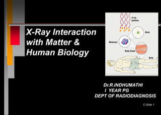
- 1. C-Slide 1 X-Ray Interaction with Matter & Human Biology Dr.R.INDHUMATHI I YEAR PG DEPT OF RADIODIAGNOSIS
- 2. INTRODUCTION n X ray photons are created by the interaction of energetic electrons with matter at atomic levels. n These photons end their lives by transferring their energy to electrons contained in matter. C-Slide 2
- 3. C-Slide 3 Interaction in The body begin at the atomic level Atoms Molecules Cells Tissues Organ structures
- 4. C-Slide 4 1. It can penetrate the section of matter without interacting. 2. It can interact with the matter and be completely absorbed by depositing its energy. 3. It can interact and be scattered or deflected from its original direction and deposit part of its energy.
- 5. C-Slide 5 Patient Interactions **Photoelectric** Classic Coherent Scatter **Compton Scattering** Pair Production Photodisintegration
- 6. C-Slide 6
- 7. Coherent Scattering n Also called: CLASSICAL SCATTERING UNMODIFIED SCATTERING ELASTIC SCATTERING C-Slide 7
- 8. COHERENT SCATTERING n It is type of interaction in which radiation undergoes a change in direction without a change in wavelength. n is a pure scattering interaction and deposits no energy in the material n It accounts for less than 5% of total interaction. C-Slide 8
- 9. PROBABILITY OF CLASSICAL SCATTERING Increases with A] low atomic number materials ( soft tissue more likely than bone ) B] lower photon energies ( 5 kev more likely than 10 kev) C-Slide 9
- 10. COHERENT SCATTERING n Low energy x-ray photons interacts with electron of the atom . Absorption of radiation by the atom n Vibration of the atom n Emission of radiation as the atom returns to its undisturbed state C-Slide 10
- 12. Coherent Scattering n The wavelength is equal to the incident x-ray n The only difference is the direction of travel n Energy in = Energy out - Only changes is direction n n No ionization takes place ( only type). C-Slide 12
- 13. SUB CLASSIFICATION THOMPSON SCATTERING: if interaction occurs with single electron. RAYLEIGH SCATTERING: if interaction occurs with all the electrons of whole atom. C-Slide 13
- 14. Use in diagnostic radiology Since its quantity is too small no value in diagnostic radiology. scattered radiation produced contributes only to film fog. C-Slide 14
- 15. Compton scattering A compton interaction is one in which only a portion of the energy is absorbed and the resultant photon is scattered with reduced energy. Occurs throughout the diagnostic imaging range Most common interaction b/w x rays and body tissues C-Slide 15
- 16. Probability of crompton scatter n 1] the probability of compton interaction is directly proportional to the number of outer shell electrons n 2] compton effect decreases with increase in photon energy. C-Slide 16
- 17. Compton Scattering an incident photon with relatively high energy Interacts with atom it ejects the outer shell electron from its orbit the incident photon thus gets deflected by electron travels in a new direction as scatter radiation. C-Slide 17
- 18. Compton Scattering this reaction produces an ion pair that is a) A positive atom b) A negative electron ( called as recoil electron) C-Slide 18
- 19. C-Slide 19
- 20. Compton Scattering n Energy of incident photon is distributed as a) part of it goes to the recoil electron as kinetic energy. b) rest is retained by the deflected photon. C-Slide 20
- 21. Compton scattering n Two factors determine the amount of energy A] its initial incident energy B] its angle of deflection off the recoil electron. C-Slide 21
- 22. Applications in diagnostic radiology n Incident energy: the higher the energy of the photons the more difficult they are to deflect n For example with x ray energies of 1Mev most scattered photons deflect in a forward direction. C-Slide 22
- 23. C-Slide 23
- 24. Compton scattering the lower the energy radiation fewer photons scatter forward and more scatter back at an angle of 180 in the diagnostic energy range upto 150 kev the photon retains most of its original energy and very little is transferred to the recoil electron. C-Slide 24
- 25. Compton scattering n Angle of deflection at narrow angles of deflection scattered photons retain almost all their original energy. a) they have an excellent chance of reaching an x ray film and producing fog C-Slide 25
- 26. Compton scattering b) they are exceedingly difficult to removed by grids because their angle of deflection is too small and also by the filters because they are too energetic this causes major radiation hazard to personnel C-Slide 26
- 27. Compton scattering n For example in flouroscopic procedure n the scattered radiation produced is almost as energetic as the primary beam n This creates a real safety hazard for the flouroscopist and other personnel in exposure rooms. C-Slide 27
- 28. Scatter from the Patient during Fluoroscopy C-Slide 28
- 29. Photoelectric interaction It is a type of photon electron interaction in which a photon transfers all of its energy to an electron located in one of the atomic shells. Most useful interaction in diagnostic radiology. C-Slide 29
- 30. Photoelectric Effect or Absorption n Inner-shell ionization n The photon is not scattered it is totally absorbed n The only disadvantage is that it exposes the patient to great deal of radiation. C-Slide 30
- 31. Photoelectric effect n An incident photon with a little more energy than the binding energy of k shell electron Ejects the electron from orbit The photon disappears giving up all its energy to electron. C-Slide 31
- 32. PHOTOELECTRIC EFFECT n The electron which is now free of its energy flies off into space as a photoelectron n The vacancy in the atom is filled by the electron from L shell n Resulting in the formation of characteristic radiation C-Slide 32
- 33. PHOTOELECTRIC EFFECT When k shell is filled by an outer shell electron from the same atom The atom is left with the deficiency of one electron Remains as a positive ion C-Slide 33
- 34. PHOTOELECTRIC EFFECT n The photoelectric effect always yields three end products 1] characteristic radiation 2] a negative ion ( photoelectron) 3] a positive ion ( an atom defecient one electron) C-Slide 34
- 35. C-Slide 35
- 36. PROBABILITY OF PE EFFECT n It depends on two factors n 1] incident photon energy n 2] atomic number C-Slide 36
- 37. Probability of photoelectric interaction n For the PE to occur incident x ray must have energy greater than binding energy of the inner shell electron. n For example the BEof k shell electron in iodine is 33 kev, hence sharp increase in interaction of photons occurs when x ray photon energy exceeds 33 kev. C-Slide 37
- 38. Binding Energy is very important C-Slide 38
- 39. PROBABILITY OF PHOTOELECTRIC EFFECT n The PE is proportional to the atomic number .the more tightly bound an electron the greater the probability of the PE if energy is greater than the BE. n The photoelectric effect is more likely to occur in absorbers of high atomic number (eg, bone, positive contrast media) C-Slide 39
- 40. APPLICATION OF PE INTERACTION n Produces radiographic images with excellent quality. n The PE does not produce scatter. n Enhances natural tissue contrast. n The PE magnifies the difference in tissue composed of different elements such as bone and soft tissue. C-Slide 40
- 41. C-Slide 41 PHOTOELECTR IC ABSORBTION IS WHAT GIVES US THE CONTRAST ON THE FILM
- 42. Pair production n It is a type of photon matter interaction in which high energy photons( energy > 1.02Mev) n Interacts with nucleus of the atom the photons diappear n Its energy is converted into matter in the form of two particles C-Slide 42
- 43. Pair production electron positron ( 0.51 Mev) (a particle with the same mass as electron but with positive charge) C-Slide 43
- 44. C-Slide 44
- 45. Probability of pair production n It happens when photon energy is in excess of 1.022 Mev. n Increases with increase in atomic number. C-Slide 45
- 46. photodisintegration In photodisintegration ,part of the nucleus of an atom is ejected by a high energy photon The ejected portion may be a neutron, a proton, an alpha particle, or a cluster of particles The photon must have sufficient energy to overcome nuclear binding energies of the order of 7 to 15Mev. C-Slide 46
- 48. Diagnostic radiology n Since pair production does not occur with photon energies less than 1.02 Mev and n Photodisintegration does not occur with energies less than 7 Mev . n Both of these interaction is of no use in diagnostic radiology because of its energy range is only upto 150 kev. C-Slide 48
- 49. Important X-ray Interactions n Of the five interactions only two are important to radiology • Photoelectric effect or photoelectric absorption • Compton scatter n Which two tube interactions are important? C-Slide 49
- 50. Compton scatter n Contributes to no useful information n Is independent of the atomic number of tissue. The probability of Compton is the same for bone atoms and for soft tissue atoms n The probability for Compton is more dependent on kVp or x-ray energy C-Slide 50
- 51. Compton Scatter n Results in image fog by optical densities not representing diagnostic information C-Slide 51
- 52. Photoelectric Absorption n Provides information to the IR because photons do not reach the IR n This represents anatomic structures with high x-ray absorption characteristics; radiopaque structures; tissue with high atomic number; or tissue with high mass density C-Slide 52
- 53. Compton vs. Photoelectric n Below 60 kVp Photoelectric absorption is predominant above 60 kVp Compton scatter begins to increase. C-Slide 53
- 54. 3 Types of x-rays are important for IMAGE FORMATION n DIFFERENTIAL ABSORPTION = the difference between those x-rays absorbed and those transmitted to the IR n Compton scatter (no useful information) n Photoelectric absorption (produces the light areas on the image) n Transmitted x-rays (produces the grey/dark areas on the image) C-Slide 54
- 55. Differential absorption n The probability of radiation interaction is a function of tissue electron density/ atomic number, tissue thickness/density, and x-ray energy (kVp). n Dense material like bone and contrast dye attenuates more X-rays from the beam than less dense material (muscle, fat, air). n The differential rate of attenuation provides the contrast necessary to form an image. n Table 10-10 C-Slide 55
- 56. Differential Absorption n Increases as the kVp is reduced n Approximately 1% of photons that interact with the patient (primary beam) reach the IR. Of that 1% approximately 0.5% interact to form the image C-Slide 56
- 57. Differential Absorption n The difference in x-ray interactions n Fundamental for image formation n Occurs because of Compton Scattering, Photoelectric absorption, and X-ray transmission C-Slide 57