
Recommended
More Related Content
Similar to radiograpicviewsforshoulderjoint-180322063116 (2).pdf
Similar to radiograpicviewsforshoulderjoint-180322063116 (2).pdf (20)
More from sudheendrapv
More from sudheendrapv (20)
Recently uploaded
Recently uploaded (20)
radiograpicviewsforshoulderjoint-180322063116 (2).pdf
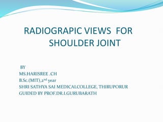
- 1. RADIOGRAPIC VIEWS FOR SHOULDER JOINT BY MS.HARISREE .CH B.Sc.(MIT),2nd year SHRI SATHYA SAI MEDICALCOLLEGE, THIRUPORUR GUIDED BY PROF.DR.I.GURUBARATH
- 2. ANATOMY OF SHOULDER JOINT
- 3. BASIC VIEWS OF SHOULDER JOINT ANTERIO-POSTERIOR (AP) SUPERIO-INFERIO(AXIAL) INFERO-SUPERIOR (REVERSE AXIAL)
- 4. ANTERIO-POSTEROR (AP) POSITIONING : The arm is supinated and slightly abducted away from the body. The medial and lateral epicondyles of the distal humerus should be parallel to the cassette The cassette is positioned so that its upper border is at least 5 cm above the shoulder to ensure that the oblique rays do not project the shoulder off the cassette.
- 5. TECHINICAL DETAILS IR SIZE : 24x30 CM FFD :100 CM GRID :YES KvP:65 ;MAS:16 CENTRAL BEAM: PERPENDICULAR TO IR CENTRAL POINT: GLENOHUMERAL JOINT, THIS IS 2.5CM BELOW THE PALPATABLE CORACOID PROCESS
- 6. RADIOGRAPHIC ANATOMY OF SHOULDER AP view
- 7. SUPERIO-INFERIOR (AXIAL) POSITIONING: • The patient is seated at the side of the table, which is lowered to waist level. • The cassette is placed on the tabletop, and the arm under examination is abducted over the cassette. • The patient leans towards the table to reduce the object-to film distance (OFD) and to ensure that the glenoid cavity is included in the image. A curved cassette, if available, can be used to reduce the OFD. • The elbow can remain flexed, but the arm should be abducted to a minimum of 45 degrees, injury permitting. If only limited abduction is possible, the cassette may be supported on pads to reduce the OFD
- 8. TECHINICAL DETAILS IRSIZE: 18X24cm GRID: NO FFD: 100 cm CENTRALBEAM: CR IS PENPENDICULAR TO IR CENTRAL POINT: CR TO SHOULDERJOINT AT AN ANGLE OF 5 TO 15 DEGREES TOWARDS THE ELBOW
- 9. RADIOGRAPIC ANATOMY OF SHOULDER AXIAL VIEW
- 10. INFERIO-SUPERIOR (REVERSE AXIAL) INTRODUCTION: This projection may be used as an alternative to the superoinferiorprojection in cases of dislocation or when the patient is supine POSITIONING: The patient lies supine, with the arm of the affected side slightly abducted and supinated without causing discomfort to the patient. The affected shoulder and arm are raised on non-opaque pads. A cassette is supported vertically against the shoulder and is pressed against the neck to include as much as possible of the scapula on the film.
- 11. TECHINICAL DETAILS IRSIZE :18X24CM GRID:NO FFD:100CM CENTRAL BEAM : CR IS PERPENDICULAR TO IR CENTRAL POINT: HORIZONTAL BEAM IS CENTRED TOWARDS THE AXILLA WITH MINIMUM ANGULATION TOWARDS TRUNK
- 12. RADIOGRAPIC ANATOMY OF SHOULDER REVERSE AXIAL VIEW
- 13. SPECIAL PROJECTIONS GARTH PROJECTION (APICAL OBLIQUE) WALLACE PROJECTION (SUPERO-INFERIOR MODIFIED) “Y” PROJECTION (ANTERIO OBLIQUE) WEST POINT PROJECTION (INFERIO-SUPERIOR) STRYKERS PROJECTION GRASHEY PROJECTION
- 14. GRATH PROJECTION INTRODUCTION : This projection is recommended as the second projection should an axial not be possible. It will more readily demonstrate Hill-Sachs lesions and glenoid rim fractures. POSITIONING: The patient is positioned erect (either standing or sitting)with their back against a vertical Bucky The patient is then rotated toward the affected side so they attain a 45 degree posterior oblique position. The elbow is usually flexed with the patient’s arm held across the chest.
- 15. TECHINICAL DETIALS IR SIZE : 24X30 CM FFD :100CM GRID: YES CENTRAL RAY: Horizontal beam is centered to the image receptor and 45 degrees caudal tube angulation is employed
- 16. Garth projection
- 17. WALLACE PROJECTION INTRODUCTION: Axillary view is an excellent method for evaluating for anterior or posterior glenohumeral subluxation or dislocation and may also be helpful in the detection of an osseous Bankart fracture involving the anterior glenoid rim. POSITION OF PATIENT: The patient sits erect with their back to the X-ray table. The torso is adjusted to bring the body of the scapula parallel with the table. The image receptor is placed flat on the tabletop immediately behind the shoulder under examination.
- 18. TECHINICAL DETIALS IR SIZE:24X30 CM FFD: 150 CM CENTRAL RAY: CENTRED TO THE MIDDLE OF THE GLENO HUMERAL JOINT USING 30 DEGREES ANGULATION FROM THE VERTICAL POSITION
- 19. Glenohumeral subluxation Osseous Bankart fracture
- 21. ‘Y’ PROJECTION INTRODUCTION: This projection is useful for differentiating the direction of a dislocation but it is less useful for demonstrating associated fractures PATIENT POSITION: The patient stands or sits with lateral aspect of the injured arm against the image receptor and is adjusted so that the axilla is in the centre of the receptor The unaffected shoulder is raised to make an angle between the trunk and the receptor approximately 60degrees. A line joining the medical and lateral borders of scapula is now at right angles to receptor.
- 22. TECHINICAL DETIALS Exposure:70 kVp 20 mAs (in Bucky) FFD :100cm Central Beam: CR perpendicular to IR Central Ray: CR directed to the scapulohumeral joint (5 - 6cm) below top of shoulder
- 23. Radiographic anatomy of shoulder Y view
- 24. WEST POINT PROJECTION INTRODUCTION: Demonstrates the anterior aspect of the glenoid rim and is useful for detecting Bankart lesions PATIENT POSITION: Patient prone on the X-ray table Abduct affected arm away from the body 90° if possible, with elbow flexed to allow forearm to hang freely over side of table Rotate the head away from the affected side Place IR against the superior surface of the affected shoulder
- 25. TECHINICAL DETAILS IR Size :18 x 24cm Gird :No Exposure:55 kVp8 mAs FFD / SID:100cm Central Ray: CR directed 25° cranially and 25° medially through the midscapulohumeral joint
- 26. Radiographic anatomy of WestPoint projection Glenoid Glenoid
- 27. STRYKER PROJECTION INTRODUCTION: • This projection is highly effective in demostrating a Hills-sachs deformity of humeral head. PATIENT POSITION: • The patient lies supine on the table. • The arm of affected side is extended fully and the elbow then flexed to allow the hand to rest on the patient head • The line joining the epicondyles of humerus remains parallel to tabletop • The center of the receptor is positioned 2.5cm superior the head of the humerus
- 28. Technical Details Exposure:65 kVp16 mAs FFD / SID: 100cm CENTRAL RAY: Vertical beam is angled 10degree cranially and centre through the centre of axilla to the head of the humerus and the centre of receptor.
- 29. Radiographic anatomy of strykers projection Hills-Sachs
- 30. GRASHEY PROJECTION INTRODUCTION: Demonstrate a clear joint space between the head of humerus and glenoid cavity. PATIENT POSITION The patient stands wit the affected shoulder against the image receptor and torso is rotted approximately 35-45 degrees toward the affected side to bring the plane of glenoidfossa perpendicular to receptor. The arm is supinated and slightly abducted away from the body.
- 31. Technical Details CR SIZE: 24X30cm FFD:100 cm CENTRE RAY: Horizontal beam is centered just below the palpable coracoid process of scapula
- 32. Radiographic anatomy of Grashey projection The glenohumeral joint is seen in profile (arrows) without overlap of the humerus and glenoid