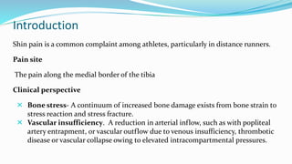
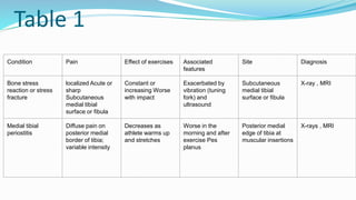
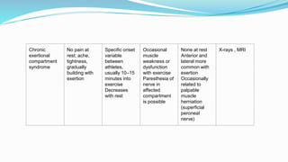
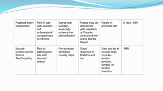
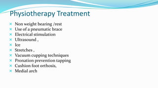
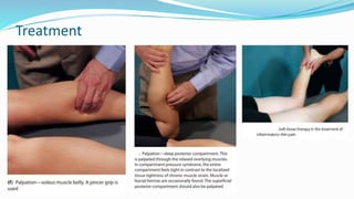
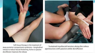
Dr. Darshita Fatnani
Introduction
Shin pain is a common complaint among athletes, particularly in distance runners.
Pain site
The pain along the medial border of the tibia
Clinical perspective
 Bone stress- A continuum of increased bone damage exists from bone strain to
stress reaction and stress fracture.
 Vascular insufficiency. A reduction in arterial inflow, such as with popliteal
artery entrapment, or vascular outflow due to venous insufficiency, thrombotic
disease or vascular collapse owing to elevated intracompartmental pressures.
 Inflammation - Develops at the insertion of muscles,
particularly the tibialis Posterior ,tibialis anterior and
soleus, and fascia to the medial border of the tibia.
 Raised intracompartmental pressure - As a result
of overuse/ inflammation, these muscle compartments
may become swollen and painful, particularly if there is
excessive fibrosis of the fascia.
 Nerve entrapment -(e.g. superficial peroneal nerve).
Role of biomechanics
Both extremes of foot type can contribute to the incidence of shin pain in athletes
A rigid, cavus foot has limited shock absorption, thus increasing the impact
pressure on the bone. In athletes with excessive pronation, the muscles of the
superficial (soleus) and deep compartments (tibialis posterior, flexor hallucis longus,
flexor digitorum longus) are placed at a relatively lengthened position and are
required to contract eccentrically harder and longer to resist pronation after heel
strike. On toe-off , these muscles must contract concentrically over a greater length
to complete the transition to a supinated foot creating a rigid lever for push off .
With fatigue, these muscles fail to provide the normal degree of shock absorption.
The athlete with excessive pronation may also develop lateral shin pain or stress
injuries of the fibula. Pronation of the fixed foot creates an obligate internal tibial
rotation. With repetitive excessive pronation, the tibia and fibula are exposed to
repetitive rotational (torque) stresses. These stresses are transferred across the fibula,
tibia, and proximal and distal tibiofibular articulations. Based on these
biomechanical stresses, overuse can lead to stress reactions or stress fracture not only
of the tibia but also of the fibula.
Ankle instability secondary to chronic ankle sprains will obligate the athlete to
overuse the peroneal tendons to compensate for ankle stability. This overuse can be
just enough to send an athlete with borderline compartment syndrome over the
brink to symptomatic complaints.
Table 1
Condition Pain Effect of exercises Associated
features
Site Diagnosis
Bone stress
reaction or stress
fracture
localized Acute or
sharp
Subcutaneous
medial tibial
surface or fibula
Constant or
increasing Worse
with impact
Exacerbated by
vibration (tuning
fork) and
ultrasound
Subcutaneous
medial tibial
surface or fibula
X-ray , MRI
Medial tibial
periostitis
Diffuse pain on
posterior medial
border of tibia;
variable intensity
Decreases as
athlete warms up
and stretches
Worse in the
morning and after
exercise Pes
planus
Posterior medial
edge of tibia at
muscular insertions
X-rays , MRI
Chronic
exertional
compartment
syndrome
No pain at
rest; ache,
tightness,
gradually
building with
exertion
Specific onset
variable
between
athletes,
usually 10–15
minutes into
exercise
Decreases
with rest
Occasional
muscle
weakness or
dysfunction
with exercise
Paresthesia of
nerve in
affected
compartment
is possible
None at rest
Anterior and
lateral more
common with
exertion
Occasionally
related to
palpable
muscle
herniation
(superficial
peroneal
nerve)
X-rays , MRI
Popliteal artery
entrapment
Pain in calf
with exertion
not
anterolateral ‘
compartment
syndrome’
Worse with
exertion,
especially
active ankle
plantarflexion
Pulses may be
diminished
with palpation
or Doppler
ultrasound with
active plantar
flexion
Rarely in
proximal calf
X-rays , MRI
Muscle–
tendon injuries
Strains
Tendinopathy
Pain at
pathological
site with
resisted
stretch
Pre-exercise
stretching
usually helps
Good
response to
NSAIDs and
ice
Pain can be at
muscle belly,
muscle–
tendon
junction,
tendon, or
tendon
insertion
MRI
Physiotherapy Treatment
 Non weight bearing /rest
 Use of a pneumatic brace
 Electrical stimulation
 Ultrasound ,
 Ice
 Stretches ,
 Vacuum cupping techniques
 Pronation prevention tapping
 Cushion foot orthosis,
 Medial arch
Treatment
Shin pain   shin splint

Shin pain shin splint

  • 1.
  • 2.
    Introduction Shin pain isa common complaint among athletes, particularly in distance runners. Pain site The pain along the medial border of the tibia Clinical perspective  Bone stress- A continuum of increased bone damage exists from bone strain to stress reaction and stress fracture.  Vascular insufficiency. A reduction in arterial inflow, such as with popliteal artery entrapment, or vascular outflow due to venous insufficiency, thrombotic disease or vascular collapse owing to elevated intracompartmental pressures.
  • 3.
     Inflammation -Develops at the insertion of muscles, particularly the tibialis Posterior ,tibialis anterior and soleus, and fascia to the medial border of the tibia.  Raised intracompartmental pressure - As a result of overuse/ inflammation, these muscle compartments may become swollen and painful, particularly if there is excessive fibrosis of the fascia.  Nerve entrapment -(e.g. superficial peroneal nerve).
  • 4.
    Role of biomechanics Bothextremes of foot type can contribute to the incidence of shin pain in athletes A rigid, cavus foot has limited shock absorption, thus increasing the impact pressure on the bone. In athletes with excessive pronation, the muscles of the superficial (soleus) and deep compartments (tibialis posterior, flexor hallucis longus, flexor digitorum longus) are placed at a relatively lengthened position and are required to contract eccentrically harder and longer to resist pronation after heel strike. On toe-off , these muscles must contract concentrically over a greater length to complete the transition to a supinated foot creating a rigid lever for push off . With fatigue, these muscles fail to provide the normal degree of shock absorption.
  • 5.
    The athlete withexcessive pronation may also develop lateral shin pain or stress injuries of the fibula. Pronation of the fixed foot creates an obligate internal tibial rotation. With repetitive excessive pronation, the tibia and fibula are exposed to repetitive rotational (torque) stresses. These stresses are transferred across the fibula, tibia, and proximal and distal tibiofibular articulations. Based on these biomechanical stresses, overuse can lead to stress reactions or stress fracture not only of the tibia but also of the fibula. Ankle instability secondary to chronic ankle sprains will obligate the athlete to overuse the peroneal tendons to compensate for ankle stability. This overuse can be just enough to send an athlete with borderline compartment syndrome over the brink to symptomatic complaints.
  • 6.
    Table 1 Condition PainEffect of exercises Associated features Site Diagnosis Bone stress reaction or stress fracture localized Acute or sharp Subcutaneous medial tibial surface or fibula Constant or increasing Worse with impact Exacerbated by vibration (tuning fork) and ultrasound Subcutaneous medial tibial surface or fibula X-ray , MRI Medial tibial periostitis Diffuse pain on posterior medial border of tibia; variable intensity Decreases as athlete warms up and stretches Worse in the morning and after exercise Pes planus Posterior medial edge of tibia at muscular insertions X-rays , MRI
  • 7.
    Chronic exertional compartment syndrome No pain at rest;ache, tightness, gradually building with exertion Specific onset variable between athletes, usually 10–15 minutes into exercise Decreases with rest Occasional muscle weakness or dysfunction with exercise Paresthesia of nerve in affected compartment is possible None at rest Anterior and lateral more common with exertion Occasionally related to palpable muscle herniation (superficial peroneal nerve) X-rays , MRI
  • 8.
    Popliteal artery entrapment Pain incalf with exertion not anterolateral ‘ compartment syndrome’ Worse with exertion, especially active ankle plantarflexion Pulses may be diminished with palpation or Doppler ultrasound with active plantar flexion Rarely in proximal calf X-rays , MRI Muscle– tendon injuries Strains Tendinopathy Pain at pathological site with resisted stretch Pre-exercise stretching usually helps Good response to NSAIDs and ice Pain can be at muscle belly, muscle– tendon junction, tendon, or tendon insertion MRI
  • 9.
    Physiotherapy Treatment  Nonweight bearing /rest  Use of a pneumatic brace  Electrical stimulation  Ultrasound ,  Ice  Stretches ,  Vacuum cupping techniques  Pronation prevention tapping  Cushion foot orthosis,  Medial arch
  • 10.