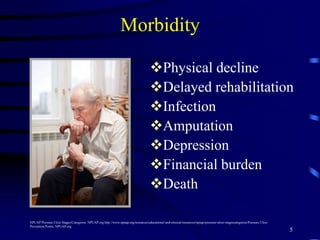
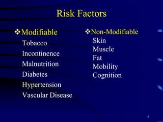
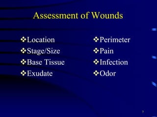
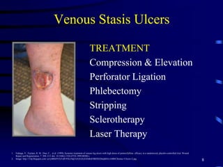
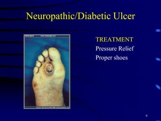
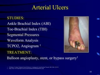
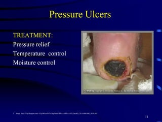
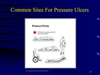
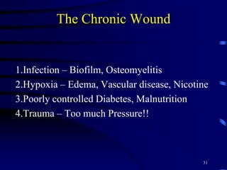
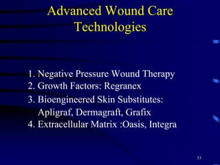
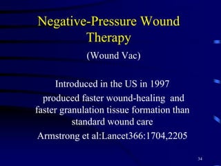
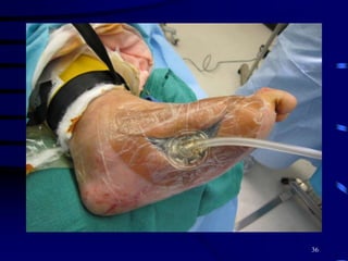
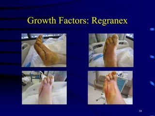
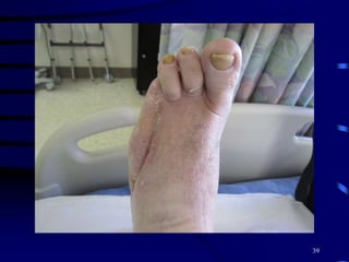
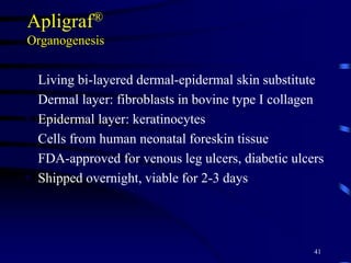
WOUNDS IN THE
GERIATRIC POPULATION
Adriana Salas, MSN, RN, CNS, ANP-BC
Ingrid Kruse, DPM
VA San Diego Healthcare
San Diego, CA
1
Outline
WHY WOUNDS ARE IMPORTANT
RISK FACTORS
ASSESSMENT
STANDARD WOUND MANAGEMENT
REASSESSMENT
ADVANCED WOUND CARE
2
Wounds are Common
ANY ULCER 12% of the population >65yrs 1
VENOUS STASIS ULCERS 1% of US
population 2
PRESSURE ULCER 0.4 – 38%
70% of pressure ulcers occur in the elderly 3,4,5
3
1. U.S. Census Bureau. Statistical abstract of the United States. 2012. http://www.census.gov/compendia/statab/2012/tables/12s0009.pdf. Accessed 9/15/14.
2. Gillespie DL. Venus ulcer diagnosis, treatment, and prevention of recurrences. J Vasc Surg. 2010: 52:8S-14.
3. Cuddigan J, Berlowitz DR, Ayello EA. Pressure ulcers in America: prevalence, incidence, and implications for the future. Reston VA: National Pressure Ulcer Advisory Panel; 2001.
4. https://www.soa.org/news-and-publications/newsroom/press-releases/society-of-actuaries/default.aspx
5. National Pressure Ulcer Advisory Panel and European Pressure Ulcer Advisory Panel. Prevention and treatment of pressure ulcers: clinical practice guideline. Washington,DC: National
Pressure Advisory Panel; 2009.
Wounds are Expensive
Foot infections are the most common reason for
hospitalization in Diabetics
PRESSURE ULCER COST
$43,180 - $151,500 per hospital stay
$9.2 -$15.6 billion total U.S. cost 1,5
LEGAL COST
2nd leading cause for litigation in long term care4
Medicare and Medicaid services stopped reimbursing acute
care facilities for treatment of pressure ulcers in 2008
4
Diabetes Care 1998;21:2161-2177 and JAPMA 98:166,2008
Morbidity
Physical decline
Delayed rehabilitation
Infection
Amputation
Depression
Financial burden
Death
5
NPUAP Pressure Ulcer Stages/Categories. NPUAP.org http://www.npuap.org/resources/educational-and-clinical-resources/npuap-pressure-ulcer-stagescategories/Pressure Ulcer
Prevention Points. NPUAP.org
Risk Factors
Modifiable
Tobacco
Incontinence
Malnutrition
Diabetes
Hypertension
Vascular Disease
6
Non-Modifiable
Skin
Muscle
Fat
Mobility
Cognition
Assessment of Wounds
Location
Stage/Size
Base Tissue
Exudate
Perimeter
Pain
Infection
Odor
7
Venous Stasis Ulcers
TREATMENT
Compression & Elevation
Perforator Ligation
Phlebectomy
Stripping
Sclerotherapy
Laser Therapy
8
1. Falanga, V., Fujitani, R. M., Diaz, C., et al (1999), Systemic treatment of venous leg ulcers with high doses of pentoxifylline: efficacy in a randomized, placebo-controlled trial. Wound
Repair and Regeneration, 7: 208–213. doi: 10.1046/j.1524-475X.1999.00208.x
2. Image: http://3.bp.blogspot.com/-ye1j4BSJ5VA/Ud9-93LC0qI/AAAAAAAABx8/IBDXEDmjhHA/s1600/Chronic+Ulcers+2.jpg
Neuropathic/Diabetic Ulcer
TREATMENT
Pressure Relief
Proper shoes
9
Arterial Ulcers
STUDIES:
Ankle Brachial Index (ABI)
Toe-Brachial Index (TBI)
Segmental Pressures
Waveform Analysis
TCPO2, Angiogram 1
10
1. D Holtman, V Gahtan. Peripheral Arterial Perfusion: is it adequate for wound healing? Wounds. 2008: 20(8): 230-235.
2. Image: http://www.angiologist.com/wp-content/uploads/2010/08/Rutherford_6.jpg
TREATMENT:
Balloon angioplasty, stent, or bypass surgery1
Pressure Ulcers
TREATMENT:
Pressure relief
Temperature control
Moisture control
11
1. Image: http://1.bp.blogspot.com/--f1gXWksxF0/TxUdgP8iwtI/AAAAAAAAA-I/O_4nc6O_CIU/s1600/IMG_0418.JPG
Common Sites For Pressure Ulcers
12
http://img.docstoccdn.com/thumb/orig/41604968.png
Stage I Pressure Ulcer:
Non-blanchable Erythema
13
NPUAP Pressure Ulcer Stages/Categories. NPUAP.org http://www.npuap.org/resources/educational-and-clinical-resources/npuap-pressure-ulcer-stagescategories/Pressure Ulcer
Prevention Points. NPUAP.org
Stage II Pressure Ulcer:
Loss of Epidermis and Dermis
14
NPUAP Pressure Ulcer Stages/Categories. NPUAP.org http://www.npuap.org/resources/educational-and-clinical-resources/npuap-pressure-ulcer-stagescategories/Pressure Ulcer
Prevention Points. NPUAP.org
Stage III Pressure Ulcer:
Subcutaneous Fat Layer
15
NPUAP Pressure Ulcer Stages/Categories. NPUAP.org http://www.npuap.org/resources/educational-and-clinical-resources/npuap-pressure-ulcer-stagescategories/Pressure Ulcer
Prevention Points. NPUAP.org
STAGE IV PRESSURE ULCER:
Exposed Bone, Tendon Or
Muscle
16
NPUAP Pressure Ulcer Stages/Categories. NPUAP.org http://www.npuap.org/resources/educational-and-clinical-resources/npuap-pressure-ulcer-stagescategories/Pressure Ulcer
Prevention Points. NPUAP.org
Unstageable:
Full Thickness, Depth Unknown
17
NPUAP Pressure Ulcer Stages/Categories. NPUAP.org http://www.npuap.org/resources/educational-and-clinical-resources/npuap-pressure-ulcer-stagescategories/Pressure Ulcer
Prevention Points. NPUAP.org
Deep Tissue Injury:
Intact Skin Or Blister
18
1. Pressure Ulcer Stages/Categories. NPUAP.org http://www.npuap.org/resources/educational-and-clinical-resources/npuap-pressure-ulcer-stNPUAP agescategories/Pressure Ulcer
Prevention Points. NPUAP.org
2. http://woundeducators.com/wp-content/uploads/2014/01/suspected-deep-tissue-injury.jpg
Mechanical Debridement
19
http://www.curezone.org/upload/_N_Forums/Natural_Heali/Wound_Packed.jpg
Enzymatic Debridement
20http://in.hartmann.info/534.php
Sharp Debridement
21
1.http://www.podiatry.com/images/desertfootsite/images/Debridement-1.jpg
Wound Maceration
TREATMENT
Paint wound edges
with betadine, avoid
occlusive dressings.
22
https://wocn.confex.com/wocn/2007AM/techprogram/images/2414-0.jpg
Hypertrophic Granulation Tissue
• TREATMENT: non occlusive
dressing, steroid cream low
potency x 5-7 days, topical
antibiotic, compression with
foam dressings, Silver nitrate
sticks.
23
1. Sephen-Haynes, J., Hampton S. Achieing effective outcomes in patients with over granulation: wound care alliance UK. www.wcauk.org/downloads/booklet_overgranulation.pdf
retr9eved 9/23/14
2. Image:https://woundcare-today.com/news/special-report/wound-care-today-special-report-overgranulation
Peri-wound Yeast Infection
Treatment: topical or oral antifungal
24
1. image: http://www.oley.org/lifeline/TubetalkMA07.html 2. http://www.monarchlabs.com/mdtdressings
Wound Infection
• SIGNS/SYMPTOMS:
induration, redness, warmth,
purulent discharge, increased
pain, fever, leukocytosis,
elevated CRP
• STUDIES: MRI, bone
biopsy, bone culture
• TREATMENT: topical, oral
or intravenous antibiotics
25
http://healthh.com/wp-content/uploads/2014/06/diabetic-foot-ulcer-
pictures-3.jpg
Wound Odor
TREATMENT:
• Dressing with activated charcoal
• Metronidazole 0.75% gel
• Frequent dressing changes
• Dakins Solution
26
Image: http://s.hswstatic.com/gif/smelly-feet-1200x800.jpg
Reassessment
MEASUREMENTS:
– Pressure Ulcer Scale for Healing (PUSH) tool 1
– Bates-Jensen Wound Assessment Tool (BWAT) 2
PICTURES: Obtain consent per facility
protocol
FREQUENCY: Daily until a working dressing
change is established, then at least weekly
27
www.npuap.org Pressure Ulcer Scale for Healing (PUSH) PUSH Tool 3.0
http://www.geronet.med.ucla.edu/centers/borun/modules/Pressure_ulcer_prevention/puBWAT.pdf
Sheehan et al. Diabetes Care 2003;26:1879-1882.
28
Wound Healing Prediction
• New Wounds that do not become 50 %
smaller in 4 weeks will likely fail to heal
and turn into a Chronic Wound!
29
The Chronic Wound
• “Non-healing Wound”
• “Stalled Wound”
• “Problem Wound”
30
The Chronic Wound
1.Infection – Biofilm, Osteomyelitis
2.Hypoxia – Edema, Vascular disease, Nicotine
3.Poorly controlled Diabetes, Malnutrition
4.Trauma – Too much Pressure!!
31
Chronic Wound
 All risk factors addressed and corrected
 But wound still not healing :
Consider using Advanced Wound Care
32
Advanced Wound Care
Technologies
1. Negative Pressure Wound Therapy
2. Growth Factors: Regranex
3. Bioengineered Skin Substitutes:
Apligraf, Dermagraft, Grafix
4. Extracellular Matrix :Oasis, Integra
33
Negative-Pressure Wound
Therapy
(Wound Vac)
Introduced in the US in 1997
produced faster wound-healing and
faster granulation tissue formation than
standard wound care
Armstrong et al:Lancet366:1704,2205
34
Negative Pressure Wound
Therapy , KCI
35
36
37
Growth Factors: Regranex
38
39
Bioengineered Skin Substitutes
Products with living cells as functional skin
equivalents
Recruitment of stem cells
Production of growth factors
Stimulation of angiogenesis
Re-epithelialization: Substrate for keratinocyte migration
Modification of inflammatory processes: Recruitment of neutrophils,
prevention of biofilms
Mansbridge J. J Biomater Sci Polymer Edn 2008;19:955-968.40
Apligraf®
Organogenesis
 Living bi-layered dermal-epidermal skin substitute
 Dermal layer: fibroblasts in bovine type I collagen
 Epidermal layer: keratinocytes
 Cells from human neonatal foreskin tissue
 FDA-approved for venous leg ulcers, diabetic ulcers
 Shipped overnight, viable for 2-3 days
41
Dermagraft®
Shire
 Bioengineered Dermal substitute
 Fibroblasts seeded on a bio-absorbable mesh
 Cells derived from human neonatal foreskin
tissue
 FDA-approved for diabetic ulcers
 Preserved at -70°C with 6-month shelf-life
42
Dermagraft® Apligraf®
Bioengineered Skin Substitutes
43
Grafix
Osiris
• Cryopreserved Placental Membrane
• Contains extracellular matrix rich in
collagen, growth factors, stem cells,
epithelial cells
44
Dermagraft
• Leg ulcer for over 1 year's duration
45
Dermagraft
46
47
48
Surgical Debridement
• In case of an abscess, incision and drainage
is ESSENTIAL with debridement of all
abscessed tissue.
Consensus Development Conference on Diabetic Foot
Care, 1999,Boston,MASS
Diabetes Care, 1999:22:1354-60 49
50
51
52
53
54
55
56
57
58
59
60
Summary
1.Diagnose and correct reversible risk factors
2.Manage wound bed
3.Assess healing after 4 weeks of standard
wound care
4.If healing<50% consider Advanced Wound
Therapies
61
Questions????
62

2015: Wounds in the Geriatric Population-Salas

  • 1.
    WOUNDS IN THE GERIATRICPOPULATION Adriana Salas, MSN, RN, CNS, ANP-BC Ingrid Kruse, DPM VA San Diego Healthcare San Diego, CA 1
  • 2.
    Outline WHY WOUNDS AREIMPORTANT RISK FACTORS ASSESSMENT STANDARD WOUND MANAGEMENT REASSESSMENT ADVANCED WOUND CARE 2
  • 3.
    Wounds are Common ANYULCER 12% of the population >65yrs 1 VENOUS STASIS ULCERS 1% of US population 2 PRESSURE ULCER 0.4 – 38% 70% of pressure ulcers occur in the elderly 3,4,5 3 1. U.S. Census Bureau. Statistical abstract of the United States. 2012. http://www.census.gov/compendia/statab/2012/tables/12s0009.pdf. Accessed 9/15/14. 2. Gillespie DL. Venus ulcer diagnosis, treatment, and prevention of recurrences. J Vasc Surg. 2010: 52:8S-14. 3. Cuddigan J, Berlowitz DR, Ayello EA. Pressure ulcers in America: prevalence, incidence, and implications for the future. Reston VA: National Pressure Ulcer Advisory Panel; 2001. 4. https://www.soa.org/news-and-publications/newsroom/press-releases/society-of-actuaries/default.aspx 5. National Pressure Ulcer Advisory Panel and European Pressure Ulcer Advisory Panel. Prevention and treatment of pressure ulcers: clinical practice guideline. Washington,DC: National Pressure Advisory Panel; 2009.
  • 4.
    Wounds are Expensive Footinfections are the most common reason for hospitalization in Diabetics PRESSURE ULCER COST $43,180 - $151,500 per hospital stay $9.2 -$15.6 billion total U.S. cost 1,5 LEGAL COST 2nd leading cause for litigation in long term care4 Medicare and Medicaid services stopped reimbursing acute care facilities for treatment of pressure ulcers in 2008 4 Diabetes Care 1998;21:2161-2177 and JAPMA 98:166,2008
  • 5.
    Morbidity Physical decline Delayed rehabilitation Infection Amputation Depression Financialburden Death 5 NPUAP Pressure Ulcer Stages/Categories. NPUAP.org http://www.npuap.org/resources/educational-and-clinical-resources/npuap-pressure-ulcer-stagescategories/Pressure Ulcer Prevention Points. NPUAP.org
  • 6.
  • 7.
    Assessment of Wounds Location Stage/Size BaseTissue Exudate Perimeter Pain Infection Odor 7
  • 8.
    Venous Stasis Ulcers TREATMENT Compression& Elevation Perforator Ligation Phlebectomy Stripping Sclerotherapy Laser Therapy 8 1. Falanga, V., Fujitani, R. M., Diaz, C., et al (1999), Systemic treatment of venous leg ulcers with high doses of pentoxifylline: efficacy in a randomized, placebo-controlled trial. Wound Repair and Regeneration, 7: 208–213. doi: 10.1046/j.1524-475X.1999.00208.x 2. Image: http://3.bp.blogspot.com/-ye1j4BSJ5VA/Ud9-93LC0qI/AAAAAAAABx8/IBDXEDmjhHA/s1600/Chronic+Ulcers+2.jpg
  • 9.
  • 10.
    Arterial Ulcers STUDIES: Ankle BrachialIndex (ABI) Toe-Brachial Index (TBI) Segmental Pressures Waveform Analysis TCPO2, Angiogram 1 10 1. D Holtman, V Gahtan. Peripheral Arterial Perfusion: is it adequate for wound healing? Wounds. 2008: 20(8): 230-235. 2. Image: http://www.angiologist.com/wp-content/uploads/2010/08/Rutherford_6.jpg TREATMENT: Balloon angioplasty, stent, or bypass surgery1
  • 11.
    Pressure Ulcers TREATMENT: Pressure relief Temperaturecontrol Moisture control 11 1. Image: http://1.bp.blogspot.com/--f1gXWksxF0/TxUdgP8iwtI/AAAAAAAAA-I/O_4nc6O_CIU/s1600/IMG_0418.JPG
  • 12.
    Common Sites ForPressure Ulcers 12 http://img.docstoccdn.com/thumb/orig/41604968.png
  • 13.
    Stage I PressureUlcer: Non-blanchable Erythema 13 NPUAP Pressure Ulcer Stages/Categories. NPUAP.org http://www.npuap.org/resources/educational-and-clinical-resources/npuap-pressure-ulcer-stagescategories/Pressure Ulcer Prevention Points. NPUAP.org
  • 14.
    Stage II PressureUlcer: Loss of Epidermis and Dermis 14 NPUAP Pressure Ulcer Stages/Categories. NPUAP.org http://www.npuap.org/resources/educational-and-clinical-resources/npuap-pressure-ulcer-stagescategories/Pressure Ulcer Prevention Points. NPUAP.org
  • 15.
    Stage III PressureUlcer: Subcutaneous Fat Layer 15 NPUAP Pressure Ulcer Stages/Categories. NPUAP.org http://www.npuap.org/resources/educational-and-clinical-resources/npuap-pressure-ulcer-stagescategories/Pressure Ulcer Prevention Points. NPUAP.org
  • 16.
    STAGE IV PRESSUREULCER: Exposed Bone, Tendon Or Muscle 16 NPUAP Pressure Ulcer Stages/Categories. NPUAP.org http://www.npuap.org/resources/educational-and-clinical-resources/npuap-pressure-ulcer-stagescategories/Pressure Ulcer Prevention Points. NPUAP.org
  • 17.
    Unstageable: Full Thickness, DepthUnknown 17 NPUAP Pressure Ulcer Stages/Categories. NPUAP.org http://www.npuap.org/resources/educational-and-clinical-resources/npuap-pressure-ulcer-stagescategories/Pressure Ulcer Prevention Points. NPUAP.org
  • 18.
    Deep Tissue Injury: IntactSkin Or Blister 18 1. Pressure Ulcer Stages/Categories. NPUAP.org http://www.npuap.org/resources/educational-and-clinical-resources/npuap-pressure-ulcer-stNPUAP agescategories/Pressure Ulcer Prevention Points. NPUAP.org 2. http://woundeducators.com/wp-content/uploads/2014/01/suspected-deep-tissue-injury.jpg
  • 19.
  • 20.
  • 21.
  • 22.
    Wound Maceration TREATMENT Paint woundedges with betadine, avoid occlusive dressings. 22 https://wocn.confex.com/wocn/2007AM/techprogram/images/2414-0.jpg
  • 23.
    Hypertrophic Granulation Tissue •TREATMENT: non occlusive dressing, steroid cream low potency x 5-7 days, topical antibiotic, compression with foam dressings, Silver nitrate sticks. 23 1. Sephen-Haynes, J., Hampton S. Achieing effective outcomes in patients with over granulation: wound care alliance UK. www.wcauk.org/downloads/booklet_overgranulation.pdf retr9eved 9/23/14 2. Image:https://woundcare-today.com/news/special-report/wound-care-today-special-report-overgranulation
  • 24.
    Peri-wound Yeast Infection Treatment:topical or oral antifungal 24 1. image: http://www.oley.org/lifeline/TubetalkMA07.html 2. http://www.monarchlabs.com/mdtdressings
  • 25.
    Wound Infection • SIGNS/SYMPTOMS: induration,redness, warmth, purulent discharge, increased pain, fever, leukocytosis, elevated CRP • STUDIES: MRI, bone biopsy, bone culture • TREATMENT: topical, oral or intravenous antibiotics 25 http://healthh.com/wp-content/uploads/2014/06/diabetic-foot-ulcer- pictures-3.jpg
  • 26.
    Wound Odor TREATMENT: • Dressingwith activated charcoal • Metronidazole 0.75% gel • Frequent dressing changes • Dakins Solution 26 Image: http://s.hswstatic.com/gif/smelly-feet-1200x800.jpg
  • 27.
    Reassessment MEASUREMENTS: – Pressure UlcerScale for Healing (PUSH) tool 1 – Bates-Jensen Wound Assessment Tool (BWAT) 2 PICTURES: Obtain consent per facility protocol FREQUENCY: Daily until a working dressing change is established, then at least weekly 27 www.npuap.org Pressure Ulcer Scale for Healing (PUSH) PUSH Tool 3.0 http://www.geronet.med.ucla.edu/centers/borun/modules/Pressure_ulcer_prevention/puBWAT.pdf
  • 28.
    Sheehan et al.Diabetes Care 2003;26:1879-1882. 28
  • 29.
    Wound Healing Prediction •New Wounds that do not become 50 % smaller in 4 weeks will likely fail to heal and turn into a Chronic Wound! 29
  • 30.
    The Chronic Wound •“Non-healing Wound” • “Stalled Wound” • “Problem Wound” 30
  • 31.
    The Chronic Wound 1.Infection– Biofilm, Osteomyelitis 2.Hypoxia – Edema, Vascular disease, Nicotine 3.Poorly controlled Diabetes, Malnutrition 4.Trauma – Too much Pressure!! 31
  • 32.
    Chronic Wound  Allrisk factors addressed and corrected  But wound still not healing : Consider using Advanced Wound Care 32
  • 33.
    Advanced Wound Care Technologies 1.Negative Pressure Wound Therapy 2. Growth Factors: Regranex 3. Bioengineered Skin Substitutes: Apligraf, Dermagraft, Grafix 4. Extracellular Matrix :Oasis, Integra 33
  • 34.
    Negative-Pressure Wound Therapy (Wound Vac) Introducedin the US in 1997 produced faster wound-healing and faster granulation tissue formation than standard wound care Armstrong et al:Lancet366:1704,2205 34
  • 35.
  • 36.
  • 37.
  • 38.
  • 39.
  • 40.
    Bioengineered Skin Substitutes Productswith living cells as functional skin equivalents Recruitment of stem cells Production of growth factors Stimulation of angiogenesis Re-epithelialization: Substrate for keratinocyte migration Modification of inflammatory processes: Recruitment of neutrophils, prevention of biofilms Mansbridge J. J Biomater Sci Polymer Edn 2008;19:955-968.40
  • 41.
    Apligraf® Organogenesis  Living bi-layereddermal-epidermal skin substitute  Dermal layer: fibroblasts in bovine type I collagen  Epidermal layer: keratinocytes  Cells from human neonatal foreskin tissue  FDA-approved for venous leg ulcers, diabetic ulcers  Shipped overnight, viable for 2-3 days 41
  • 42.
    Dermagraft® Shire  Bioengineered Dermalsubstitute  Fibroblasts seeded on a bio-absorbable mesh  Cells derived from human neonatal foreskin tissue  FDA-approved for diabetic ulcers  Preserved at -70°C with 6-month shelf-life 42
  • 43.
  • 44.
    Grafix Osiris • Cryopreserved PlacentalMembrane • Contains extracellular matrix rich in collagen, growth factors, stem cells, epithelial cells 44
  • 45.
    Dermagraft • Leg ulcerfor over 1 year's duration 45
  • 46.
  • 47.
  • 48.
  • 49.
    Surgical Debridement • Incase of an abscess, incision and drainage is ESSENTIAL with debridement of all abscessed tissue. Consensus Development Conference on Diabetic Foot Care, 1999,Boston,MASS Diabetes Care, 1999:22:1354-60 49
  • 50.
  • 51.
  • 52.
  • 53.
  • 54.
  • 55.
  • 56.
  • 57.
  • 58.
  • 59.
  • 60.
  • 61.
    Summary 1.Diagnose and correctreversible risk factors 2.Manage wound bed 3.Assess healing after 4 weeks of standard wound care 4.If healing<50% consider Advanced Wound Therapies 61
  • 62.