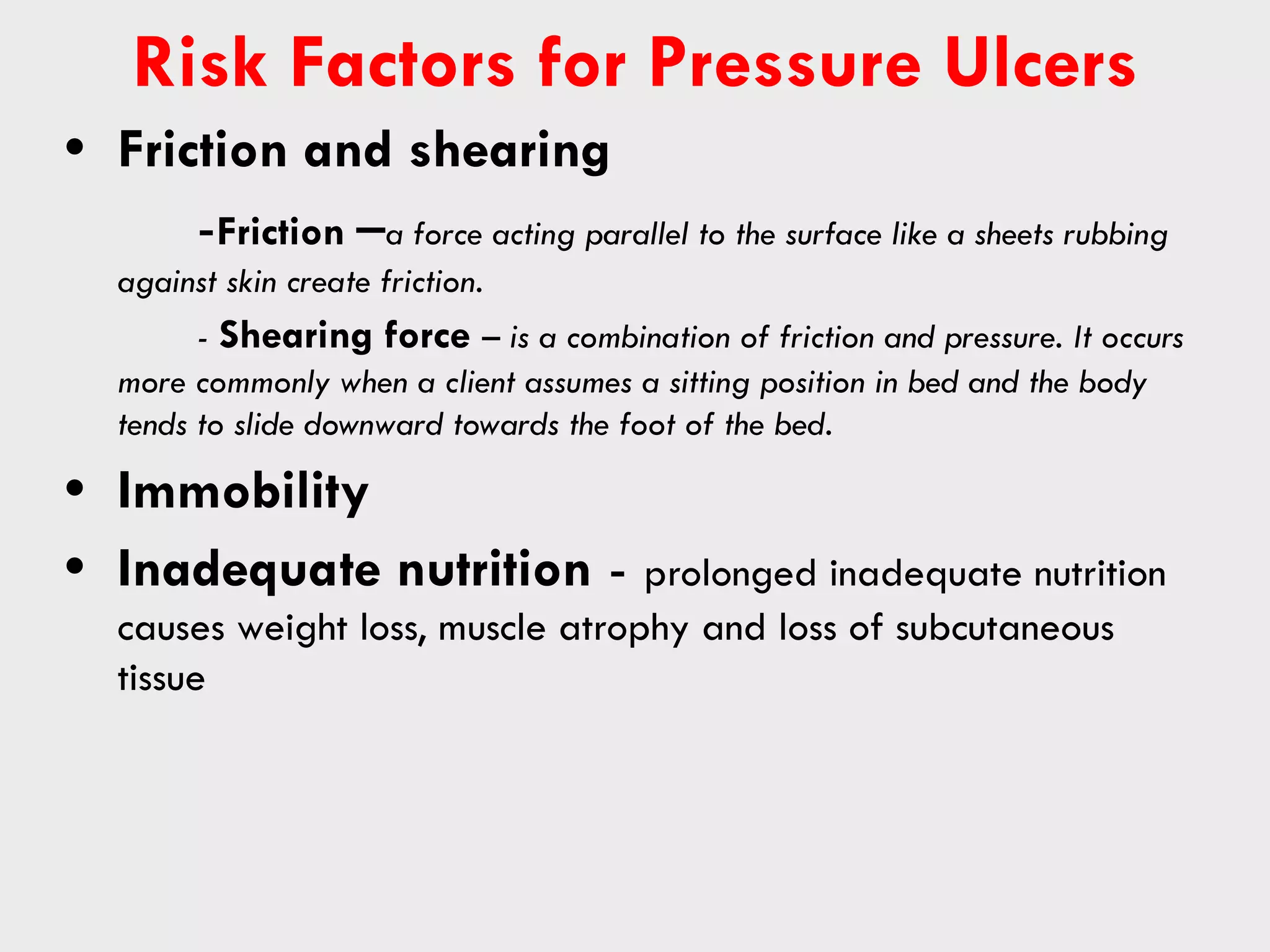
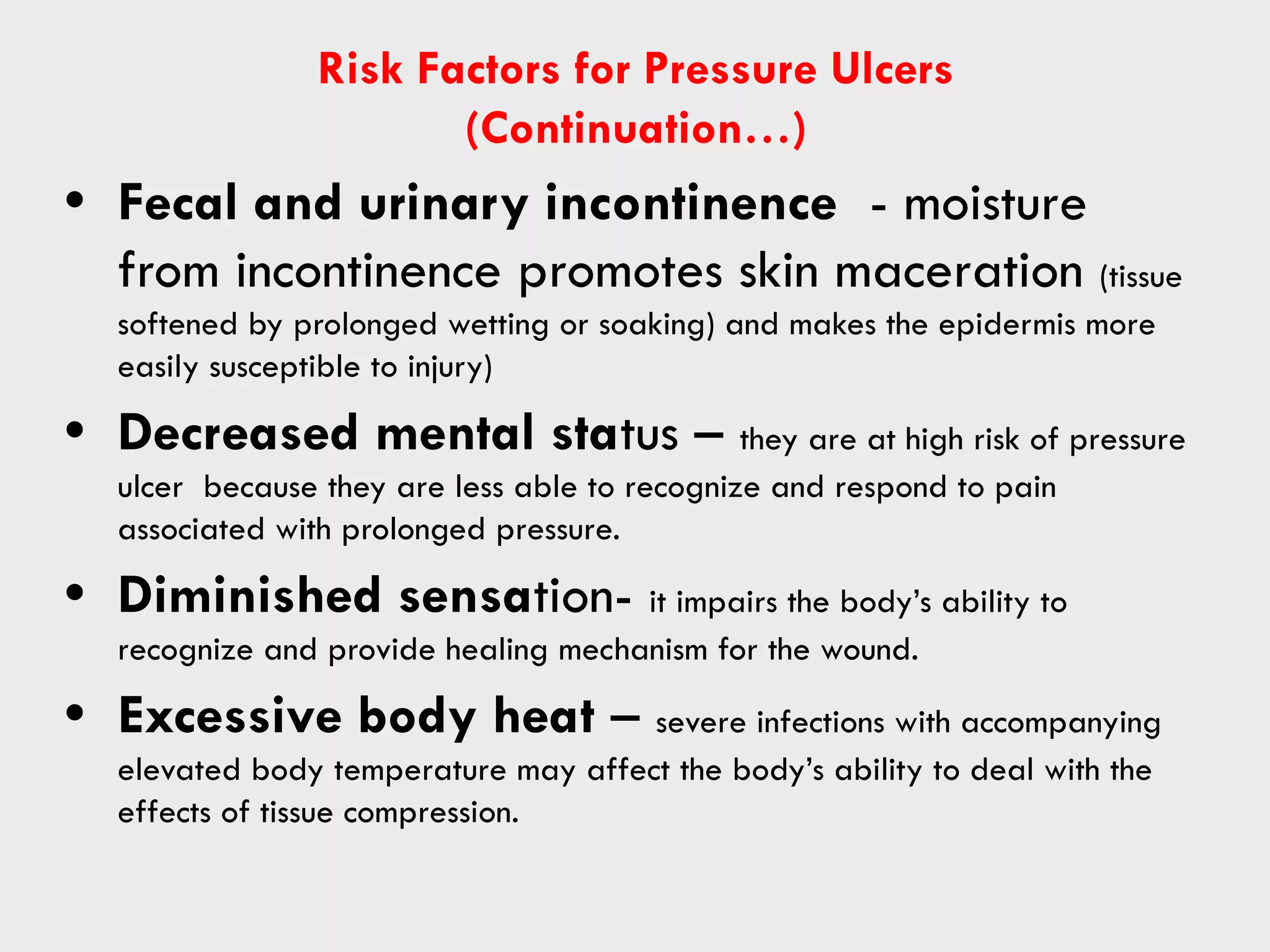
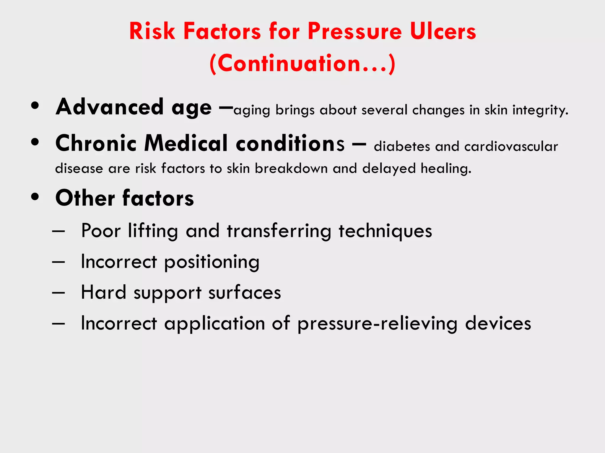
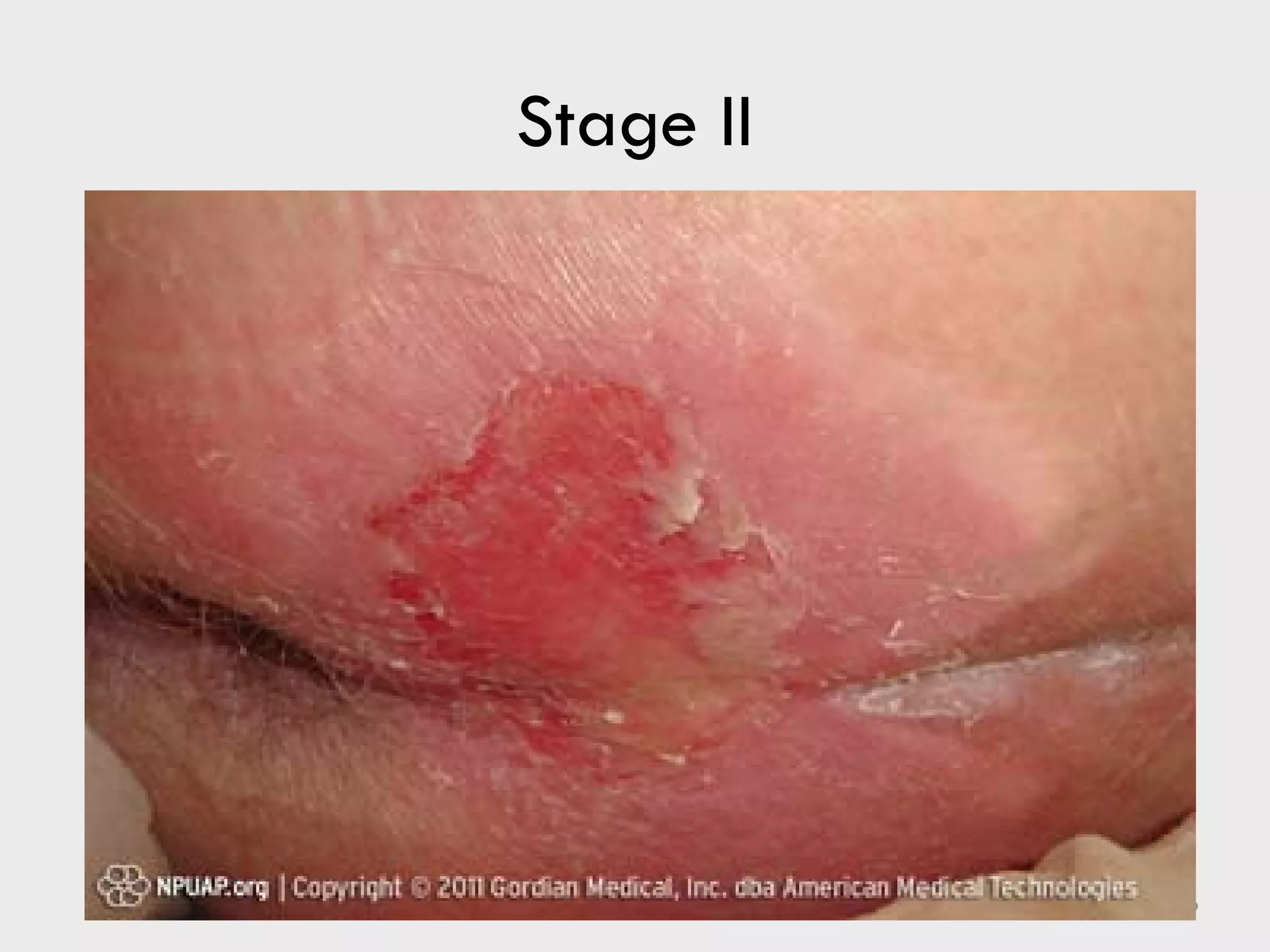
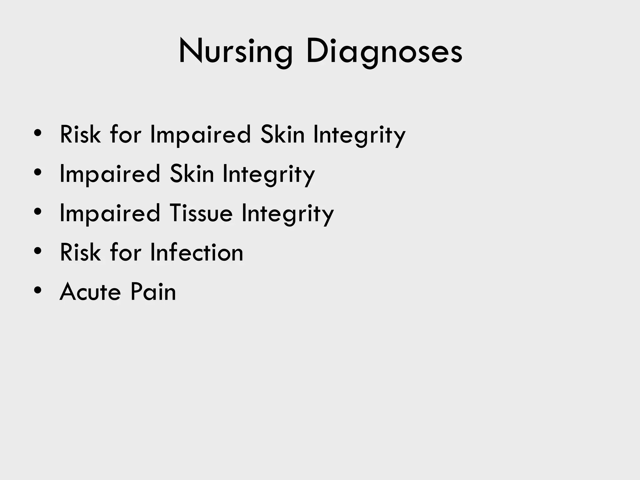
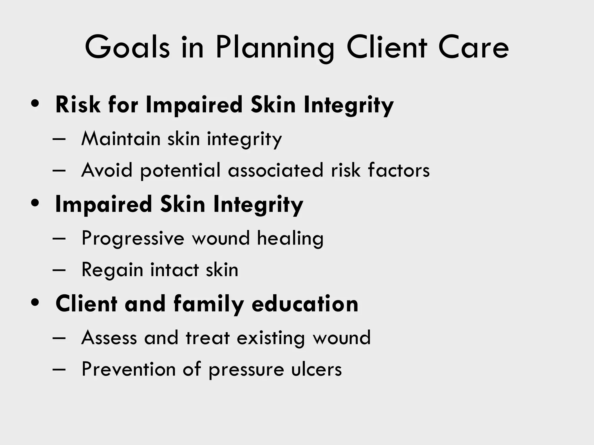
University of Tabuk
Faculty of Applied Medical Sciences
Department of Nursing
Fundamentals of Nursing 2Theory
A.Y. 2022- 2023
Skin Integrity and Wound Care
Factors Affecting Skin
Integrity
• Genetics and heredity (determines many aspect of a person’s skin)
• Age (skin of very young and very old are more fragile)
• Chronic illnesses and their treatments
• Medications (Example: corticosteroids causes thinning of the skin)
• Poor nutrition (can interfere with appearance and function of normal skin)
PRESSURE ULCERS
PRESSURE ULCERS – consists of injury of the skin
and /or underlying tissue, usually over a bony
prominence, as a result of force alone or in
combination with movement.(Black et, al., 2007)
- were previously called decubitus ulcers,
pressure sores, or bedsores.
Department of Nursing 3
Risk Factors for Pressure Ulcers
• Friction and shearing
-Friction –a force acting parallel to the surface like a sheets rubbing
against skin create friction.
- Shearing force – is a combination of friction and pressure. It occurs
more commonly when a client assumes a sitting position in bed and the body
tends to slide downward towards the foot of the bed.
• Immobility
• Inadequate nutrition - prolonged inadequate nutrition
causes weight loss, muscle atrophy and loss of subcutaneous
tissue
Risk Factors for Pressure Ulcers
(Continuation…)
• Fecal and urinary incontinence - moisture
from incontinence promotes skin maceration (tissue
softened by prolonged wetting or soaking) and makes the epidermis more
easily susceptible to injury)
• Decreased mental status – they are at high risk of pressure
ulcer because they are less able to recognize and respond to pain
associated with prolonged pressure.
• Diminished sensation- it impairs the body’s ability to
recognize and provide healing mechanism for the wound.
• Excessive body heat – severe infections with accompanying
elevated body temperature may affect the body’s ability to deal with the
effects of tissue compression.
Risk Factors for Pressure Ulcers
(Continuation…)
• Advanced age –aging brings about several changes in skin integrity.
• Chronic Medical conditions – diabetes and cardiovascular
disease are risk factors to skin breakdown and delayed healing.
• Other factors
– Poor lifting and transferring techniques
– Incorrect positioning
– Hard support surfaces
– Incorrect application of pressure-relieving devices
Stages of Pressure Ulcers
• Stage I: nonblanchable erythema signaling potential
ulceration
• Stage II: partial-thickness skin loss involving epidermis
and possibly dermis
• Stage III: full-thickness skin loss involving damage or
necrosis of subcutaneous tissue that may extend down to,
but not through, underlying fascia.
• Stage IV: full-thickness skin loss with tissue
necrosis or damage to muscle, bone, or
supporting structures such as tendon and joint
capsule.
Stage I
Department of Nursing 8
Stage II
Department of Nursing 9
Stage III
Department of Nursing 10
Stage IV
Department of Nursing 11
WOUND HEALING
- Healing is also referred to as regeneration.
2 Types of healing influenced by the amount of
tissue loss:
1. Primary intention healing
2. Secondary intention healing
3. Tertiary intention
Department of Nursing 12
Primary Intention Healing
• Occurs when the tissue surfaces have been
approximated (closed) and there is minimal or
tissue loss.
• Characterized by formation of granulation
tissue and scarring.
• Also called primary union or first intention
healing.
Secondary Intention Healing
• A wound that is extensive and involves
considerable tissue loss and in which edges
cannot be approximated.
• Repair time is longer
• The scarring is greater.
• Susceptibility to infection is greater.
Tertiary Intention Healing
(Delayed Primary Intention)
• Wounds that are left open for 3-5 days to
allow edema or infection to resolve or exudate
to drain.
• They are closed with sutures, staples or
adhesive skin closures.
Phases of Wound Healing
• Inflammatory Phase
– Initiated immediately after injury and lasts 3 to 6
days
– 2 major processes occurs during this phase:
• Hemostasis (cessation of bleeding) .
• Phagocytosis (macrophages engulf microorganism and
cellular debris on the injury)
Phases of Wound Healing
• Proliferative phase
– The second phase in healing, extends from day 3
or 4 until day 21 post injury.
– Collagen synthesis occurs ( Collagen – is a whitish protein
substance that adds tensile strength to the wound.)
– Granulation tissue formation (capillaries grow across the
wound, increasing blood supply.)
Phases of Wound Healing
• Maturation phase
– Begins on about day 21 and can extend 1 to 3
years after the injury
– Fibroblast continues to synthesize collagen.
– There may be keloid formation in some individuals
• Keloid – these are hypertrophic scars, which is due to an
abnormal amount of collagen.)
Phases of wound healing
Department of Nursing 19
Keloid formation
Department of Nursing 20
A keloid scar is a thick raised scar. It can occur wherever
you have a skin injury but usually forms on earlobes,
shoulders, cheeks or the chest. If you're prone to developing
keloids, you might get them in more than one place.
A keloid scar isn't harmful to your physical health, but it can
cause emotional distress.
Types of Wound Exudate
• Exudate - is material, such as fluid and cells, that
have escaped from blood vessels during inflammatory
process and is deposited in the tissue or tissue surfaces.
• Three (3) major types:
– Serous
– Purulent
– Sanguineous (hemorrhagic)
Serous Exudate
• Consists mostly of serum
(the clear portion of the blood.)
• Derived from blood and
the serous membranes of
the body.
• Looks watery, few cells
• An example is a fluid in
a blister from a burn.
Department of Nursing 22
Purulent Exudate
• Thicker than serous
exudates.
• Presence of pus
• Color varies with
causative organism
Department of Nursing 23
Sanguineous Exudate
• Consists of large amount of
red blood cells indicating
damage to capillaries that
is severe enough to allow
the escape of red blood
cells in the plasma.
• Frequently seen in open
wounds.
• A serosanguinous exudate
is frequently seen in
surgical incision.
Department of Nursing 24
Mixed Exudate
• Serosanguineous
- Consisting o clear and blood-tinged drainage
• Purosanguineous
– Consisting of Pus and blood
Complications of Wound Healing
• Hemorrhage - a profuse discharge of blood, as from a
ruptured blood vessel
- massive bleeding.
• Infection – contamination of the wound surface with
microorganism.
• Dehiscence – partial or total rupturing of a sutured wound
• Evisceration- the protrusion of an internal viscera through
an incision.
Hemorrhage
Department of Nursing 27
Infection
Department of Nursing 28
Dehiscence
Department of Nursing 29
Evisceration
Department of Nursing 30
Factors Affecting Wound Healing
• Age (healthy children and adult often heal more quickly than older adults.)
• Nutritional status- wound healing requires good nutrition---
diet rich in protein, carbohydrates, lipids, Vitamins A and C, and
minerals (iron, zinc and copper)
• Lifestyle- exercise brings good circulation, which leads to
quick healing.
• Medications- anti inflammatory drugs and anti-neoplastic
drugs interfere with healing.
Nursing Process: Assessment
• Nursing history
– Review of systems
– Information regarding skin diseases
– Previous bruising
– General skin condition
– Skin lesions
– Usual healing of sores
Assessment Data
• Inspection and palpation
– Skin color distribution
– Skin turgor
– Presence of edema
– Characteristics of any skin lesions
– Particular attention paid to areas that are most
likely to break down
Assessment Data
• Untreated wounds
– Location
– Extent of tissue damage
– Wound length, width, and depth
– Bleeding
– Foreign bodies
– Associated injuries
– Last tetanus toxoid injection
Assessment Data
• Treated wounds
– Appearance
– Size
– Drainage
– Presence of swelling
– Pain
– Status of drains or tubes
Assessment of Pressure Ulcers
• Location of the ulcer related to a bony prominence
• Size of ulcer in centimeters including length (head
to toe), width (side to side), and depth
• Presence of undermining or sinus tracts (if these
are located in the head)
• Stage of the ulcer
• Color of the wound bed and location of necrosis
(dead tissue)
Assessment of Pressure Ulcers (cont'd)
• Condition of the wound margins
• Integrity of surrounding skin
• Clinical signs of infection (such as redness,
warmth, swelling, pain, odor and exudate - note
for the color of the exudate)
Figure 36-6 Wound/skin documentation
sheet.
Laboratory Data
- The laboratory data can often support the nurse’s clinical
assessment of the wound’s progress in healing.
• Leukocyte count – decreased leukocyte count can delay healing and
increase possibility of infection.
• Hemoglobin level- a low level of hemoglobin indicates poor oxygen
delivery to the tissues.
• Blood coagulation studies- prolonged coagulation can result in
excessive blood loss.
• Serum protein analysis
• Albumin level which is low indicates poor nutrition.
• Results of wound culture and sensitivities – can confirm
or rule out infection.
Nursing Diagnoses
• Risk for Impaired Skin Integrity
• Impaired Skin Integrity
• Impaired Tissue Integrity
• Risk for Infection
• Acute Pain
Goals in Planning Client Care
• Risk for Impaired Skin Integrity
– Maintain skin integrity
– Avoid potential associated risk factors
• Impaired Skin Integrity
– Progressive wound healing
– Regain intact skin
• Client and family education
– Assess and treat existing wound
– Prevention of pressure ulcers
Client Teaching Skin Integrity
• Maintaining intact skin
• Adequate nutrition
• Appropriate positions for pressure relief
• Establish a turning or repositioning schedule
• Providing supportive devices
• Instruct to report persistent reddened areas
• Identify potential sources of skin trauma
Preventing Pressure Ulcers
✔ Check patients skin several times each day -
Check for red skin over bony areas. Use a mirror if you have trouble seeing
certain areas, or ask another person to look.
✔ Conduct a pressure assessment for all clients .
✔ Change clients position often.
- change the patients position every 2 hours/Turn patients every 2
hours.
✔ Protect the skin over bony areas
- Use pillows or foam wedges to keep bony
areas from touching one another.
Preventing Pressure Ulcers (con’t.)..
✔ Keep skin clean, dry, and moisturized
- Use mild soaps and warm (not hot) water
to clean your skin. Be gentle. Do not rub hard
or use force when you wash your skin. Do not
use soaps and other products that contain
alcohol, because they can dry out your skin.
✔ Change wet bedding and clothes
- Change sheets, pads, and bedclothes
right away if they get wet.
✔ Advise patient to eat healthy foods—Provide
Nutrition
Maintaining Skin Hygiene
• Minimize the force and friction
• Mild cleansing agents
• Avoid hot water
• Moisturizing lotions/skin protection
• Skin should be clean and dry
• Free of irritation and maceration
Avoiding Skin Trauma
• Smooth, firm, and wrinkle free foundation on
which to sit or lie helps prevent skin trauma
• Frequent shifts in position
• Exercise and ambulation to stimulate blood
circulation
• Lifting devices should be used to avoid dragging
the client.
• Reposition every 2 hours, depending on the
clients need.
Providing Supportive Devices
• Mattresses
• Beds
• Wedges, pillows
• Miscellaneous devices
Treating Pressure Ulcers
• Minimize direct pressure
• Reposition the client at least every 2 hours
• Schedule and record position changes
• Clean and dress the ulcer using surgical asepsis
• Never use alcohol or hydrogen peroxide.
• Obtain culture and sensitivity (C&S), if infected
• Teach the client to move frequently
• Provide range of motion exercises.
RYB Color Guide for
Wound Care
• Red (protect)
• Yellow (cleanse)
• Black (debride)
Preventing Complications
• Prevent entry of microorganisms
• Prevent transmission of pathogens
Irrigating a Wound
Irrigating a wound is done to:
- clean the area of the wound.
- Apply heat and hasten the healing process
- Apply an antimicrobial solution.
Department of Nursing 51
Types of Bandages
-A bandage is a strip of cloth used to wrap some
parts of the body.
• Gauze
– Retains dressings on wounds
– Bandage fingers, hands, toes, and feet
• Elasticized
– Provide pressure to an area
– Improve venous circulation in legs
Gauze bandage
Department of Nursing 53
Elasticized bandage
Department of Nursing 54
Heat and Cold Applications
✔ Local Effects of Heat
- Heat is an old remedy for aches and pains and
provides comfort and relief.
- application of heat promotes soft tissue healing.
✔ Local Effects of Cold
- cold lowers the temperature of the skin and causes
vasoconstriction.
- it is most often used in sport injuries (sprain, strain and
fractures) to limit post injury healing and bleeding.
Department of Nursing 55
Indications for Cold Application
• Sprains - A sprain is an injury to the ligaments around a
joint.
• Strains- occurs when a muscle is stretched too much and
tears. It is also called a pulled muscle. A strain is a painful
injury.
• Fractures- a break in the continuity of a bone.
• Post injury swelling and bleeding
Physiological Effects of
Heat and Cold
HEAT COLD
Vasodilation Vasoconstriction
Increased capillary permeability Decreased capillary
permeability
Increased cellular metabolism Decreased cellular metabolism
Increases inflammation Slows bacterial growth and
decreases inflammation
Sedative effect Local anesthetic effect
Department of Nursing 57
Methods for Applying
Dry and Moist Heat
• Dry heat
– Hot water bottle
– Aquathermia pad
– Disposable heat pack
– Electric pad
Disposable heat pack
Department of Nursing 59
Methods for Applying
Dry and Moist Heat (cont'd)
• Moist heat
– Compress
– Hot pack
– Soak
Methods for Applying Dry
and Moist Cold
• Dry cold
– Cold pack
– Ice bag
– Ice glove
– Ice collar
• Moist cold
– Compress
– Cooling sponge bath
Ice bag
Department of Nursing 62

4 SKIN INTEGRITY AND WOUND CARE 2.pdf

  • 1.
    University of Tabuk Facultyof Applied Medical Sciences Department of Nursing Fundamentals of Nursing 2Theory A.Y. 2022- 2023 Skin Integrity and Wound Care
  • 2.
    Factors Affecting Skin Integrity •Genetics and heredity (determines many aspect of a person’s skin) • Age (skin of very young and very old are more fragile) • Chronic illnesses and their treatments • Medications (Example: corticosteroids causes thinning of the skin) • Poor nutrition (can interfere with appearance and function of normal skin)
  • 3.
    PRESSURE ULCERS PRESSURE ULCERS– consists of injury of the skin and /or underlying tissue, usually over a bony prominence, as a result of force alone or in combination with movement.(Black et, al., 2007) - were previously called decubitus ulcers, pressure sores, or bedsores. Department of Nursing 3
  • 4.
    Risk Factors forPressure Ulcers • Friction and shearing -Friction –a force acting parallel to the surface like a sheets rubbing against skin create friction. - Shearing force – is a combination of friction and pressure. It occurs more commonly when a client assumes a sitting position in bed and the body tends to slide downward towards the foot of the bed. • Immobility • Inadequate nutrition - prolonged inadequate nutrition causes weight loss, muscle atrophy and loss of subcutaneous tissue
  • 5.
    Risk Factors forPressure Ulcers (Continuation…) • Fecal and urinary incontinence - moisture from incontinence promotes skin maceration (tissue softened by prolonged wetting or soaking) and makes the epidermis more easily susceptible to injury) • Decreased mental status – they are at high risk of pressure ulcer because they are less able to recognize and respond to pain associated with prolonged pressure. • Diminished sensation- it impairs the body’s ability to recognize and provide healing mechanism for the wound. • Excessive body heat – severe infections with accompanying elevated body temperature may affect the body’s ability to deal with the effects of tissue compression.
  • 6.
    Risk Factors forPressure Ulcers (Continuation…) • Advanced age –aging brings about several changes in skin integrity. • Chronic Medical conditions – diabetes and cardiovascular disease are risk factors to skin breakdown and delayed healing. • Other factors – Poor lifting and transferring techniques – Incorrect positioning – Hard support surfaces – Incorrect application of pressure-relieving devices
  • 7.
    Stages of PressureUlcers • Stage I: nonblanchable erythema signaling potential ulceration • Stage II: partial-thickness skin loss involving epidermis and possibly dermis • Stage III: full-thickness skin loss involving damage or necrosis of subcutaneous tissue that may extend down to, but not through, underlying fascia. • Stage IV: full-thickness skin loss with tissue necrosis or damage to muscle, bone, or supporting structures such as tendon and joint capsule.
  • 8.
  • 9.
  • 10.
  • 11.
  • 12.
    WOUND HEALING - Healingis also referred to as regeneration. 2 Types of healing influenced by the amount of tissue loss: 1. Primary intention healing 2. Secondary intention healing 3. Tertiary intention Department of Nursing 12
  • 13.
    Primary Intention Healing •Occurs when the tissue surfaces have been approximated (closed) and there is minimal or tissue loss. • Characterized by formation of granulation tissue and scarring. • Also called primary union or first intention healing.
  • 14.
    Secondary Intention Healing •A wound that is extensive and involves considerable tissue loss and in which edges cannot be approximated. • Repair time is longer • The scarring is greater. • Susceptibility to infection is greater.
  • 15.
    Tertiary Intention Healing (DelayedPrimary Intention) • Wounds that are left open for 3-5 days to allow edema or infection to resolve or exudate to drain. • They are closed with sutures, staples or adhesive skin closures.
  • 16.
    Phases of WoundHealing • Inflammatory Phase – Initiated immediately after injury and lasts 3 to 6 days – 2 major processes occurs during this phase: • Hemostasis (cessation of bleeding) . • Phagocytosis (macrophages engulf microorganism and cellular debris on the injury)
  • 17.
    Phases of WoundHealing • Proliferative phase – The second phase in healing, extends from day 3 or 4 until day 21 post injury. – Collagen synthesis occurs ( Collagen – is a whitish protein substance that adds tensile strength to the wound.) – Granulation tissue formation (capillaries grow across the wound, increasing blood supply.)
  • 18.
    Phases of WoundHealing • Maturation phase – Begins on about day 21 and can extend 1 to 3 years after the injury – Fibroblast continues to synthesize collagen. – There may be keloid formation in some individuals • Keloid – these are hypertrophic scars, which is due to an abnormal amount of collagen.)
  • 19.
    Phases of woundhealing Department of Nursing 19
  • 20.
    Keloid formation Department ofNursing 20 A keloid scar is a thick raised scar. It can occur wherever you have a skin injury but usually forms on earlobes, shoulders, cheeks or the chest. If you're prone to developing keloids, you might get them in more than one place. A keloid scar isn't harmful to your physical health, but it can cause emotional distress.
  • 21.
    Types of WoundExudate • Exudate - is material, such as fluid and cells, that have escaped from blood vessels during inflammatory process and is deposited in the tissue or tissue surfaces. • Three (3) major types: – Serous – Purulent – Sanguineous (hemorrhagic)
  • 22.
    Serous Exudate • Consistsmostly of serum (the clear portion of the blood.) • Derived from blood and the serous membranes of the body. • Looks watery, few cells • An example is a fluid in a blister from a burn. Department of Nursing 22
  • 23.
    Purulent Exudate • Thickerthan serous exudates. • Presence of pus • Color varies with causative organism Department of Nursing 23
  • 24.
    Sanguineous Exudate • Consistsof large amount of red blood cells indicating damage to capillaries that is severe enough to allow the escape of red blood cells in the plasma. • Frequently seen in open wounds. • A serosanguinous exudate is frequently seen in surgical incision. Department of Nursing 24
  • 25.
    Mixed Exudate • Serosanguineous -Consisting o clear and blood-tinged drainage • Purosanguineous – Consisting of Pus and blood
  • 26.
    Complications of WoundHealing • Hemorrhage - a profuse discharge of blood, as from a ruptured blood vessel - massive bleeding. • Infection – contamination of the wound surface with microorganism. • Dehiscence – partial or total rupturing of a sutured wound • Evisceration- the protrusion of an internal viscera through an incision.
  • 27.
  • 28.
  • 29.
  • 30.
  • 31.
    Factors Affecting WoundHealing • Age (healthy children and adult often heal more quickly than older adults.) • Nutritional status- wound healing requires good nutrition--- diet rich in protein, carbohydrates, lipids, Vitamins A and C, and minerals (iron, zinc and copper) • Lifestyle- exercise brings good circulation, which leads to quick healing. • Medications- anti inflammatory drugs and anti-neoplastic drugs interfere with healing.
  • 32.
    Nursing Process: Assessment •Nursing history – Review of systems – Information regarding skin diseases – Previous bruising – General skin condition – Skin lesions – Usual healing of sores
  • 33.
    Assessment Data • Inspectionand palpation – Skin color distribution – Skin turgor – Presence of edema – Characteristics of any skin lesions – Particular attention paid to areas that are most likely to break down
  • 34.
    Assessment Data • Untreatedwounds – Location – Extent of tissue damage – Wound length, width, and depth – Bleeding – Foreign bodies – Associated injuries – Last tetanus toxoid injection
  • 35.
    Assessment Data • Treatedwounds – Appearance – Size – Drainage – Presence of swelling – Pain – Status of drains or tubes
  • 36.
    Assessment of PressureUlcers • Location of the ulcer related to a bony prominence • Size of ulcer in centimeters including length (head to toe), width (side to side), and depth • Presence of undermining or sinus tracts (if these are located in the head) • Stage of the ulcer • Color of the wound bed and location of necrosis (dead tissue)
  • 37.
    Assessment of PressureUlcers (cont'd) • Condition of the wound margins • Integrity of surrounding skin • Clinical signs of infection (such as redness, warmth, swelling, pain, odor and exudate - note for the color of the exudate)
  • 38.
    Figure 36-6 Wound/skindocumentation sheet.
  • 39.
    Laboratory Data - Thelaboratory data can often support the nurse’s clinical assessment of the wound’s progress in healing. • Leukocyte count – decreased leukocyte count can delay healing and increase possibility of infection. • Hemoglobin level- a low level of hemoglobin indicates poor oxygen delivery to the tissues. • Blood coagulation studies- prolonged coagulation can result in excessive blood loss. • Serum protein analysis • Albumin level which is low indicates poor nutrition. • Results of wound culture and sensitivities – can confirm or rule out infection.
  • 40.
    Nursing Diagnoses • Riskfor Impaired Skin Integrity • Impaired Skin Integrity • Impaired Tissue Integrity • Risk for Infection • Acute Pain
  • 41.
    Goals in PlanningClient Care • Risk for Impaired Skin Integrity – Maintain skin integrity – Avoid potential associated risk factors • Impaired Skin Integrity – Progressive wound healing – Regain intact skin • Client and family education – Assess and treat existing wound – Prevention of pressure ulcers
  • 42.
    Client Teaching SkinIntegrity • Maintaining intact skin • Adequate nutrition • Appropriate positions for pressure relief • Establish a turning or repositioning schedule • Providing supportive devices • Instruct to report persistent reddened areas • Identify potential sources of skin trauma
  • 43.
    Preventing Pressure Ulcers ✔Check patients skin several times each day - Check for red skin over bony areas. Use a mirror if you have trouble seeing certain areas, or ask another person to look. ✔ Conduct a pressure assessment for all clients . ✔ Change clients position often. - change the patients position every 2 hours/Turn patients every 2 hours. ✔ Protect the skin over bony areas - Use pillows or foam wedges to keep bony areas from touching one another.
  • 44.
    Preventing Pressure Ulcers(con’t.).. ✔ Keep skin clean, dry, and moisturized - Use mild soaps and warm (not hot) water to clean your skin. Be gentle. Do not rub hard or use force when you wash your skin. Do not use soaps and other products that contain alcohol, because they can dry out your skin. ✔ Change wet bedding and clothes - Change sheets, pads, and bedclothes right away if they get wet. ✔ Advise patient to eat healthy foods—Provide Nutrition
  • 45.
    Maintaining Skin Hygiene •Minimize the force and friction • Mild cleansing agents • Avoid hot water • Moisturizing lotions/skin protection • Skin should be clean and dry • Free of irritation and maceration
  • 46.
    Avoiding Skin Trauma •Smooth, firm, and wrinkle free foundation on which to sit or lie helps prevent skin trauma • Frequent shifts in position • Exercise and ambulation to stimulate blood circulation • Lifting devices should be used to avoid dragging the client. • Reposition every 2 hours, depending on the clients need.
  • 47.
    Providing Supportive Devices •Mattresses • Beds • Wedges, pillows • Miscellaneous devices
  • 48.
    Treating Pressure Ulcers •Minimize direct pressure • Reposition the client at least every 2 hours • Schedule and record position changes • Clean and dress the ulcer using surgical asepsis • Never use alcohol or hydrogen peroxide. • Obtain culture and sensitivity (C&S), if infected • Teach the client to move frequently • Provide range of motion exercises.
  • 49.
    RYB Color Guidefor Wound Care • Red (protect) • Yellow (cleanse) • Black (debride)
  • 50.
    Preventing Complications • Prevententry of microorganisms • Prevent transmission of pathogens
  • 51.
    Irrigating a Wound Irrigatinga wound is done to: - clean the area of the wound. - Apply heat and hasten the healing process - Apply an antimicrobial solution. Department of Nursing 51
  • 52.
    Types of Bandages -Abandage is a strip of cloth used to wrap some parts of the body. • Gauze – Retains dressings on wounds – Bandage fingers, hands, toes, and feet • Elasticized – Provide pressure to an area – Improve venous circulation in legs
  • 53.
  • 54.
  • 55.
    Heat and ColdApplications ✔ Local Effects of Heat - Heat is an old remedy for aches and pains and provides comfort and relief. - application of heat promotes soft tissue healing. ✔ Local Effects of Cold - cold lowers the temperature of the skin and causes vasoconstriction. - it is most often used in sport injuries (sprain, strain and fractures) to limit post injury healing and bleeding. Department of Nursing 55
  • 56.
    Indications for ColdApplication • Sprains - A sprain is an injury to the ligaments around a joint. • Strains- occurs when a muscle is stretched too much and tears. It is also called a pulled muscle. A strain is a painful injury. • Fractures- a break in the continuity of a bone. • Post injury swelling and bleeding
  • 57.
    Physiological Effects of Heatand Cold HEAT COLD Vasodilation Vasoconstriction Increased capillary permeability Decreased capillary permeability Increased cellular metabolism Decreased cellular metabolism Increases inflammation Slows bacterial growth and decreases inflammation Sedative effect Local anesthetic effect Department of Nursing 57
  • 58.
    Methods for Applying Dryand Moist Heat • Dry heat – Hot water bottle – Aquathermia pad – Disposable heat pack – Electric pad
  • 59.
  • 60.
    Methods for Applying Dryand Moist Heat (cont'd) • Moist heat – Compress – Hot pack – Soak
  • 61.
    Methods for ApplyingDry and Moist Cold • Dry cold – Cold pack – Ice bag – Ice glove – Ice collar • Moist cold – Compress – Cooling sponge bath
  • 62.