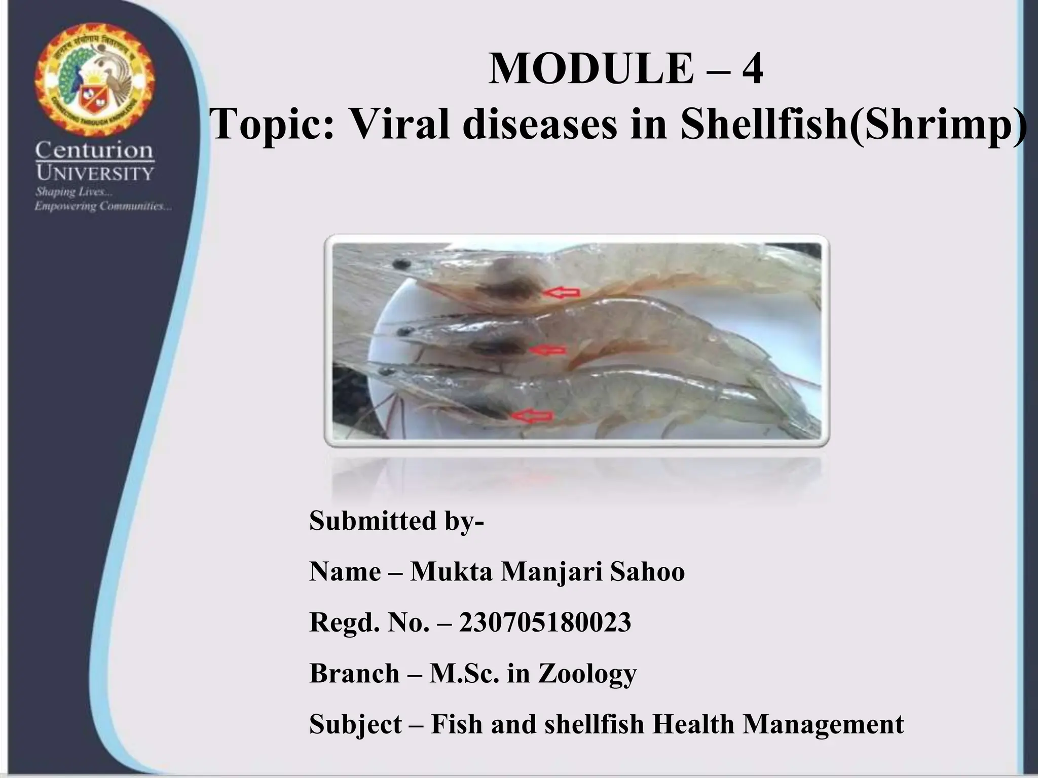
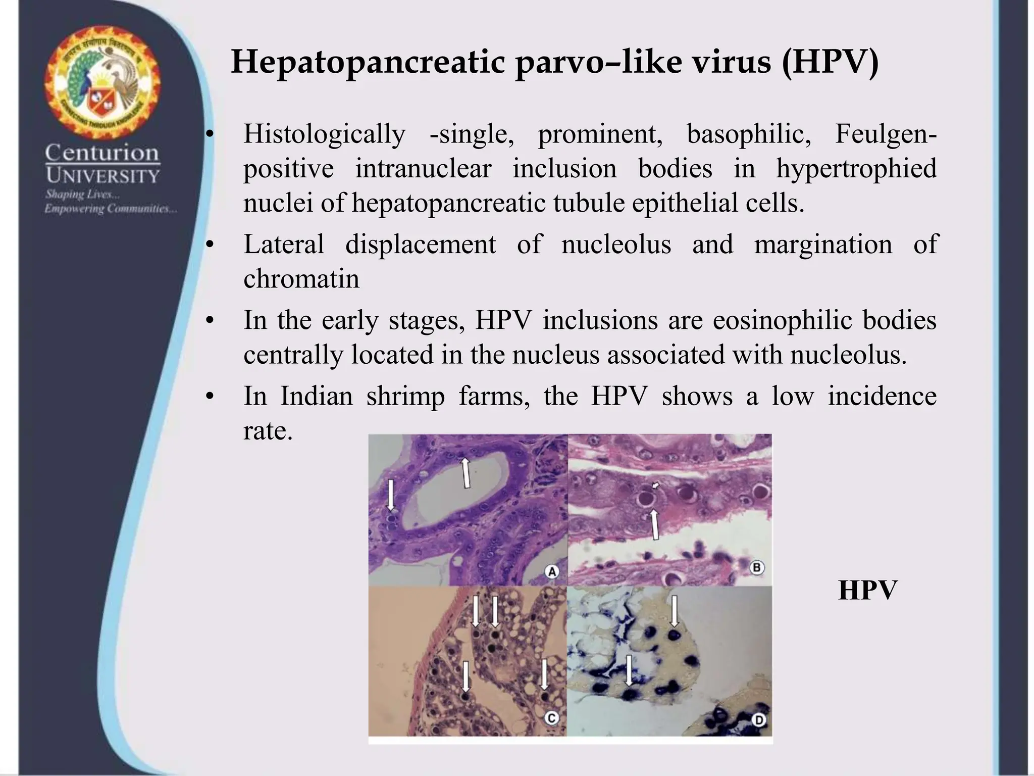
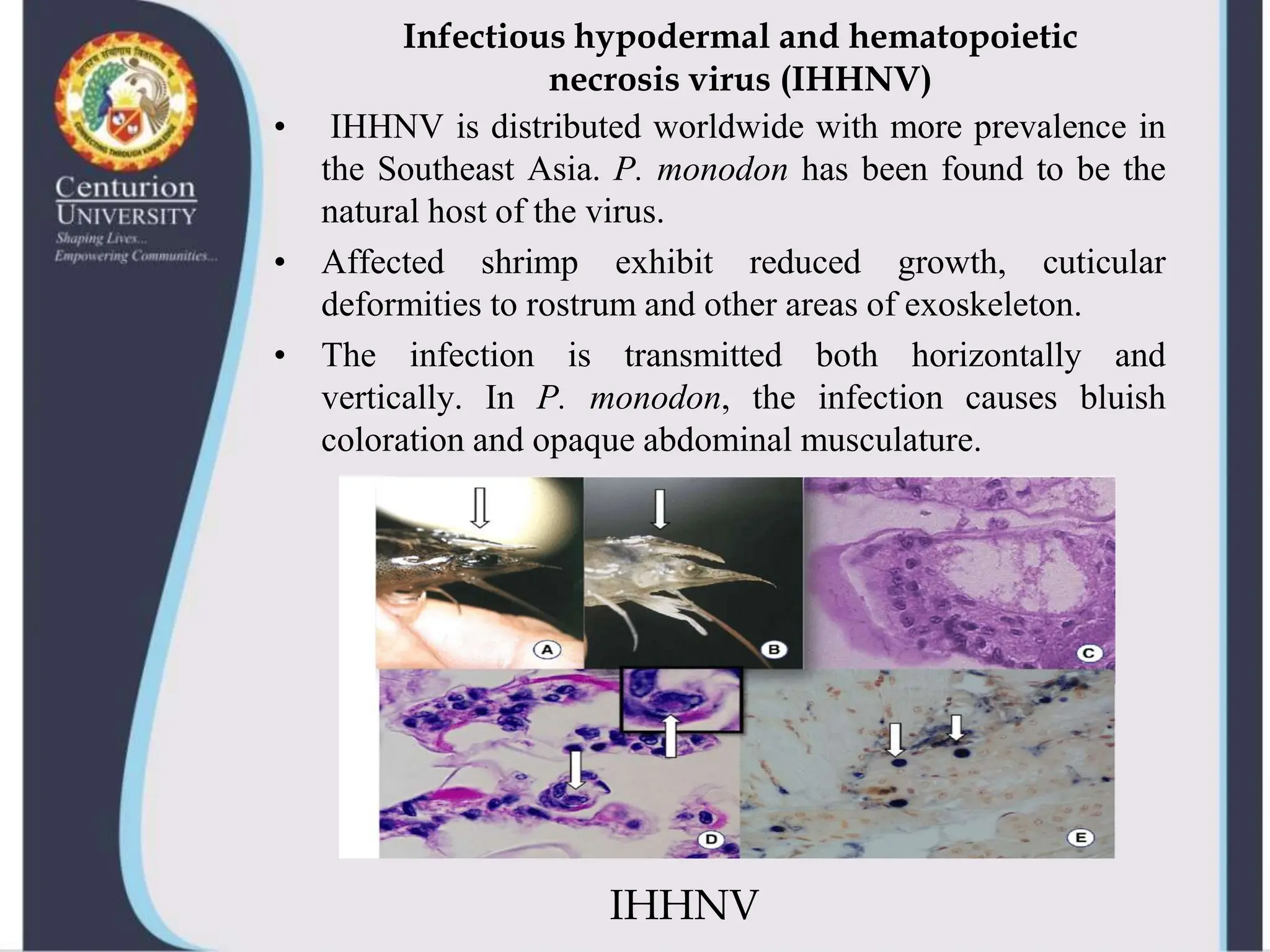
MODULE – 4
Topic: Viral diseases in Shellfish(Shrimp)
Submitted by-
Name – Mukta Manjari Sahoo
Regd. No. – 230705180023
Branch – M.Sc. in Zoology
Subject – Fish and shellfish Health Management
INTRODUCTION
• Viral diseases are a growing concern for the shellfish industry,
causing significant economic losses and posing a potential
threat to human health.
• Shellfish, such as oysters, clams, mussels, and scallops, are
filter feeders that can accumulate viruses from the surrounding
water.
• Viruses are considered to be the most important pathogens in
shrimp.
• Currently, at least 14 virus diseases of cultured shrimp are
recognised.
• The major virus families present in the crustaceans include
Parvoviridae, Baculoviridae, Picornaviridae, Reoviridae,
Togaviridae, Cornaviridae.
How do we know?
• SIGNS AND SYMPTOMS
• ANOREXIA
• ANTENNAE CUT
• CHANGE IN COLOUR
• CHANGE IN SHAPE
• POOR GROWTH
• SIZE VARIATION
• SURFACING
• MORTALITY
• OPAQUENESS
• LOOSE SHELL
• MELANIZED LESIONS
• PARTIAL MOULTING
• DELAYED BLOOD
CLOTTING
• WHITE FECAL MATTER
• WHITE GUT
• TAIL ROT
• EDEMA
• BLISTERS
• GILL COLOUR
• WHITE SPOTS ON
CARAPACE
Caridina cantonensis
Hepatopancreatic parvo–like virus (HPV)
• Histologically -single, prominent, basophilic, Feulgen-
positive intranuclear inclusion bodies in hypertrophied
nuclei of hepatopancreatic tubule epithelial cells.
• Lateral displacement of nucleolus and margination of
chromatin
• In the early stages, HPV inclusions are eosinophilic bodies
centrally located in the nucleus associated with nucleolus.
• In Indian shrimp farms, the HPV shows a low incidence
rate.
HPV
Infectious hypodermal and hematopoietic
necrosis virus (IHHNV)
• IHHNV is distributed worldwide with more prevalence in
the Southeast Asia. P. monodon has been found to be the
natural host of the virus.
• Affected shrimp exhibit reduced growth, cuticular
deformities to rostrum and other areas of exoskeleton.
• The infection is transmitted both horizontally and
vertically. In P. monodon, the infection causes bluish
coloration and opaque abdominal musculature.
IHHNV
Lymphoid organ parvo-like virus (LOPV)
• LOPV was found in cultured shrimps (P. monodon, P.
merguiensis and P. esculentus) in Australia.
• Giant cell nuclei had intranuclear inclusion bodies
containing DNA. Electron microscopic studies revealed
the presence of 25-30 mm diameter viruslike particles.
There is a speculation that IHHNV and LOPV are the
same agent.
Reolike viruses
• Reo-like viruses are reported from P. japonicus, P.
monodon, P. chinensis and P. vannamei. Hepatopancreas
has been suggested as the principal target for both the viral
strains.
• Reovirus infections were always reported in mixed
infections. Hence the role of reovirus as pathogens is not
completely clear.
REO-VIRUS580
Baculoviruses
• (1) Type A occlusion body forming viruses BP and MBV
and
• (2) Type C nonoccluded baculoviruses BMN, TCBV,
Owen’s hemocyte–infecting baculovirus and WSDV.
• Transmission: horizontal, some transmit vertically
• In hatcheries, BP and BMN often cause serious epizootics
in larvae and postlarvae (PL)
• MBV produce serious infections and mortalities in the late
PL and juvenile stages of hosts.
BP Type baculoviruses
• BP (Baculovirus penaei): diagnosis: demonstration of
prominent tetrahedral occlusion bodies in unstained squash
preparations of hepatopancreas, midgut or faeces and also
in histological sections.
• In histological sections, occlusion bodies are found in
single or multiple, eosinophilic, usually triangular within
hypertrophied nuclei of hepatopancreatic tubule epithelial
cells or in midgut epithelial cells.
Monodon type baculoviruses
• MBV enjoy a world-wide distribution. These are type A
baculoviruses measuring 75 x 324 mm.
• Diagnosis: presence of single or multiple, generally
spherical intranuclear occlusion bodies in hepatopancreas
and midgut epithelial cells. Squash preparation (0.05%
aqueous malachite green), epifluorescence microscopy
and acridine orange staining visualizes MBV occlusions
• MBV was first discovered in a quarantined population of
P. monodon that had originated from Taiwan.
• Despite the world distribution, MBV is not a highly
virulent pathogen of P. monodon. MBV is found in
healthy prawns and in disease epizootics, P. monodon has
been found to frequently have mixed infections by MBV
and other viral, bacterial or protozoan pathogens.
MBV574
White spot syndrome virus (WSSV)
• WSSV, formerly known as SEMBV is a nonoccluded
baculovirus – like agent
• Epizootic of white spot disease cause mortalities ranging up
to 80  100% in 2  7 days
susceptible species: Ongrowing juvenile shrimp of many
species of all ages but mostly from 1 - 3 months old in the
grow-out ponds.
• WSSV outbreak occurs in all types of farming systems
irrespective of stocking density, water quality and salinity.
Diagnosis: Broken antennae, white spots of 1 mm size in the
cuticle and / or reddish discoloration, empty guts and
cuticular epibiont fouling and lymphoid organ swelling.
WHITE SPOT DISEASE
Yellow Head Virus (YHV)
• YHV is an RNA virus reported only from P. monodon in
Thailand.
• All ages of juveniles could be infected and mass mortalities
up to 100% are observed within 3 - 5 days
• Diagnosis: Pale body colour with yellowish gills and
hepatopancreas. It affects many tissues such as gills,
lymphoid organ, hemocytes and connective tissue.
• Histology: Degenerative changes in nuclei and presence of
cytoplasmic basophilic inclusion bodies.
PREVENTION
•Use clean water. Shrimp farmers should use clean,
pathogen-free water in their ponds
•Use Specific pathogen-free (SPF) shrimp. SPF shrimp
are shrimp that have been breed to be free of certain
pathogens.
•Practice good biosecurity. Shrimp farmers should take
steps to prevent the introduction of pathogens into their
ponds, such as disinfecting equipment and clothing.
•Maintain good water quality. Maintaining good water
quality in shrimp ponds can help to reduce stress on shrimp
and make them less susceptible to disease.
1. Chang, P.H., Kuo, S. T., Lai, S. H. et al. (2005). Herpes-
like virus infection causing mortality of cultured abalone
Halitosis diversicolor supertexta in Taiwan. Diseases of
Aquatic organisms: 65, 23-27
2. Lees, D. (2000). Viruses and bivalve shellfish:
International Journal of Food Microbiology: 59, 81-116.
3. Owens, L., I. G. Anderson, M. Kenway, L. Trott, and J. A.
H. Benzie (1992): Infectious hypodermal and
haematopoietic necrosis virus (IHHNV) in an interspecies
hybrid pended prawn from tropical Area: 14,219-228.
REFERENCES
Thank you
Thank you
.
.

Viral diseases in shellfish--shrimp-virus

  • 1.
    MODULE – 4 Topic:Viral diseases in Shellfish(Shrimp) Submitted by- Name – Mukta Manjari Sahoo Regd. No. – 230705180023 Branch – M.Sc. in Zoology Subject – Fish and shellfish Health Management
  • 2.
    INTRODUCTION • Viral diseasesare a growing concern for the shellfish industry, causing significant economic losses and posing a potential threat to human health. • Shellfish, such as oysters, clams, mussels, and scallops, are filter feeders that can accumulate viruses from the surrounding water. • Viruses are considered to be the most important pathogens in shrimp. • Currently, at least 14 virus diseases of cultured shrimp are recognised. • The major virus families present in the crustaceans include Parvoviridae, Baculoviridae, Picornaviridae, Reoviridae, Togaviridae, Cornaviridae.
  • 3.
    How do weknow? • SIGNS AND SYMPTOMS • ANOREXIA • ANTENNAE CUT • CHANGE IN COLOUR • CHANGE IN SHAPE • POOR GROWTH • SIZE VARIATION • SURFACING • MORTALITY • OPAQUENESS • LOOSE SHELL • MELANIZED LESIONS • PARTIAL MOULTING • DELAYED BLOOD CLOTTING • WHITE FECAL MATTER • WHITE GUT • TAIL ROT • EDEMA • BLISTERS • GILL COLOUR • WHITE SPOTS ON CARAPACE Caridina cantonensis
  • 4.
    Hepatopancreatic parvo–like virus(HPV) • Histologically -single, prominent, basophilic, Feulgen- positive intranuclear inclusion bodies in hypertrophied nuclei of hepatopancreatic tubule epithelial cells. • Lateral displacement of nucleolus and margination of chromatin • In the early stages, HPV inclusions are eosinophilic bodies centrally located in the nucleus associated with nucleolus. • In Indian shrimp farms, the HPV shows a low incidence rate. HPV
  • 5.
    Infectious hypodermal andhematopoietic necrosis virus (IHHNV) • IHHNV is distributed worldwide with more prevalence in the Southeast Asia. P. monodon has been found to be the natural host of the virus. • Affected shrimp exhibit reduced growth, cuticular deformities to rostrum and other areas of exoskeleton. • The infection is transmitted both horizontally and vertically. In P. monodon, the infection causes bluish coloration and opaque abdominal musculature. IHHNV
  • 6.
    Lymphoid organ parvo-likevirus (LOPV) • LOPV was found in cultured shrimps (P. monodon, P. merguiensis and P. esculentus) in Australia. • Giant cell nuclei had intranuclear inclusion bodies containing DNA. Electron microscopic studies revealed the presence of 25-30 mm diameter viruslike particles. There is a speculation that IHHNV and LOPV are the same agent. Reolike viruses • Reo-like viruses are reported from P. japonicus, P. monodon, P. chinensis and P. vannamei. Hepatopancreas has been suggested as the principal target for both the viral strains. • Reovirus infections were always reported in mixed infections. Hence the role of reovirus as pathogens is not completely clear.
  • 7.
  • 8.
    Baculoviruses • (1) TypeA occlusion body forming viruses BP and MBV and • (2) Type C nonoccluded baculoviruses BMN, TCBV, Owen’s hemocyte–infecting baculovirus and WSDV. • Transmission: horizontal, some transmit vertically • In hatcheries, BP and BMN often cause serious epizootics in larvae and postlarvae (PL) • MBV produce serious infections and mortalities in the late PL and juvenile stages of hosts. BP Type baculoviruses • BP (Baculovirus penaei): diagnosis: demonstration of prominent tetrahedral occlusion bodies in unstained squash preparations of hepatopancreas, midgut or faeces and also in histological sections. • In histological sections, occlusion bodies are found in single or multiple, eosinophilic, usually triangular within hypertrophied nuclei of hepatopancreatic tubule epithelial cells or in midgut epithelial cells.
  • 9.
    Monodon type baculoviruses •MBV enjoy a world-wide distribution. These are type A baculoviruses measuring 75 x 324 mm. • Diagnosis: presence of single or multiple, generally spherical intranuclear occlusion bodies in hepatopancreas and midgut epithelial cells. Squash preparation (0.05% aqueous malachite green), epifluorescence microscopy and acridine orange staining visualizes MBV occlusions • MBV was first discovered in a quarantined population of P. monodon that had originated from Taiwan. • Despite the world distribution, MBV is not a highly virulent pathogen of P. monodon. MBV is found in healthy prawns and in disease epizootics, P. monodon has been found to frequently have mixed infections by MBV and other viral, bacterial or protozoan pathogens.
  • 10.
  • 11.
    White spot syndromevirus (WSSV) • WSSV, formerly known as SEMBV is a nonoccluded baculovirus – like agent • Epizootic of white spot disease cause mortalities ranging up to 80  100% in 2  7 days susceptible species: Ongrowing juvenile shrimp of many species of all ages but mostly from 1 - 3 months old in the grow-out ponds. • WSSV outbreak occurs in all types of farming systems irrespective of stocking density, water quality and salinity. Diagnosis: Broken antennae, white spots of 1 mm size in the cuticle and / or reddish discoloration, empty guts and cuticular epibiont fouling and lymphoid organ swelling.
  • 12.
  • 13.
    Yellow Head Virus(YHV) • YHV is an RNA virus reported only from P. monodon in Thailand. • All ages of juveniles could be infected and mass mortalities up to 100% are observed within 3 - 5 days • Diagnosis: Pale body colour with yellowish gills and hepatopancreas. It affects many tissues such as gills, lymphoid organ, hemocytes and connective tissue. • Histology: Degenerative changes in nuclei and presence of cytoplasmic basophilic inclusion bodies.
  • 14.
    PREVENTION •Use clean water.Shrimp farmers should use clean, pathogen-free water in their ponds •Use Specific pathogen-free (SPF) shrimp. SPF shrimp are shrimp that have been breed to be free of certain pathogens. •Practice good biosecurity. Shrimp farmers should take steps to prevent the introduction of pathogens into their ponds, such as disinfecting equipment and clothing. •Maintain good water quality. Maintaining good water quality in shrimp ponds can help to reduce stress on shrimp and make them less susceptible to disease.
  • 15.
    1. Chang, P.H.,Kuo, S. T., Lai, S. H. et al. (2005). Herpes- like virus infection causing mortality of cultured abalone Halitosis diversicolor supertexta in Taiwan. Diseases of Aquatic organisms: 65, 23-27 2. Lees, D. (2000). Viruses and bivalve shellfish: International Journal of Food Microbiology: 59, 81-116. 3. Owens, L., I. G. Anderson, M. Kenway, L. Trott, and J. A. H. Benzie (1992): Infectious hypodermal and haematopoietic necrosis virus (IHHNV) in an interspecies hybrid pended prawn from tropical Area: 14,219-228. REFERENCES
  • 16.