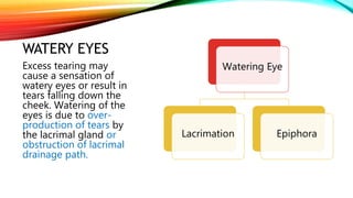
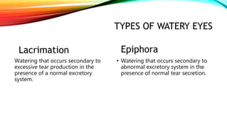
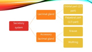
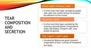
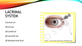
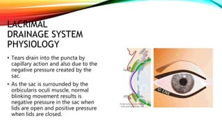
WATERY EYE
CAUSES &
TREATMENT
By: Qurat-ul-ain
Ophthalmic Medical
Technologist/ MBA Health &
Hospital management
WATERY EYES
Watering Eye
Lacrimation Epiphora
Excess tearing may
cause a sensation of
watery eyes or result in
tears falling down the
cheek. Watering of the
eyes is due to over-
production of tears by
the lacrimal gland or
obstruction of lacrimal
drainage path.
TYPES OF WATERY EYES
Lacrimation
Watering that occurs secondary to
excessive tear production in the
presence of a normal excretory
system.
Epiphora
• Watering that occurs secondary to
abnormal excretory system in the
presence of normal tear secretion.
SECRETORY SYSTEM
Normal tear secretion is necessary to keep the ocular surface
moist.
Secretory
system
Lacrimal gland
Orbital part (2/3
part)
Palpebral part
(1/3 part)
Accessory
lacrimal gland
Krause
Wolfring
TEAR
COMPOSITION
AND
SECRETION
• It is the inner most layer, secreted by goblet
cells, alters the surface tension & increases
the adhesion to the cornea.
Mucin Layer/ Mucous Layer
• It is the most thick layer, secreted by the
accessory lacrimal glands. Composed of
water, electrolytes, inorganic salts, urea,
protein & glucose.
Aqueous Layer
• Secreted by Meibomian gland, prevents
evaporation of tears. Consists of cholesterol
and lipids.
Oily Layer/ Lipid Layer
LACRIMAL
SYSTEM
Consists of:
 Puncta
 Canaliculi
 Lacrimal sac
 Nasolacrimal duct
LACRIMAL
DRAINAGE SYSTEM
PHYSIOLOGY
• Tears drain into the puncta by
capillary action and also due to the
negative pressure created by the
sac.
• As the sac is surrounded by the
orbicularis oculi muscle, normal
blinking movement results is
negative pressure in the sac when
lids are open and positive pressure
when lids are closed.
PATHOLOGY OF
THE TEAR FILM
• Kerato-conjunctivitis Sicca
Deficiency of the aqueous layer
• Paradoxical watering
Deficiency of the mucous layer (compensatory
excessive aqueous secretion)
CAUSES OF
LACRIMATION:
• Secondary to ocular inflammation or surface
disease
• Emotional distress
• Irritation of the eyes (smoke, dust, foreign
body and injury)
Hypersecretion
CAUSES OF
EPIPHORA
• Malposition of the lacrimal puncta (e.g.
ectropion)
• Obstruction (e.g. anywhere along the
drainage system)
• Lacrimal pump failure (e.g. lower lid laxity or
weakness of the orbicularis oculi muscle)
Defective drainage
ACQUIRED
OBSTRUCTION
• Primary punctal stenosis (chr. Blepharitis,
idiopathic, herpetic infection eye-lid,
irradiation, cicatrizing conjunctivitis,
trachoma, cytotoxic drugs)
• Secondary punctal stenosis (punctal
eversion)
• Canalicular obstruction (cong. Trauma,
herpetic infection, drugs)
• NLDO (idiopathic, previous surgery,
granulomatous disease, tumors)
• Dacryolithiasis
CONGENITAL
OBSTRUCTION • NLDO
• Congenital dacryocele
EVALUATION OF
AN ADULT
PATIENT
• Pseudo epiphora
• Lacrimation
• Dry eye
• Blepharitis
• True epiphora
EVALUATION
• External examination
• Fluorescein disappearance test
• Probing & irrigation
• Jones dye testing
• Contrast dacryo-cystography
• Nuclear lacrimal scintigraphy
TREATMENT
Primary punctal stenosis  dilatation with
nettle-ship dilatator, puncto-plasty
Secondary punctal stenosis  Ziegler
cautery, medial conjunctivo-plasty, lower
lid tightening
Canalicular obstruction:
• Partial: intubation
• Total: canaliculi-dacryo-cysto-rhino-
stomy and intubation or CDCR & the
insertion of Lester Jones tube
Lacrimation  usually medical
Dry eye  treatment consists of
topical lubricants
Blepharitis  treatment consists
of lid scrubs, oral tetracycline,
topical lubricants
Ectropion & entropion  lid
repair
CONTI..
• Nasolacrimal duct obstruction
• Infants: massage , probing
• Adults: DCR, TCL-DCR, intubation, stents, balloon dilatation
• Dacryolithiasis  DCR
• Congenital dacryocele  probing
• Lacrimal surgery  conventional dacryo-cysto-rhino-stomy
• Lester Jones tube
• Endonasal surgery
• Endocanlicular laser DCR
• Balloon dacryocystoplasty
CONCLUSION
• The proper diagnosis and treatment of watering eye is important due to its
impact on the outcome of many other ocular procedures.
• Undiagnosed and untreated watering eye substantially decreases the
patient’s quality of life, visual acuity and impairs social contact.
RESOURCES:
• https://www.medindia.net/patients/patientinfo/watery-eyes.htm
• Harley’s Pediatric Ophthalmology, 6th edition by Leonard B. Nelson, Scott E. Olitsky
• Comprehensive Ophthalmology by Dr. Nasir Chaudhry
THANK YOU!

5. Watery eye.pptx

  • 1.
    WATERY EYE CAUSES & TREATMENT By:Qurat-ul-ain Ophthalmic Medical Technologist/ MBA Health & Hospital management
  • 2.
    WATERY EYES Watering Eye LacrimationEpiphora Excess tearing may cause a sensation of watery eyes or result in tears falling down the cheek. Watering of the eyes is due to over- production of tears by the lacrimal gland or obstruction of lacrimal drainage path.
  • 3.
    TYPES OF WATERYEYES Lacrimation Watering that occurs secondary to excessive tear production in the presence of a normal excretory system. Epiphora • Watering that occurs secondary to abnormal excretory system in the presence of normal tear secretion.
  • 4.
    SECRETORY SYSTEM Normal tearsecretion is necessary to keep the ocular surface moist.
  • 5.
    Secretory system Lacrimal gland Orbital part(2/3 part) Palpebral part (1/3 part) Accessory lacrimal gland Krause Wolfring
  • 6.
    TEAR COMPOSITION AND SECRETION • It isthe inner most layer, secreted by goblet cells, alters the surface tension & increases the adhesion to the cornea. Mucin Layer/ Mucous Layer • It is the most thick layer, secreted by the accessory lacrimal glands. Composed of water, electrolytes, inorganic salts, urea, protein & glucose. Aqueous Layer • Secreted by Meibomian gland, prevents evaporation of tears. Consists of cholesterol and lipids. Oily Layer/ Lipid Layer
  • 7.
    LACRIMAL SYSTEM Consists of:  Puncta Canaliculi  Lacrimal sac  Nasolacrimal duct
  • 8.
    LACRIMAL DRAINAGE SYSTEM PHYSIOLOGY • Tearsdrain into the puncta by capillary action and also due to the negative pressure created by the sac. • As the sac is surrounded by the orbicularis oculi muscle, normal blinking movement results is negative pressure in the sac when lids are open and positive pressure when lids are closed.
  • 9.
    PATHOLOGY OF THE TEARFILM • Kerato-conjunctivitis Sicca Deficiency of the aqueous layer • Paradoxical watering Deficiency of the mucous layer (compensatory excessive aqueous secretion)
  • 10.
    CAUSES OF LACRIMATION: • Secondaryto ocular inflammation or surface disease • Emotional distress • Irritation of the eyes (smoke, dust, foreign body and injury) Hypersecretion
  • 11.
    CAUSES OF EPIPHORA • Malpositionof the lacrimal puncta (e.g. ectropion) • Obstruction (e.g. anywhere along the drainage system) • Lacrimal pump failure (e.g. lower lid laxity or weakness of the orbicularis oculi muscle) Defective drainage
  • 12.
    ACQUIRED OBSTRUCTION • Primary punctalstenosis (chr. Blepharitis, idiopathic, herpetic infection eye-lid, irradiation, cicatrizing conjunctivitis, trachoma, cytotoxic drugs) • Secondary punctal stenosis (punctal eversion) • Canalicular obstruction (cong. Trauma, herpetic infection, drugs) • NLDO (idiopathic, previous surgery, granulomatous disease, tumors) • Dacryolithiasis
  • 13.
  • 14.
    EVALUATION OF AN ADULT PATIENT •Pseudo epiphora • Lacrimation • Dry eye • Blepharitis • True epiphora
  • 15.
    EVALUATION • External examination •Fluorescein disappearance test • Probing & irrigation • Jones dye testing • Contrast dacryo-cystography • Nuclear lacrimal scintigraphy
  • 16.
    TREATMENT Primary punctal stenosis dilatation with nettle-ship dilatator, puncto-plasty Secondary punctal stenosis  Ziegler cautery, medial conjunctivo-plasty, lower lid tightening Canalicular obstruction: • Partial: intubation • Total: canaliculi-dacryo-cysto-rhino- stomy and intubation or CDCR & the insertion of Lester Jones tube Lacrimation  usually medical Dry eye  treatment consists of topical lubricants Blepharitis  treatment consists of lid scrubs, oral tetracycline, topical lubricants Ectropion & entropion  lid repair
  • 17.
    CONTI.. • Nasolacrimal ductobstruction • Infants: massage , probing • Adults: DCR, TCL-DCR, intubation, stents, balloon dilatation • Dacryolithiasis  DCR • Congenital dacryocele  probing • Lacrimal surgery  conventional dacryo-cysto-rhino-stomy • Lester Jones tube • Endonasal surgery • Endocanlicular laser DCR • Balloon dacryocystoplasty
  • 18.
    CONCLUSION • The properdiagnosis and treatment of watering eye is important due to its impact on the outcome of many other ocular procedures. • Undiagnosed and untreated watering eye substantially decreases the patient’s quality of life, visual acuity and impairs social contact.
  • 19.
    RESOURCES: • https://www.medindia.net/patients/patientinfo/watery-eyes.htm • Harley’sPediatric Ophthalmology, 6th edition by Leonard B. Nelson, Scott E. Olitsky • Comprehensive Ophthalmology by Dr. Nasir Chaudhry
  • 20.