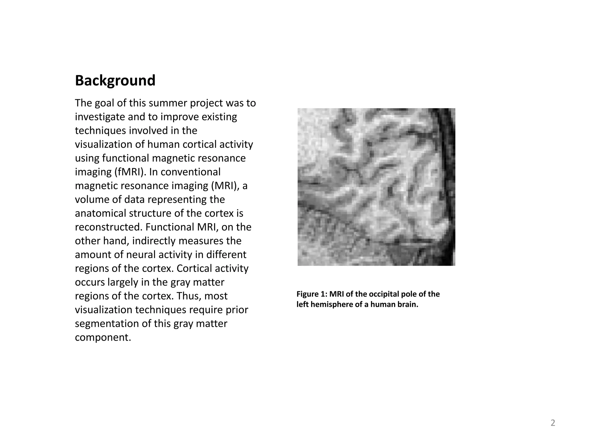
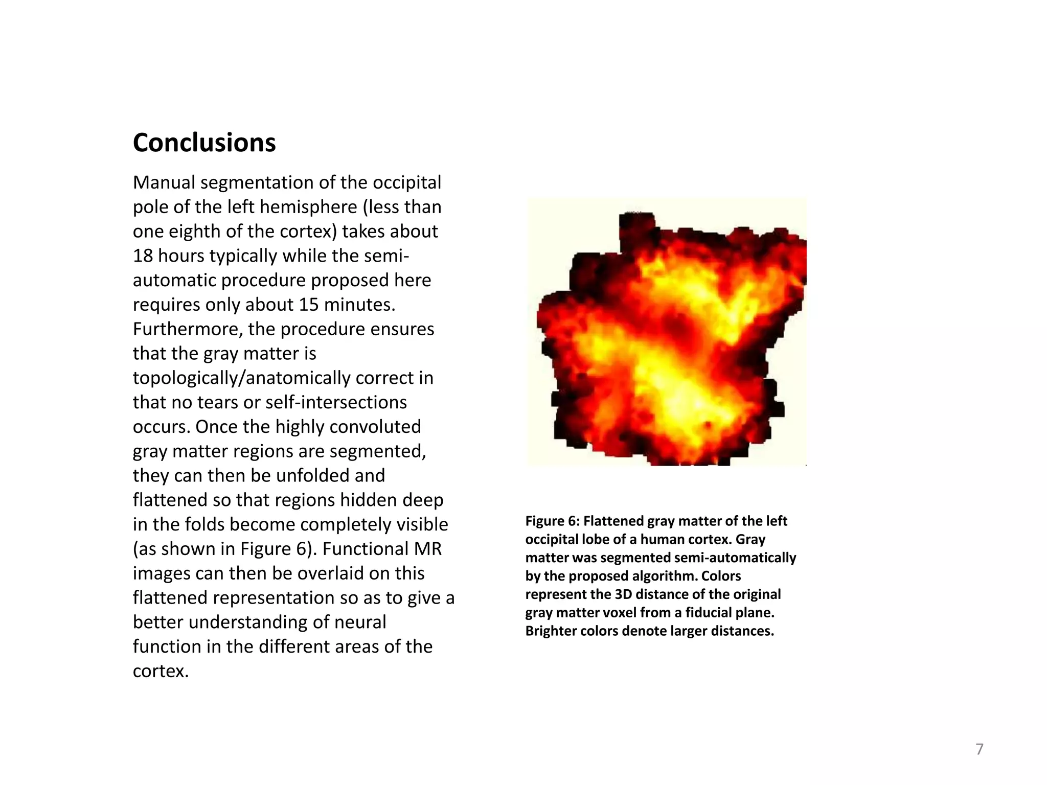
Visualization of Cortical Activity Using fMRI

                    Patrick Teo, Guillermo Sapiro, and Brian Wandell*
            Imaging Technology Department, HP Labs, and Stanford University*

                                                    Summer 1996

      Sponsored by the Hewlett Packard Labs Grassroots Basic Research Program

   Acknowledgements: Hagit Hel-Or (Stanford) for the original version of the segmentation tool. Steve Engel (Stanford) for
      the brain unfolding software. Geoff Boynton and Jon Demb (Stanford) for expertise on anatomy and f/MRI. Tom
      Malzbender (HP Labs) for discussions on graphics and visualization. Daniel Lee, Alex Drukarev, and the Grassroots
                            Research Program Committee (HP Labs) for supporting this research.


                                                                                                                             1
Background
The goal of this summer project was to
investigate and to improve existing
techniques involved in the
visualization of human cortical activity
using functional magnetic resonance
imaging (fMRI). In conventional
magnetic resonance imaging (MRI), a
volume of data representing the
anatomical structure of the cortex is
reconstructed. Functional MRI, on the
other hand, indirectly measures the
amount of neural activity in different
regions of the cortex. Cortical activity
occurs largely in the gray matter
regions of the cortex. Thus, most          Figure 1: MRI of the occipital pole of the
                                           left hemisphere of a human brain.
visualization techniques require prior
segmentation of this gray matter
component.


                                                                                        2
Background (cont’d)
                                             Gray matter segmentation is difficult
                                             because gray matter forms only a thin
                                             (2 mm or 2 pixel) layer on the surface
                                             of the cortex, which itself is inundated
                                             with numerous ridges and folds (as
                                             shown in Figure 2). Currently, much of
                                             gray matter segmentation is
                                             performed manually. This process is
                                             tedious and requires several days even
                                             for a trained person. On the other
                                             hand, most automatic gray matter
                                             segmentation methods that have been
                                             proposed do not consider anatomical
                                             constraints and often produce
                                             segmentations that are anatomically
                                             incorrect. We propose a new semi-
Figure 2: Photograph of a cross section of   automatic method of gray matter
an actual human cortex.                      segmentation that takes into
                                             consideration these anatomical
                                             requirements.


                                                                                        3
Method (Step 1)
Our proposed method consists of four
steps. In the first step, white matter is
segmented. White matter is
segmented because it is less noisy and
suffers less from partial volume
effects. Figure 3 shows the results of
segmenting the white matter in Figure
1. The maximum a posteriori
probability (MAP) estimate coupled
with a structural prior is used to
determine segmentation. The
structural prior is applied using a novel
technique involving anisotropic
smoothing.
                                            Figure 3: Segmentation results of white
                                            matter.


                                                                                      4
Method (Step 2)
                                         In the second step, the white matter
                                         connected component corresponding
                                         to the cortex is selected using a 3D
                                         connected components algorithm.
                                         After this single connected component
                                         is extracted, the topology of that
                                         component is verified in the third step.
                                         Since, in the anatomy, gray matter
                                         borders on white matter, the topology
                                         of gray matter places constraints on
                                         the topology of white matter.
Figure 4: Segmentation results of gray   Specifically, because gray matter is a
matter.                                  sheet, the white matter component
                                         cannot have cavities or handles.
                                         Cavities are identified using a flood-
                                         filling algorithm. Handles are detected
                                         by computing the Euler characteristic
                                         of the white matter.


                                                                                    5
Method (Step 3)
In the final step, the gray matter
classification is grown out from the
boundary of the white matter
component (as shown in Figures 4 and
5). Since gray matter is at most 2 mm
(or 2 pixel) thick in this direction, only
two layers of gray matter classification
are grown. Connectivity between gray
matter voxels are determined from the
connectivity of their parents from
which they were grown. Thus, gray
matter voxels may be adjacent in the
volume but not connected on the gray
matter surface.
                                             Figure 5: Segmentation results of gray
                                             matter overlaid on original MR image.


                                                                                      6
Conclusions
Manual segmentation of the occipital
pole of the left hemisphere (less than
one eighth of the cortex) takes about
18 hours typically while the semi-
automatic procedure proposed here
requires only about 15 minutes.
Furthermore, the procedure ensures
that the gray matter is
topologically/anatomically correct in
that no tears or self-intersections
occurs. Once the highly convoluted
gray matter regions are segmented,
they can then be unfolded and
flattened so that regions hidden deep
in the folds become completely visible     Figure 6: Flattened gray matter of the left
                                           occipital lobe of a human cortex. Gray
(as shown in Figure 6). Functional MR      matter was segmented semi-automatically
images can then be overlaid on this        by the proposed algorithm. Colors
flattened representation so as to give a   represent the 3D distance of the original
                                           gray matter voxel from a fiducial plane.
better understanding of neural             Brighter colors denote larger distances.
function in the different areas of the
cortex.


                                                                                         7
Publications


•   Anisotropic Smoothing of Posterior Probabilities.
    Patrick C. Teo and Guillermo Sapiro and Brian A. Wandell.
    Dynamical Systems, Control, Coding, Computer Vision : New Trends, Interfaces, and Interplay, Giorgio Picci
    and David S. Gilliam, Editors, Progress in Systems and Control Theory, vol. 25, 1999.

•   Creating connected representations of cortical gray matter for functional MRI visualization.
    Patrick C. Teo and Guillermo Sapiro and Brian A. Wandell.
    IEEE Transactions on Medical Imaging, vol. 16, no. 6, pp. 852-863, Dec. 1997.


                                                                                                             8

Visualization of Cortical Activity using fMRI - Summary

  • 1.
    Visualization of CorticalActivity Using fMRI Patrick Teo, Guillermo Sapiro, and Brian Wandell* Imaging Technology Department, HP Labs, and Stanford University* Summer 1996 Sponsored by the Hewlett Packard Labs Grassroots Basic Research Program Acknowledgements: Hagit Hel-Or (Stanford) for the original version of the segmentation tool. Steve Engel (Stanford) for the brain unfolding software. Geoff Boynton and Jon Demb (Stanford) for expertise on anatomy and f/MRI. Tom Malzbender (HP Labs) for discussions on graphics and visualization. Daniel Lee, Alex Drukarev, and the Grassroots Research Program Committee (HP Labs) for supporting this research. 1
  • 2.
    Background The goal ofthis summer project was to investigate and to improve existing techniques involved in the visualization of human cortical activity using functional magnetic resonance imaging (fMRI). In conventional magnetic resonance imaging (MRI), a volume of data representing the anatomical structure of the cortex is reconstructed. Functional MRI, on the other hand, indirectly measures the amount of neural activity in different regions of the cortex. Cortical activity occurs largely in the gray matter regions of the cortex. Thus, most Figure 1: MRI of the occipital pole of the left hemisphere of a human brain. visualization techniques require prior segmentation of this gray matter component. 2
  • 3.
    Background (cont’d) Gray matter segmentation is difficult because gray matter forms only a thin (2 mm or 2 pixel) layer on the surface of the cortex, which itself is inundated with numerous ridges and folds (as shown in Figure 2). Currently, much of gray matter segmentation is performed manually. This process is tedious and requires several days even for a trained person. On the other hand, most automatic gray matter segmentation methods that have been proposed do not consider anatomical constraints and often produce segmentations that are anatomically incorrect. We propose a new semi- Figure 2: Photograph of a cross section of automatic method of gray matter an actual human cortex. segmentation that takes into consideration these anatomical requirements. 3
  • 4.
    Method (Step 1) Ourproposed method consists of four steps. In the first step, white matter is segmented. White matter is segmented because it is less noisy and suffers less from partial volume effects. Figure 3 shows the results of segmenting the white matter in Figure 1. The maximum a posteriori probability (MAP) estimate coupled with a structural prior is used to determine segmentation. The structural prior is applied using a novel technique involving anisotropic smoothing. Figure 3: Segmentation results of white matter. 4
  • 5.
    Method (Step 2) In the second step, the white matter connected component corresponding to the cortex is selected using a 3D connected components algorithm. After this single connected component is extracted, the topology of that component is verified in the third step. Since, in the anatomy, gray matter borders on white matter, the topology of gray matter places constraints on the topology of white matter. Figure 4: Segmentation results of gray Specifically, because gray matter is a matter. sheet, the white matter component cannot have cavities or handles. Cavities are identified using a flood- filling algorithm. Handles are detected by computing the Euler characteristic of the white matter. 5
  • 6.
    Method (Step 3) Inthe final step, the gray matter classification is grown out from the boundary of the white matter component (as shown in Figures 4 and 5). Since gray matter is at most 2 mm (or 2 pixel) thick in this direction, only two layers of gray matter classification are grown. Connectivity between gray matter voxels are determined from the connectivity of their parents from which they were grown. Thus, gray matter voxels may be adjacent in the volume but not connected on the gray matter surface. Figure 5: Segmentation results of gray matter overlaid on original MR image. 6
  • 7.
    Conclusions Manual segmentation ofthe occipital pole of the left hemisphere (less than one eighth of the cortex) takes about 18 hours typically while the semi- automatic procedure proposed here requires only about 15 minutes. Furthermore, the procedure ensures that the gray matter is topologically/anatomically correct in that no tears or self-intersections occurs. Once the highly convoluted gray matter regions are segmented, they can then be unfolded and flattened so that regions hidden deep in the folds become completely visible Figure 6: Flattened gray matter of the left occipital lobe of a human cortex. Gray (as shown in Figure 6). Functional MR matter was segmented semi-automatically images can then be overlaid on this by the proposed algorithm. Colors flattened representation so as to give a represent the 3D distance of the original gray matter voxel from a fiducial plane. better understanding of neural Brighter colors denote larger distances. function in the different areas of the cortex. 7
  • 8.
    Publications • Anisotropic Smoothing of Posterior Probabilities. Patrick C. Teo and Guillermo Sapiro and Brian A. Wandell. Dynamical Systems, Control, Coding, Computer Vision : New Trends, Interfaces, and Interplay, Giorgio Picci and David S. Gilliam, Editors, Progress in Systems and Control Theory, vol. 25, 1999. • Creating connected representations of cortical gray matter for functional MRI visualization. Patrick C. Teo and Guillermo Sapiro and Brian A. Wandell. IEEE Transactions on Medical Imaging, vol. 16, no. 6, pp. 852-863, Dec. 1997. 8