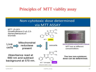
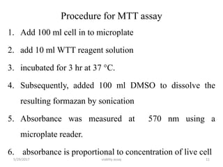
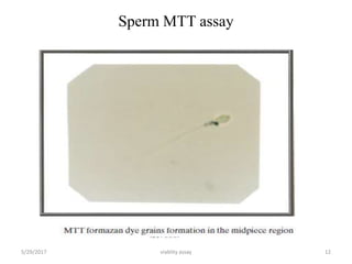
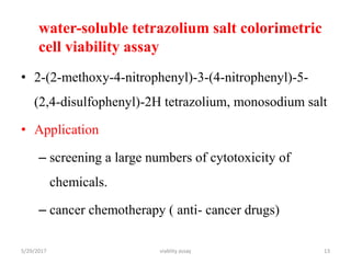
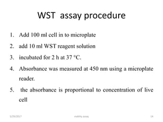
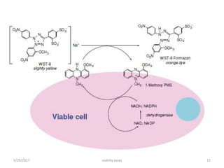
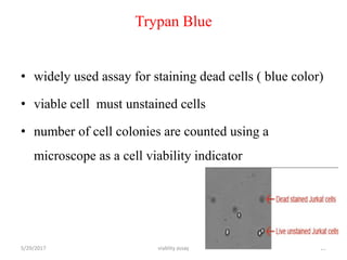
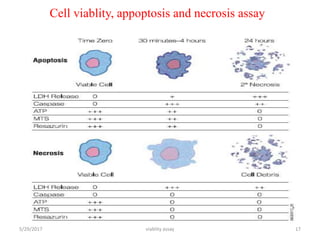
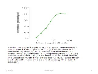
This document discusses viability assays, which determine if cells are alive or dead. It defines viability assays and explains they are based on cell functions like enzyme activity and membrane permeability. Common viability assays described include MTT, WST, trypan blue, and LDH assays. The MTT and WST assays work by measuring the reduction of tetrazolium salts by metabolically active cells, indicating the number of living cells. Trypan blue distinguishes live from dead cells by staining only dead cells blue. The document classifies viability assays and explains their applications in screening drugs, diagnosing diseases, and measuring cell survival.
The MTT assay and the MTS assay are colorimetric assays for measuring the activity of enzymes that reduce MTT or close dyes (XTT, MTS, WSTs) to formazan dyes, giving a purple color The main application allows to assess the viability (cell counting) and the proliferation of cells (cell culture assays)
It can also be used to determine cytotoxicity of potential medicinal agents and toxic materials, since those agents would stimulate or inhibit cell viability and growth
Biology and characterization of the cell cultureKAUSHAL SAHU
Introduction
History
Important terminology
Biology of culture cell
Characterization of culture cell
Application of animal culture
Conclusion
References
Equipments used , types of culture and media, subculturing, secondary culture, finite & continuous cell lines, cryopreservation and applications of cell culture
A brief presentation on cell counting and cell viability assays. For cell cytotoxicity assays, you can check my profile where I have uploaded a separate file.
Prepared in July 2015
Principles of cell viability assays by surendra.pptxSurendra Chowdary
1.DYE EXCLUSION ASSAYS:
Dye exclusion assays are the simplest methods that are based on utilization of different dyes such as trypan blue, eosin, congo red, and erythrosine B, which are excluded by the living cells, but not by dead cells.
For these assays, although staining procedure is quite straightforward, experimental procedure may be time-consuming in case of large sample sizes.
a. Trypan blue stain assay:
Trypan blue stain assay has initially been developed in 1975 to measure viable cell count and is still used as a confirmatory test for measuring changes in viable cell number caused by a drug or toxin.
Trypan blue stain, a large negatively charged molecule, is one of the simplest assays that are used to determine the number of viable cells in a cell suspension.
Principle:
The principle of this assay is that living cells have intact cell membranes that exclude the trypan blue stain, whereas dead cells do not.
Cell suspension is mixed with the trypan blue stain and examined visually under light microscopy to determine whether cells include or exclude the stain.
A viable cell will have a clear cytoplasm, whereas a nonviable cell will have a blue cytoplasm.
Reagent preparation:
To perform the trypan blue stain assay, 0.4% trypan blue stain and phosphate- buffered saline (PBS) or serum-free medium are obtained.
Trypan blue stain should be stored in dark and filtered after prolonged storage.
As trypan blue stain binds to serum proteins and causing misleading results, serum-free medium should be used to obtain reliable results.
Assay Protocol:
The cell suspension to be tested is centrifuged at 100 g for 5 min.
The supernatant is discarded and the pellet is resuspended in 1-ml PBS solution or serum-free medium.
Then, one portion of this cell suspension is mixed with one portion of trypan blue stain.
The mixture is allowed to stay at room temperature for 3 min. It is important to note that the cells should be counted within 3–5 min of mixing with trypan blue, as longer incubation periods will lead to cell death and hence reduced viability counts.
Following the incubation, a drop of the mixture is applied to a hemocytometer, which is placed on the stage of a binocular microscope.
Viable cells will remain unstained, and nonviable cells will stain, in the hemocytometer and these cells are counted separately.
.
Calculation:
After counting viable and nonviable cells, the total number of viable cells per milliliter of aliquot is determined by multiplying the total number of viable cells by 2, which is the dilution factor for trypan blue.
Similarly, total number of cells per milliliter of aliquot is determined by addition of number of viable and nonviable cells and multiplying it by 2.
Then, the percentage of viable cells is calculated using the following equation.
% Viable cells = Total number of viable cells per milliliter of aliquot × 100.
Total number of cells per milliliter of aliquot
2.COLORIMETRIC ASSAYS:
Colorimetric assays
IOSR Journal of Pharmacy and Biological Sciences(IOSR-JPBS) is an open access international journal that provides rapid publication (within a month) of articles in all areas of Pharmacy and Biological Science. The journal welcomes publications of high quality papers on theoretical developments and practical applications in Pharmacy and Biological Science. Original research papers, state-of-the-art reviews, and high quality technical notes are invited for publications.
The Main Advantage
The main advantages of flow cytometry over histology and IHC is the possibility to precisely measure the quantities of antigens and the possibility to stain each cell with multiple antibodies-fluorophores, in current laboratories around 10 antibodies can be bound to each cell. This is much less than mass cytometer where up to 40 can be currently measured, but at a higher and slower pace.
Aquatic research
In aquatic systems, flow cytometry is used for the analysis of autofluorescing cells or cells that are fluorescently-labeled with added stains.
This research started in 1981 when Clarice Yentsch used flow cytometry to measure the fluorescence in a red tide producing dinoflagellates
Marine scientists use the sorting ability of flow cytometers to make discrete measurements of cellular activity and diversity, to conduct investigations into the mutualistic relationships between microorganisms that live in close proximity,and to measure biogeochemical rates of multiple processes in the ocean
Cell Proliferation assay
Cell proliferation is the major function in the immune system. Often it is required to analyse the proliferative nature of the cells in order to make some conclusions. One such assay to determine the cell proliferation is the tracking dye carboxyfluorescein diacetate succinimidyl ester (CFSE). It helps to monitor proliferative cells. This assay gives quantitative as well as qualitative data during time-series experiments
Cell counting
Cell sorting
Determining cell characteristics and function
Detecting microorganisms
Biomarker detection
Protein engineering detection
Diagnosis of health disorders such as blood cancers
Flow cytometry can be used for cell cycle analysis to estimate the percentages of a cell population in the different phases of the cell cycle, or it can be used with other reagents to analyze just the S phase.
Why flow cytometry is ideal for cell cycle analysis
Live-cell cycle analysis stains—Vybrant DyeCycle stains
Classic DNA cell cycle stains such as Hoechst 33342 and DRAQ5 for cell cycle analysis, but most of these have limitations that have to be considered when using them in an experiment which is why the Invitrogen Vybrant DyeCycle stains for live-cell cycle analysis were developed.
Fixed-cell cycle analysis stains FxCycle reagents
We offer classic DNA cell cycle stains such as DAPI, PI, and 7-AAD for fixed cell cycle analysis, but these reagents do not cover the full spectrum of laser excitation available.
The FxCycle reagents offer options for the 405 nm (violet) and 633 nm (red) laser thereby increasing the ability to multiplex by freeing up the 488 nm and 633 nm lasers for other cellular analyses such as immunophenotyping, apoptosis analysis, and dead cell discrimination.
Precise—Accurate cell cycle analysis in living cells
Safe—Low cytotoxicity for combining with additional live cell experiments
Cell sort compatible—Easily sort cells based on phase of the cell cycle
In vitro methods for the assessment of general cellular toxicity,
End-points for the assessment of general cellular toxicity
Specialized cells commonly used in toxicology
This presentation details the definition of cell cytotoxicity and cell viability, the difference between the two term and methods of assessment of cells in culture for presence and absence of cytotoxic chemicals or metabolites.
Assignment on Toxicokinetics- Toxicokinetic evaluation in preclinical studies, saturation kinetics Importance and applications of toxicokinetic studies. Alternative methods to animal toxicity testing.
Title: Sense of Smell
Presenter: Dr. Faiza, Assistant Professor of Physiology
Qualifications:
MBBS (Best Graduate, AIMC Lahore)
FCPS Physiology
ICMT, CHPE, DHPE (STMU)
MPH (GC University, Faisalabad)
MBA (Virtual University of Pakistan)
Learning Objectives:
Describe the primary categories of smells and the concept of odor blindness.
Explain the structure and location of the olfactory membrane and mucosa, including the types and roles of cells involved in olfaction.
Describe the pathway and mechanisms of olfactory signal transmission from the olfactory receptors to the brain.
Illustrate the biochemical cascade triggered by odorant binding to olfactory receptors, including the role of G-proteins and second messengers in generating an action potential.
Identify different types of olfactory disorders such as anosmia, hyposmia, hyperosmia, and dysosmia, including their potential causes.
Key Topics:
Olfactory Genes:
3% of the human genome accounts for olfactory genes.
400 genes for odorant receptors.
Olfactory Membrane:
Located in the superior part of the nasal cavity.
Medially: Folds downward along the superior septum.
Laterally: Folds over the superior turbinate and upper surface of the middle turbinate.
Total surface area: 5-10 square centimeters.
Olfactory Mucosa:
Olfactory Cells: Bipolar nerve cells derived from the CNS (100 million), with 4-25 olfactory cilia per cell.
Sustentacular Cells: Produce mucus and maintain ionic and molecular environment.
Basal Cells: Replace worn-out olfactory cells with an average lifespan of 1-2 months.
Bowman’s Gland: Secretes mucus.
Stimulation of Olfactory Cells:
Odorant dissolves in mucus and attaches to receptors on olfactory cilia.
Involves a cascade effect through G-proteins and second messengers, leading to depolarization and action potential generation in the olfactory nerve.
Quality of a Good Odorant:
Small (3-20 Carbon atoms), volatile, water-soluble, and lipid-soluble.
Facilitated by odorant-binding proteins in mucus.
Membrane Potential and Action Potential:
Resting membrane potential: -55mV.
Action potential frequency in the olfactory nerve increases with odorant strength.
Adaptation Towards the Sense of Smell:
Rapid adaptation within the first second, with further slow adaptation.
Psychological adaptation greater than receptor adaptation, involving feedback inhibition from the central nervous system.
Primary Sensations of Smell:
Camphoraceous, Musky, Floral, Pepperminty, Ethereal, Pungent, Putrid.
Odor Detection Threshold:
Examples: Hydrogen sulfide (0.0005 ppm), Methyl-mercaptan (0.002 ppm).
Some toxic substances are odorless at lethal concentrations.
Characteristics of Smell:
Odor blindness for single substances due to lack of appropriate receptor protein.
Behavioral and emotional influences of smell.
Transmission of Olfactory Signals:
From olfactory cells to glomeruli in the olfactory bulb, involving lateral inhibition.
Primitive, less old, and new olfactory systems with different path
- Video recording of this lecture in English language: https://youtu.be/lK81BzxMqdo
- Video recording of this lecture in Arabic language: https://youtu.be/Ve4P0COk9OI
- Link to download the book free: https://nephrotube.blogspot.com/p/nephrotube-nephrology-books.html
- Link to NephroTube website: www.NephroTube.com
- Link to NephroTube social media accounts: https://nephrotube.blogspot.com/p/join-nephrotube-on-social-media.html
Flu Vaccine Alert in Bangalore Karnatakaaddon Scans
As flu season approaches, health officials in Bangalore, Karnataka, are urging residents to get their flu vaccinations. The seasonal flu, while common, can lead to severe health complications, particularly for vulnerable populations such as young children, the elderly, and those with underlying health conditions.
Dr. Vidisha Kumari, a leading epidemiologist in Bangalore, emphasizes the importance of getting vaccinated. "The flu vaccine is our best defense against the influenza virus. It not only protects individuals but also helps prevent the spread of the virus in our communities," he says.
This year, the flu season is expected to coincide with a potential increase in other respiratory illnesses. The Karnataka Health Department has launched an awareness campaign highlighting the significance of flu vaccinations. They have set up multiple vaccination centers across Bangalore, making it convenient for residents to receive their shots.
To encourage widespread vaccination, the government is also collaborating with local schools, workplaces, and community centers to facilitate vaccination drives. Special attention is being given to ensuring that the vaccine is accessible to all, including marginalized communities who may have limited access to healthcare.
Residents are reminded that the flu vaccine is safe and effective. Common side effects are mild and may include soreness at the injection site, mild fever, or muscle aches. These side effects are generally short-lived and far less severe than the flu itself.
Healthcare providers are also stressing the importance of continuing COVID-19 precautions. Wearing masks, practicing good hand hygiene, and maintaining social distancing are still crucial, especially in crowded places.
Protect yourself and your loved ones by getting vaccinated. Together, we can help keep Bangalore healthy and safe this flu season. For more information on vaccination centers and schedules, residents can visit the Karnataka Health Department’s official website or follow their social media pages.
Stay informed, stay safe, and get your flu shot today!
Basavarajeeyam is an important text for ayurvedic physician belonging to andhra pradehs. It is a popular compendium in various parts of our country as well as in andhra pradesh. The content of the text was presented in sanskrit and telugu language (Bilingual). One of the most famous book in ayurvedic pharmaceutics and therapeutics. This book contains 25 chapters called as prakaranas. Many rasaoushadis were explained, pioneer of dhatu druti, nadi pareeksha, mutra pareeksha etc. Belongs to the period of 15-16 century. New diseases like upadamsha, phiranga rogas are explained.
ARTIFICIAL INTELLIGENCE IN HEALTHCARE.pdfAnujkumaranit
Artificial intelligence (AI) refers to the simulation of human intelligence processes by machines, especially computer systems. It encompasses tasks such as learning, reasoning, problem-solving, perception, and language understanding. AI technologies are revolutionizing various fields, from healthcare to finance, by enabling machines to perform tasks that typically require human intelligence.
HOT NEW PRODUCT! BIG SALES FAST SHIPPING NOW FROM CHINA!! EU KU DB BK substit...GL Anaacs
Contact us if you are interested:
Email / Skype : kefaya1771@gmail.com
Threema: PXHY5PDH
New BATCH Ku !!! MUCH IN DEMAND FAST SALE EVERY BATCH HAPPY GOOD EFFECT BIG BATCH !
Contact me on Threema or skype to start big business!!
Hot-sale products:
NEW HOT EUTYLONE WHITE CRYSTAL!!
5cl-adba precursor (semi finished )
5cl-adba raw materials
ADBB precursor (semi finished )
ADBB raw materials
APVP powder
5fadb/4f-adb
Jwh018 / Jwh210
Eutylone crystal
Protonitazene (hydrochloride) CAS: 119276-01-6
Flubrotizolam CAS: 57801-95-3
Metonitazene CAS: 14680-51-4
Payment terms: Western Union,MoneyGram,Bitcoin or USDT.
Deliver Time: Usually 7-15days
Shipping method: FedEx, TNT, DHL,UPS etc.Our deliveries are 100% safe, fast, reliable and discreet.
Samples will be sent for your evaluation!If you are interested in, please contact me, let's talk details.
We specializes in exporting high quality Research chemical, medical intermediate, Pharmaceutical chemicals and so on. Products are exported to USA, Canada, France, Korea, Japan,Russia, Southeast Asia and other countries.
Tom Selleck Health: A Comprehensive Look at the Iconic Actor’s Wellness Journeygreendigital
Tom Selleck, an enduring figure in Hollywood. has captivated audiences for decades with his rugged charm, iconic moustache. and memorable roles in television and film. From his breakout role as Thomas Magnum in Magnum P.I. to his current portrayal of Frank Reagan in Blue Bloods. Selleck's career has spanned over 50 years. But beyond his professional achievements. fans have often been curious about Tom Selleck Health. especially as he has aged in the public eye.
Follow us on: Pinterest
Introduction
Many have been interested in Tom Selleck health. not only because of his enduring presence on screen but also because of the challenges. and lifestyle choices he has faced and made over the years. This article delves into the various aspects of Tom Selleck health. exploring his fitness regimen, diet, mental health. and the challenges he has encountered as he ages. We'll look at how he maintains his well-being. the health issues he has faced, and his approach to ageing .
Early Life and Career
Childhood and Athletic Beginnings
Tom Selleck was born on January 29, 1945, in Detroit, Michigan, and grew up in Sherman Oaks, California. From an early age, he was involved in sports, particularly basketball. which played a significant role in his physical development. His athletic pursuits continued into college. where he attended the University of Southern California (USC) on a basketball scholarship. This early involvement in sports laid a strong foundation for his physical health and disciplined lifestyle.
Transition to Acting
Selleck's transition from an athlete to an actor came with its physical demands. His first significant role in "Magnum P.I." required him to perform various stunts and maintain a fit appearance. This role, which he played from 1980 to 1988. necessitated a rigorous fitness routine to meet the show's demands. setting the stage for his long-term commitment to health and wellness.
Fitness Regimen
Workout Routine
Tom Selleck health and fitness regimen has evolved. adapting to his changing roles and age. During his "Magnum, P.I." days. Selleck's workouts were intense and focused on building and maintaining muscle mass. His routine included weightlifting, cardiovascular exercises. and specific training for the stunts he performed on the show.
Selleck adjusted his fitness routine as he aged to suit his body's needs. Today, his workouts focus on maintaining flexibility, strength, and cardiovascular health. He incorporates low-impact exercises such as swimming, walking, and light weightlifting. This balanced approach helps him stay fit without putting undue strain on his joints and muscles.
Importance of Flexibility and Mobility
In recent years, Selleck has emphasized the importance of flexibility and mobility in his fitness regimen. Understanding the natural decline in muscle mass and joint flexibility with age. he includes stretching and yoga in his routine. These practices help prevent injuries, improve posture, and maintain mobilit
Lung Cancer: Artificial Intelligence, Synergetics, Complex System Analysis, S...Oleg Kshivets
RESULTS: Overall life span (LS) was 2252.1±1742.5 days and cumulative 5-year survival (5YS) reached 73.2%, 10 years – 64.8%, 20 years – 42.5%. 513 LCP lived more than 5 years (LS=3124.6±1525.6 days), 148 LCP – more than 10 years (LS=5054.4±1504.1 days).199 LCP died because of LC (LS=562.7±374.5 days). 5YS of LCP after bi/lobectomies was significantly superior in comparison with LCP after pneumonectomies (78.1% vs.63.7%, P=0.00001 by log-rank test). AT significantly improved 5YS (66.3% vs. 34.8%) (P=0.00000 by log-rank test) only for LCP with N1-2. Cox modeling displayed that 5YS of LCP significantly depended on: phase transition (PT) early-invasive LC in terms of synergetics, PT N0—N12, cell ratio factors (ratio between cancer cells- CC and blood cells subpopulations), G1-3, histology, glucose, AT, blood cell circuit, prothrombin index, heparin tolerance, recalcification time (P=0.000-0.038). Neural networks, genetic algorithm selection and bootstrap simulation revealed relationships between 5YS and PT early-invasive LC (rank=1), PT N0—N12 (rank=2), thrombocytes/CC (3), erythrocytes/CC (4), eosinophils/CC (5), healthy cells/CC (6), lymphocytes/CC (7), segmented neutrophils/CC (8), stick neutrophils/CC (9), monocytes/CC (10); leucocytes/CC (11). Correct prediction of 5YS was 100% by neural networks computing (area under ROC curve=1.0; error=0.0).
CONCLUSIONS: 5YS of LCP after radical procedures significantly depended on: 1) PT early-invasive cancer; 2) PT N0--N12; 3) cell ratio factors; 4) blood cell circuit; 5) biochemical factors; 6) hemostasis system; 7) AT; 8) LC characteristics; 9) LC cell dynamics; 10) surgery type: lobectomy/pneumonectomy; 11) anthropometric data. Optimal diagnosis and treatment strategies for LC are: 1) screening and early detection of LC; 2) availability of experienced thoracic surgeons because of complexity of radical procedures; 3) aggressive en block surgery and adequate lymph node dissection for completeness; 4) precise prediction; 5) adjuvant chemoimmunoradiotherapy for LCP with unfavorable prognosis.
NVBDCP.pptx Nation vector borne disease control programSapna Thakur
NVBDCP was launched in 2003-2004 . Vector-Borne Disease: Disease that results from an infection transmitted to humans and other animals by blood-feeding arthropods, such as mosquitoes, ticks, and fleas. Examples of vector-borne diseases include Dengue fever, West Nile Virus, Lyme disease, and malaria.
Title: Sense of Taste
Presenter: Dr. Faiza, Assistant Professor of Physiology
Qualifications:
MBBS (Best Graduate, AIMC Lahore)
FCPS Physiology
ICMT, CHPE, DHPE (STMU)
MPH (GC University, Faisalabad)
MBA (Virtual University of Pakistan)
Learning Objectives:
Describe the structure and function of taste buds.
Describe the relationship between the taste threshold and taste index of common substances.
Explain the chemical basis and signal transduction of taste perception for each type of primary taste sensation.
Recognize different abnormalities of taste perception and their causes.
Key Topics:
Significance of Taste Sensation:
Differentiation between pleasant and harmful food
Influence on behavior
Selection of food based on metabolic needs
Receptors of Taste:
Taste buds on the tongue
Influence of sense of smell, texture of food, and pain stimulation (e.g., by pepper)
Primary and Secondary Taste Sensations:
Primary taste sensations: Sweet, Sour, Salty, Bitter, Umami
Chemical basis and signal transduction mechanisms for each taste
Taste Threshold and Index:
Taste threshold values for Sweet (sucrose), Salty (NaCl), Sour (HCl), and Bitter (Quinine)
Taste index relationship: Inversely proportional to taste threshold
Taste Blindness:
Inability to taste certain substances, particularly thiourea compounds
Example: Phenylthiocarbamide
Structure and Function of Taste Buds:
Composition: Epithelial cells, Sustentacular/Supporting cells, Taste cells, Basal cells
Features: Taste pores, Taste hairs/microvilli, and Taste nerve fibers
Location of Taste Buds:
Found in papillae of the tongue (Fungiform, Circumvallate, Foliate)
Also present on the palate, tonsillar pillars, epiglottis, and proximal esophagus
Mechanism of Taste Stimulation:
Interaction of taste substances with receptors on microvilli
Signal transduction pathways for Umami, Sweet, Bitter, Sour, and Salty tastes
Taste Sensitivity and Adaptation:
Decrease in sensitivity with age
Rapid adaptation of taste sensation
Role of Saliva in Taste:
Dissolution of tastants to reach receptors
Washing away the stimulus
Taste Preferences and Aversions:
Mechanisms behind taste preference and aversion
Influence of receptors and neural pathways
Impact of Sensory Nerve Damage:
Degeneration of taste buds if the sensory nerve fiber is cut
Abnormalities of Taste Detection:
Conditions: Ageusia, Hypogeusia, Dysgeusia (parageusia)
Causes: Nerve damage, neurological disorders, infections, poor oral hygiene, adverse drug effects, deficiencies, aging, tobacco use, altered neurotransmitter levels
Neurotransmitters and Taste Threshold:
Effects of serotonin (5-HT) and norepinephrine (NE) on taste sensitivity
Supertasters:
25% of the population with heightened sensitivity to taste, especially bitterness
Increased number of fungiform papillae
Recomendações da OMS sobre cuidados maternos e neonatais para uma experiência pós-natal positiva.
Em consonância com os ODS – Objetivos do Desenvolvimento Sustentável e a Estratégia Global para a Saúde das Mulheres, Crianças e Adolescentes, e aplicando uma abordagem baseada nos direitos humanos, os esforços de cuidados pós-natais devem expandir-se para além da cobertura e da simples sobrevivência, de modo a incluir cuidados de qualidade.
Estas diretrizes visam melhorar a qualidade dos cuidados pós-natais essenciais e de rotina prestados às mulheres e aos recém-nascidos, com o objetivo final de melhorar a saúde e o bem-estar materno e neonatal.
Uma “experiência pós-natal positiva” é um resultado importante para todas as mulheres que dão à luz e para os seus recém-nascidos, estabelecendo as bases para a melhoria da saúde e do bem-estar a curto e longo prazo. Uma experiência pós-natal positiva é definida como aquela em que as mulheres, pessoas que gestam, os recém-nascidos, os casais, os pais, os cuidadores e as famílias recebem informação consistente, garantia e apoio de profissionais de saúde motivados; e onde um sistema de saúde flexível e com recursos reconheça as necessidades das mulheres e dos bebês e respeite o seu contexto cultural.
Estas diretrizes consolidadas apresentam algumas recomendações novas e já bem fundamentadas sobre cuidados pós-natais de rotina para mulheres e neonatos que recebem cuidados no pós-parto em unidades de saúde ou na comunidade, independentemente dos recursos disponíveis.
É fornecido um conjunto abrangente de recomendações para cuidados durante o período puerperal, com ênfase nos cuidados essenciais que todas as mulheres e recém-nascidos devem receber, e com a devida atenção à qualidade dos cuidados; isto é, a entrega e a experiência do cuidado recebido. Estas diretrizes atualizam e ampliam as recomendações da OMS de 2014 sobre cuidados pós-natais da mãe e do recém-nascido e complementam as atuais diretrizes da OMS sobre a gestão de complicações pós-natais.
O estabelecimento da amamentação e o manejo das principais intercorrências é contemplada.
Recomendamos muito.
Vamos discutir essas recomendações no nosso curso de pós-graduação em Aleitamento no Instituto Ciclos.
Esta publicação só está disponível em inglês até o momento.
Prof. Marcus Renato de Carvalho
www.agostodourado.com
New Directions in Targeted Therapeutic Approaches for Older Adults With Mantl...i3 Health
i3 Health is pleased to make the speaker slides from this activity available for use as a non-accredited self-study or teaching resource.
This slide deck presented by Dr. Kami Maddocks, Professor-Clinical in the Division of Hematology and
Associate Division Director for Ambulatory Operations
The Ohio State University Comprehensive Cancer Center, will provide insight into new directions in targeted therapeutic approaches for older adults with mantle cell lymphoma.
STATEMENT OF NEED
Mantle cell lymphoma (MCL) is a rare, aggressive B-cell non-Hodgkin lymphoma (NHL) accounting for 5% to 7% of all lymphomas. Its prognosis ranges from indolent disease that does not require treatment for years to very aggressive disease, which is associated with poor survival (Silkenstedt et al, 2021). Typically, MCL is diagnosed at advanced stage and in older patients who cannot tolerate intensive therapy (NCCN, 2022). Although recent advances have slightly increased remission rates, recurrence and relapse remain very common, leading to a median overall survival between 3 and 6 years (LLS, 2021). Though there are several effective options, progress is still needed towards establishing an accepted frontline approach for MCL (Castellino et al, 2022). Treatment selection and management of MCL are complicated by the heterogeneity of prognosis, advanced age and comorbidities of patients, and lack of an established standard approach for treatment, making it vital that clinicians be familiar with the latest research and advances in this area. In this activity chaired by Michael Wang, MD, Professor in the Department of Lymphoma & Myeloma at MD Anderson Cancer Center, expert faculty will discuss prognostic factors informing treatment, the promising results of recent trials in new therapeutic approaches, and the implications of treatment resistance in therapeutic selection for MCL.
Target Audience
Hematology/oncology fellows, attending faculty, and other health care professionals involved in the treatment of patients with mantle cell lymphoma (MCL).
Learning Objectives
1.) Identify clinical and biological prognostic factors that can guide treatment decision making for older adults with MCL
2.) Evaluate emerging data on targeted therapeutic approaches for treatment-naive and relapsed/refractory MCL and their applicability to older adults
3.) Assess mechanisms of resistance to targeted therapies for MCL and their implications for treatment selection
New Directions in Targeted Therapeutic Approaches for Older Adults With Mantl...
Viablity assay
1. DEPARTMENT OF IMMUNOLOGYAND MOLECULAR
BIOLOGY
VIABLITY ASSAY
By :- Birhanu Ayelign
1
viablity assay
UNIVERSITY OF GONDAR
COLLEGE OF MEDICINE AND HEALTH SCIENCES
SCHOOL OF BIOMEDICALAND LABORATORY SCIENCES
5/29/2017
2. Objective
At the end of this portion you will be able to :-
Define viability assay?
What are the classification of viability assay?
What are the application of viability assay?
Discuses some of the common viability assay?
5/29/2017 2viablity assay
3. Introduction To Viability Assay
A viability assay
• is an assay to determine viability of organism or cell
• Viability can be distinguished live to died cell
5/29/2017 viablity assay 3
4. Features of viable cell
• They are based on various cell functions such as
– enzyme activity,
– cell membrane permeability,
– cell adherence,
– ATP production,
– co-enzyme production, and
– nucleotide uptake activity.
5/29/2017 viablity assay 4
5. Introduction To Viability Assay con…
Used to measure a
• markers that indicate the number of
– dead cells (cytotoxicity assay),
– the number of live cells(viability assay),
• the total number of cells or
• the mechanism of cell death (e.g., apoptosis).
5/29/2017 viablity assay 5
6. Application of viability assay
• To detect cytotoxic or growth inhibitory lymphokines
• To detect mammalian cell survival and proliferations
• To diagnose disease
• To diagnose male infertility
• To screen drugs
5/29/2017 viablity assay 6
7. Classification of viability assays
A. Cytolysis or membrane leakage assays
B. Mitochondrial activity or caspase assays
C. Functional assays
D. Genomic and proteomic assays
5/29/2017 viablity assay 7
8. Common methods used in viablity assay
MTT cell viability assay
WST methods
Trypan Blue
Lactate dehydrogenase (LDH)
Flow cytometry
PCR ???????????
5/29/2017 viablity assay 8
9. MTT CELL VIABILITYASSAY
• 3-(4,5-Dimethyl-2-thiazolyl)-2,5-diphenyl-2H tetrazolium
bromide
• Application
– screening a large numbers of cytotoxicity of chemicals.
– cancer chemotherapy ( anti- cancer drugs)
– Sperm analysis
– Cell proliferation
5/29/2017 viablity assay 9
11. Procedure for MTT assay
1. Add 100 ml cell in to microplate
2. add 10 ml WTT reagent solution
3. incubated for 3 hr at 37 °C.
4. Subsequently, added 100 ml DMSO to dissolve the
resulting formazan by sonication
5. Absorbance was measured at 570 nm using a
microplate reader.
6. absorbance is proportional to concentration of live cell
5/29/2017 viablity assay 11
13. water-soluble tetrazolium salt colorimetric
cell viability assay
• 2-(2-methoxy-4-nitrophenyl)-3-(4-nitrophenyl)-5-
(2,4-disulfophenyl)-2H tetrazolium, monosodium salt
• Application
– screening a large numbers of cytotoxicity of
chemicals.
– cancer chemotherapy ( anti- cancer drugs)
5/29/2017 viablity assay 13
14. WST assay procedure
1. Add 100 ml cell in to microplate
2. add 10 ml WST reagent solution
3. incubated for 2 h at 37 °C.
4. Absorbance was measured at 450 nm using a microplate
reader.
5. the absorbance is proportional to concentration of live
cell
5/29/2017 viablity assay 14
16. Trypan Blue
• widely used assay for staining dead cells ( blue color)
• viable cell must unstained cells
• number of cell colonies are counted using a
microscope as a cell viability indicator
5/29/2017 viablity assay 16
19. ACKNOWLEGMENT
I would like to express my deepest and heartfelt gratitude
to our instructor Mr. Demeke G. & Mr. Fitsumbrhan
Tajebew give me an opportunity to do this assignment .
Finally, I would like to thank immunology and molecular
biology, school of biomedical and laboratory science,
Gondar university.
5/29/2017 viablity assay 19
20. Reference
I. Green LM, Reade JL, Ware CF. Rapid colormetric assay
for cell viability: application to the quantitation of
cytotoxic and growth inhibitory lymphokines. Journal of
immunological methods. 1984 May 25;70(2):257-68.
II. Molecular genetics of bacteria: Jermey W.Dale &
Simone F. Park, 5th edition.
III. Geneomes : T.A. Br own, 2th edition.
viablity assay 205/29/2017
OFT,,,, hereditary spherocytosis To maintain viable cells in vivo/vitro the cells must be suspended in a solution with an osmotic concentration relatively equal to their interior osmotic concentration: ISOTONIC :0.85% NaCl solution.
T. pallidium,,,, immobilization test
LDH,,,,, myocardiac infraction, hemolysis, liver disease
Male fertility by sperm analysis
Use Premixed WST-1 to measure cell proliferation and viability, or to measure cytotoxicity and growth inhibition. A mitochondrial enzyme in viable cells converts WST-1 to a dye…simply measure its absorbance.When cells are proliferating, dye accumulates.
When cells are not proliferating, dye levels drop.
Cytolysis or membrane leakage assays: This category includes the lactate dehydrogenase assay, a stable enzyme common in all cells which can be readily detected when cell membranes are no longer intact. Examples:Propidium iodide,Trypan blue7-Aminoactinomycin D
Mitochondrial activity or caspase assays: Resazurin and Formazan (MTT/XTT) can assay for various stages in the apoptosis process that foreshadow cell death.
Functional assays: Assays of cell function will be highly specific to the types of cells being assayed. For example, motility is a widely used assay of sperm cell function. Gamete survival can be used to assay fertility, in general. Red blood cells have been assayed in terms of deformability, osmotic fragility, hemolysis, ATP level, and hemoglobin content. For transplantable whole organs the ultimate assay is the ability to sustain life after transplantation, an assay which is not helpful in preventing transplantation of non-functional organs.
Genomic and proteomic assays: Cells can be assayed for activation of stress pathways using DNA microarrays and protein chips.
In order to address this limitation, alternative PCR-based strategies have been developed. This article reviews two complementary strategies. One strategy, termed viability PCR, or vPCR, correlates viability with cell envelope impermeability (9, 10). In viability PCR, microbes in samples are incubated with a membrane-impermeative reagent such as propidium monoazide (PMA). Upon photoactivation, PMA binds tightly to exposed DNA and interferes with PCR amplification. Nonviable cells with damaged membranes, and free nucleic acids, are not protected from the reagent, and their amplification is inhibited after the reagent-DNA complex is photoactivated. In contrast, viable cells with intact cell membranes exclude PMA, enabling strong quantitative PCR (qPCR) signals in the presence of the reagent. The operating principle is similar to microscopy-based live/dead staining, in which a membrane-impermeative DNA stain (typically, propidium iodide [PI]) is excluded from intact cells but penetrates and stains the DNA of membrane-compromised cells. In live/dead staining, inactivated cells are quantified relative to total cell counts by fluorescence microscopy or flow cytometry (11) rather than by PCR.
The second strategy, termed “molecular viability testing” (MVT), correlates viability with the ability to rapidly synthesize a macromolecule (a species-specific rRNA precursor, or pre-rRNA) in response to a brief nutritional stimulus (12,–14). Pre-rRNA synthesis upon nutritional stimulation is detected by reverse transcriptase-qPCR (RT-qPCR) measurement of species-specific pre-rRNA molecules. Pre-rRNAs in inactivated cells, and free nucleic acids, do not increase in numbers upon nutritional stimulation and therefore are excluded.
sperm MTT viability assay as a new diagnostic test for the discriminationof live sperm from dead sperm.
Formation ofMTT formazan granules or spikes around the spermmidpiece show that sperm mitochondria
contains dehydrogenase,which is capable of converting MTT toMTT formazan, and the presence of MTT
formazangranules in the midpiece region identifies that sperm
as a live sperm.
Trypan Blue
Trypan Blue is one of several stains recommended for use in dye exclusion procedures for viable cell counting. This method is based on the principle that live (viable) cells do not take up certain dyes, whereas dead (non-viable) cells do. Staining facilitates the visualization of cell morphology.
NOTE: Trypan Blue has a greater affinity for serum proteins than for cellular protein. If the background is too dark, cells should be pelleted and resuspended in protein-free medium or salt solution prior to counting.
Protocol for Viable Cell Counting using Trypan Blue
Prepare a cell suspension in a balanced salt solution (e.g., Hanks' Balanced Salts [HBSS], Cat. No. H9269).
Transfer 0.5 ml of 0.4% Trypan Blue solution (w/v) to a test tube. Add 0.3 ml of HBSS and 0.2 ml of the cell suspension (dilution factor = 5) and mix thoroughly. Allow to stand for 5 to 15 minutes.Note: If cells are exposed to Trypan Blue for extended periods of time, viable cells, as well as non-viable cells, may begin to take up dye.
With the cover-slip in place, use a Pasteur pipette or other suitable device to transfer a small amount of Trypan Blue-cell suspension mixture to both chambers of the hemacytometer. Carefully touch the edge of the cover-slip with the pipette tip and allow each chamber to fill by capillary action. Do not overfill or underfill the chambers.
Starting with chamber 1 of the hemacytometer, count all the cells in the 1 mm center square and four 1 mm corner squares (see Diagram I). Non-viable cells will stain blue. Keep a separate count of viable and non-viable cells.Note: Count cells on top and left touching middle line of the perimeter of each square. Do not count cells touching the middle line at bottom and right sides (see Diagram II).
Repeat this procedure for chamber 2.Note: If greater than 10% of the cells appear clustered, repeat entire procedure making sure the cells are dispersed by vigorous pipetting in the original cell suspension as well as the Trypan Blue-cell suspension mixture. If less than 200 or greater than 500 cells (i.e., 20–50 cells/square) are observed in the 10 squares, repeat the procedure adjusting to an appropriate dilution factor.
Withdraw a second sample and repeat count procedure to ensure accuracy.
Calculations
Cell Counts – Each square of the hemacytometer, with cover-slip in place, represents a total volume of 0.1 mm3 or 10-4 cm3. Since 1 cm3 is equivalent to approximately 1 ml, the subsequent cell concentration per ml (and the total number of cells) will be determined using the following calculations:
Cells Per mL = the average count per square × dilution factor × 104 (count 10 squares)
Example: If the average count per square is 45 cells × 5 × 104 = 2.25 × 10⁶ cells/ml.
Total Cells = cells per ml × the original volume of fluid from which cell sample was removed.
Example: 2.25 × 10⁶ (cells/ml) × 10 ml (original volume) = 2.25 × 10⁷ total cells.
Cell Viability (%) = total viable cells (unstained) ÷ total cells (stained and unstained) × 100.
Example: If the average count per square of unstained (viable) cells is 37.5, the total viable cells = [37.5 × 5 × 104] viable cells/ml × 10 ml (original volume) = 1.875 × 10⁷ viable cells. Cell viability (%) = 1.875 × 10⁷ (viable cells) ÷ 2.25 × 10⁷ (total cells) × 100 = 83% viability
Apoptosis is a complex processes involving a cascade mechanism that employs many proteins. However, the key enzymes in this process are the caspases, a family of cysteine proteases that control and mediate the apoptotic response. Virtually all animal cells contain caspases, but they occur as inactive zymogens that can do no harm. There are various triggers that can lead to their activation, which usually occurs through proteolytic processing of the zymogen at conserved aspartic acid residues. Needless to say, their activation and suicidal function is highly regulated. Once activated caspases act as cysteine proteases, using a cysteine side chain for catalysing peptide bond cleavage at aspartyl residues in their substrates. The name “caspase” denotes their function: Cysteine-dependentASPartyl-specific proteASE. There are many such caspases within an organism, which work together in a proteolytic cascade to activate themselves and one other. Cascades are effective means of amplifying a signal to give a much larger response than could be achieved through a single enzymatic reaction.
Easy, Colorimetric Test for Cell Death
The easiest way to measure cell death is by measuring lactate dehydrogenase (LDH), a stable cytoplasmic enzyme which is present in all cells but only released when the plasma membrane is damaged. The LDH Cytotoxicity Detection Kit provides a simple and precise colorimetric assay for LDH activity: a two-step enzymatic reaction creates a formazan dye that is easily measured by A492.
Why Use the LDH Cytotoxicity Detection Kit?
Sensitive assay: Detect as few as 2,000 dead or damaged cells per well.
Accurate results: The amount of formazan dye you measure is directly proportional to the number of dead or damaged cells.
Cost-effective: Get your LDH assay for a better price.
Fast, Simple LDH Assay Protocol
The LDH assay does not require prelabeling or washing steps. The entire procedure can be performed in a single 96-well plate.