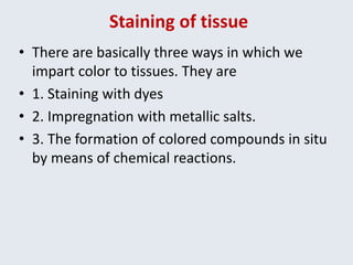
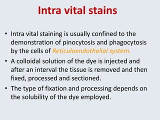
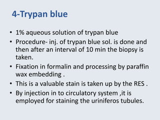
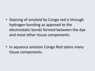
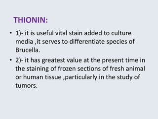
SlideShare a Scribd company logo
Vital stains
-Dr. Anil Kr
Introduction
• Most oral cancers are preceded by precancerous lesions
and early cancers that can be identified by visual inspection
of the oral cavity.
Conventional oral examination is useful in the discovery of
some oral lesions but it does not identify all potentially
premalignant lesions, as some are not readily apparent to
visual inspection alone .
-Adjunctive techniques have emerged that may facilitate early
detection of oral premalignant and malignant lesions
Staining of tissue
• There are basically three ways in which we
impart color to tissues. They are
• 1. Staining with dyes
• 2. Impregnation with metallic salts.
• 3. The formation of colored compounds in situ
by means of chemical reactions.
Vital stains
• Vital staining is defined as staining of the cells
or tissues in a living state
• Classified as-
Intra vital staining :staining done in the body (in
vivo) by injecting colloidal solution of dyes.
Supra vital staining : staining of living cells out
side the body (in vitro) usually applied to slide
preparation of detached cells.
Intravital and supra vital stains
• 1- Trypan Blue
• 2- Vital New Red
• 3- Indian Ink
• 4- Congo Red
• 5- Toluidine Blue
• 6- Lugol’s Iodine
• 7- Methylene Blue
• 8- Acetic Acid
• 9- Rose Bengal
• 1-Ehrlich’s Methylene Blue
• 2-Janus Green
• 3-Neutral Red
• 4-Tetrazolium Salt
• 5-Fluorochrome
• 6-Pinacynol
• 7-Acridine Orange
• 8-Thionin
• 9-Nile Blue, Azo Blue
Intra vital stains
• Intra vital staining is usually confined to the
demonstration of pinocytosis and phagocytosis
by the cells of Reticuloendothelial system.
• A colloidal solution of the dye is injected and
after an interval the tissue is removed and then
fixed, processed and sectioned.
• The type of fixation and processing depends on
the solubility of the dye employed.
Intravital staining
1-Toluidine Blue
• Toluidine blue may be used as an additional
aid to assess at risk of cancer lesions.
• The mechanism of increase uptake in
dysplastic and malignant lesions may be due
to increase DNA synthesis in malignant cells by
greater diffusion through increased
intercellular channels in tumor cells.
• The assessment of dye uptake depends on
clinical judgment and experience.
• Composition:
100 ml of 1% TB contains-
• 1 gm of toluidine blue powder,
• 10 ml of 1% acetic acid,
• 4.19 ml of absolute alcohol and
• 86 ml of distilled water,
• pH maintained at 4.5.
TISSUE STAIN APPLICATION SEQUENCE-
Application of 1% acetic with Q-tip (20 sec)
(to remove soapy saliva)
Rinse in water
Apply toluidine blue 1% with Q-tip (10-20 sec)
Decolorize with 2% acetic acid (to reduce
mechanically stained stains)
Toluidine blue staining
ADVANTAGES
• Helps to determine the extend of biopsy
site.
• Easy to perform.
• Non invasive.
• Inexpensive.
• Helps to monitor treated cancer patients
for recurrence.
• Sensitivity of 72-100%
• Specificity of 45-93%
DISADVANTAGES
• Not recommended for patients with physical or
mental disability.
• Acetic acid may irritate the mucosa.
• Equivocal dye retention.
• Variable mode of application.
• 30% false positive results noted.
2-LUGOL’s IODINE
• Lugol’s iodine consists of iodine , potassium iodide and
distilled water in contrast to TB.
• Lugol’s iodine is retained in normal squamous
epithelial cells but not in dysplastic or malignant
epithelial cells.
• As there is enhanced glycolysis in cancer cells, do not
promote the iodine–starch reaction.
• Lugol’s iodine solution produces a brown black stain
by reaction of the iodine with glycogen.
• Normal mucosa contains higher amount of glycogen than
abnormal mucosa and produces brown black stain.
Lesions after being stained with Lugol's
iodine solution
3- Acetic acid staining
• In the past, 3-5% acetic acid was used as a vital
staining for the detection of oral cancers.
Composition: Of 1% acetic acid rinse –
1 ml of glacial acetic acid and 99 ml distilled water
• Application of acetic acid causes reversible
coagulation of cellular proteins particularly in
abnormal squamous epithelial areas.
• Aceto-whitening is seen distinctly as compared with
the normal pinkish color of the surrounding normal
squamous epithelium.
(a)Before application of the vital stain. (b) After
application of vital stain, asterisk showing biopsy site.
Acetic acid staining in lateral
border of tongue
formazan
4-Trypan blue
• 1% aqueous solution of trypan blue
• Procedure- inj. of trypan blue sol. is done and
then after an interval of 10 min the biopsy is
taken.
• Fixation in formalin and processing by paraffin
wax embedding .
• This is a valuable stain is taken up by the RES .
• By injection in to circulatory system ,it is
employed for staining the uriniferos tubules.
Trypan blue
A) Trypan blue exclusion test shows unstained live cells (L) and
dark-blue stained dead cells (D)
uses
• Trypan blue is commonly used in microscopy (for cell
counting) and in laboratory mice for assessment of
tissue viability.
• The method cannot distinguish
between necrotic and apoptotic cells.
• It may be used to observe fungal hyphae.
• Trypan blue is also used in ophthalmic cataract
surgery.
5- Congo red test for amyloidosis
• Bennhold in 1922
• Highly selective affinity for amyloid and subsequent
green birefringence when viewed with polarizing
microscope.
• After intravenous injection of the dye all the amyloid
deposition in the body are stained bright red.
• More than 80% retention aft 1-4 hrs.
• Staining of amyloid by Congo red is through
hydrogen bonding as apposed to the
electrostatic bonds formed between the dye
and most other tissue components.
• In aqueous solution Congo Red stains many
tissue components.
Localized amyloidosis of the upper gingiva
Vital staining of macrophages and
osteoclasts:
• The identification of macrophages under
experimental conditions were greatly
facilitated by the development of vital staining
which as it names implies ,means a method in
which living cells become stained because of
some vital activity ( Phagocytosis)
• Dye trypan blue injected either directly in to
CT or administered IV or into peritoneal cavity
• Dye phagocytized by macrophages and be
seen as blue material in their cytoplasm.
• Such vital stains are colloidal dyes and it is
because they are in the form of
macromolecular aggregates that they are
phagocytized.
• Colloidal silver and diluted Indian ink are also
frequently employed for vital staining of
macrophages.
Osteoclasts and vital staining
• When the method of vital staining was first
developed ,many experiments were performed
not to determine the origin of osteoclasts but to
see if they were phagocytic.
• Whereas it was shown readily that the cells we
term macrophages would take up vital stains such
as trypan blue or particulate matter such as
Indian ink , osteoclast examined in same animals
showed no significant uptake.
• Since at that time osteoclasts were generally
believed to develop from osteogenic cells or
osteoblasts and since neither of these cell
types was shown to be phagocytic
• it caused little surprise that osteoclasts
evidenced no phagocytic abilities when tested
by this method.
• In 1963 Jee and Neelson using bone charcoal as
suitable particulate matter for vital staining
showed that material could be osteoclasts but
only after enough time had elapsed for
macrophages that had taken up the markers by
phagocytosis to have fused and formed
osteoclasts
• This proven some proof of two concept –
• -the osteoclasts were derived from macrophages
• -The osteoclasts themselves were not phagocytic.
6-Vital new red
• 5% aqueous solution of the dye is used. Tissue
may be fixed in saturated aqueous solution of
mercuric chloride ,processed by paraffin wax
tech.
7-Indian ink- commercial Indian ink is
diluted with distilled water and after filtration
sterilized in autoclave it with stands routine
fixation and processing in paraffin wax.
SUPRA VITAL STAINING
• Staining of living cells after removal from
organism( in vitro) usually applied to slide
preparation of detached cells.
eg.
• Ehrlich’s methylene blue ,Janus green stain.
• Mechanism –supravital staining involves the
application of specific dyes that penetrate all
the cells and color certain cellular tissue
components,
• A dye used for supra vital work should be able not only to
enter the cell but also diffuse through the protoplasm
without the killing the cell and to color pre-exiting cell
inclusions, distinctively. or color the whole cytoplasm of
the particular cell strongly enough that these cells stand
out prominently from surrounding inter cellular material
and other cells
• Vital dyes stains blood cells in vitro while they are still
alive.
• A number of these stains are employed in hematology.
1)Ehrlich’s methylene blue technique
• By this method nerve endings in dissociated
tissue (e.g. muscles) can be demonstrated.
• Method- tissue is teased out in warm saline
solution in a watch glass.
• Small pieces are transferred to dilute (0.025-
0.25%)solution of methylene blue in normal
saline solution either on a slide or in another
watch glass for 10-15 min.
• Having the watch glass inside a dish containing a
piece of wet cotton wool controls evaporation.
• The preparation should be examined under the
microscope from time to time .
• if staining is sufficient or absent after 45 min ,the
strength of the stain should be increased and the
technique repeated..
• Note- the staining fades quite quickly if the
preparation is covered (oxygen is excluded ) treatment
with saturated ammonium molybdate for 2 hrs. before
mounting in glycerin helps to preserve the color.
• RESULTS-Nerve fibers and nerve ending –blue
other tissue constituents- colorless to pale blue
2-Supra vital staining of leucocytes or
other living cells:
• A solution of Janus green and Neutral red are
used as a vital stain in hematological lab for
the stains of leukocytes to differentiate
between acute leukemia of the blastic type.
• RESULT– mitochondria in myeloblasts are
small and tend to aggregate whereas in
lymphoblast they are large and diffuse.
• The technique is based on the affinity of Janus
green B for mitochondria and neutral red for
the neutral red vacuoles.
• Both dyes are toxic and their strength must be
carefully controlled.
• Slide preparations are used and must be
maintained at a constant temperature of 37oC.
Whitby & Hynes method: for supra vital staining
with Jasus green and neutral Red stain.
Mechanism- the selective staining of the
mitochondria and of intra cytoplasmic granules
is probably the result of their enzymatic activity
.
Stock solution –
Neutral red- 0.2 gm. in 50 ml absolute
alcohol
Janus green -0.25 gm. in 50 ml absolute
alcohol.
working solution-
Janus green solution- .25 ml (.025%)
Neutral red solution -0.8 ml (0.4%)
Absolute alcohol -5 ml
A clean microscopic slide is flooded with stain
and dries in horizontal position one small drop
of blood not anti coagulated or bone marrow
placed on the slide and covered with a cover
slip .this will allow specimen to form a thin
film ,allow to stain for 20 min.
RESULT-
Intra cytoplasmic granules and vacuoles
stain deep purple to orange.
Mitochondria-green
Cytoplasm- pale yellow to pale green blue
Nucleus- colorless
Neutrophils- yellow ,
Eosinophils- orange
Basophils- maroon
3- Tetrazolium salt-
• For the demonstration of many oxidative
enzymes, tetrazolium salts are used as a
hydrogen acceptor.
• on reduction they will form colored deposit.
• Two tetrazolium salts are in common use at
the present time -monotetrazolium and
ditetrazolium.
• Ditetrazolium salt ,ditetrazolium chloride –nitro
known as NBT (nitro blue tetrazolium) produces
highly colored formazan deposit that is insoluble
in lipid.
• Formazan dyes are artificial chromogenic
products of the reduction of tetrazolium salts by
dehydrogenases and reductases.
• monotetrazolium produces a finely granular
formazan which is soluble in lipids.
• The soluble yellowish ,oxidized, Nitro Blue
Tetrazolium (NBT) salt is reduced to dark blue
insoluble formazan when normal phagocytic
cells( granulocytes and monocytes ) are
incubated with NBT.
• In healthy individuals less than 10 % of
phagocytic cells are NBT positive.
• A much higher percentage of positive cells
results if bacteria stimulate the cells in vitro or
in vivo and then incubated with (NBT).
• Since granulocytes exposed to bacteria have a
markedly stimulated metabolic activity.
formazan
4- Fluorochrome:
• They are basic dyes ,that fluorescence when
excited by UV light. The stained living cells are
examined by a combination of fluorescence
and phase microscopy.
• Fluorochromes can be used to identify
cytoplasmic inclusions e.g. mitochondria,
Golgi apparatus, DNA staining.
• Also used to demonstrate acid fast bacteria in
smears and stains.
Fluorochromes for DNA Staining and
Quantitation
5-Pinacyanol and Neutral red stain
• Pinacynol stains the mitochondria deep blue, has the
advantage over Janus green ,that it does not fade for
many hours and can successfully be combined with
neutral red.
• REAGENT :
• Stock solution- Pinacyanol 0.03 % in absolute alcohol.
Neutral red 0.4% in absolute alcohol.
• Working sol: 1 ml of each solution added to 5 ml of
absolute alcohol.
• RESULT- Mitochondria-deep blue
6- Acridine Orange supravital stain:
• Tech- Living cells are stained with acridine orange
and are examined under fluorescence
microscope.
• Reagent- Acridine Orange 0.001 % in N saliva
• pH -6-8
• Procedure- 1 drop of fresh blood or bone marrow
in several drops of stain on a slide is added ,cover
slip applied and slide allowed to incubate at room
temp. for 3 min and examined under
fluorescence microscope.
stained with acridine orange
• RESULT-
• living nuclei (DNA) – fluorescence yellow green
• Nuclear +cytoplasmic RNA – fluorescence orange
red
• PMNL granules- Bright red
• Eosinophilic granules- orange
• Mast cell - Red
• Dead Nuclei - dull red
7- Supra Vital staining of reticulocytes
• Reticulocytes are large nuclear cells in bone
marrow when stained with vital dye (new
methylene blue) seen to contain a network of
bluish granules(ppt ribosomes) responsible for
the name.
Advantage of vital stain:
• 1)- Elimination of staining artifacts (since living)
• 2)- Undamaged cells are examined.
Disadvantage of vital stains:
• 1)- the preparation is not permanent and must be
examined fresh before they dry out.
• 2)- the living cells are extremely fragile and
therefore any error in technique is detrimental.
• 3)- the stain fades after short time
Limitations of vital staining
• 1)- Only a few specific elements may be
demonstrated .
• 2)- Nuclear material not stained.
• 3)- the strength of dye is very critical
• Too weak- there is little or no staining
• Too strong- the cells are killed.
• .
• 4)- Intra vital staining is mostly confined to the
demonstration of the Reticuloendothelial
system.
• Dye particles from a colloidal solution are
ingested by the phagocytic cells and are
subsequently found loose in the cytoplasm ,no
specific elements being colored.
• 5)- some of the method (ex. Ehrlich’s )are not
truly vital in that staining will take place after
the death of tissue.
• 6)- vital staining dye like methylene blue will
retain their color only if there is excess of
oxygen. and too bright light fade the color.
• 7)- temperature of the solution is also critical
,inta vital and supra vital stain must be done
under controlled temp conditions.
• 8)- vital staining has limited application in
electron microscopy as it can lead to
alterations in metabolism ,which will be
manifest as change in fine structure .
THIONIN:
• 1)- it is useful vital stain added to culture
media ,it serves to differentiate species of
Brucella.
• 2)- it has greatest value at the present time in
the staining of frozen sections of fresh animal
or human tissue ,particularly in the study of
tumors.
NILE BLUE:
Used to stain hydrae ,protozoa ,yeasts and for
staining bone sections. It stains lipid vesicles in the
cell and gives red colour under flouro microscopy
AZO BLUE:
Vital staining of protozoa .it can replace Indian ink
in demonstration of capsule of bacteria.
ALIZARIN RED:
1)- Vital Stain for nervous tissue
2)- An important application is for the gross staining
of skeletons especially in fetus.
Vital staining ppt notes

More Related Content

What's hot

Special stains in histopathology
Special stains in histopathologySpecial stains in histopathology
Special stains in histopathologyEkta Jajodia
 
Principles of Staining
Principles of StainingPrinciples of Staining
Principles of Staining
Charlotte McLean
 
Staining ( rouine and special in cytology) rajiv kumar
Staining ( rouine and special in cytology) rajiv kumarStaining ( rouine and special in cytology) rajiv kumar
Staining ( rouine and special in cytology) rajiv kumar
rajusehrawat
 
Pas staining
Pas staining Pas staining
Fixatives in Histopathology
Fixatives in HistopathologyFixatives in Histopathology
Fixatives in HistopathologyIshwar9
 
Decalcification
Decalcification  Decalcification
Decalcification
Layal Fahad
 
Tissue processing
Tissue  processingTissue  processing
Tissue processing
Sowmya Srinivas
 
Mounting media
Mounting mediaMounting media
Mounting media
MAHMOUD IBRAHIM
 
Connective Tissue
Connective TissueConnective Tissue
Connective Tissue
MAHMOUD IBRAHIM
 
Fixation & fixatives in histopathology, dr naveen reddy
Fixation & fixatives in histopathology, dr naveen reddyFixation & fixatives in histopathology, dr naveen reddy
Fixation & fixatives in histopathology, dr naveen reddy
Naveen Parvathareddy
 
Special stains by sowmya
Special stains by sowmyaSpecial stains by sowmya
Special stains by sowmya
Sowmya Srinivas
 
Frozen section
Frozen sectionFrozen section
Frozen section
Sizan Thapa
 
Prothrombin time
Prothrombin timeProthrombin time
Prothrombin time
rajexh777
 
PAS STAIN.pptx
PAS STAIN.pptxPAS STAIN.pptx
PAS STAIN.pptx
ShahzebHUSSAIN5
 
Demonstration of le cells
Demonstration of le cellsDemonstration of le cells
Demonstration of le cells
SHRUTHI VASAN
 
Staining by hematoxylin and eosin
Staining by hematoxylin and eosinStaining by hematoxylin and eosin
Staining by hematoxylin and eosin
Sowmya Srinivas
 
Embedding
EmbeddingEmbedding
Differential leukocyte count
Differential leukocyte countDifferential leukocyte count
Differential leukocyte count
Dr. Varughese George
 
Histopatholgy staining by suchit kumar
Histopatholgy staining by suchit kumarHistopatholgy staining by suchit kumar
Histopatholgy staining by suchit kumar
suchitkumar24
 

What's hot (20)

Special stains in histopathology
Special stains in histopathologySpecial stains in histopathology
Special stains in histopathology
 
Principles of Staining
Principles of StainingPrinciples of Staining
Principles of Staining
 
Staining ( rouine and special in cytology) rajiv kumar
Staining ( rouine and special in cytology) rajiv kumarStaining ( rouine and special in cytology) rajiv kumar
Staining ( rouine and special in cytology) rajiv kumar
 
Pas staining
Pas staining Pas staining
Pas staining
 
Fixatives in Histopathology
Fixatives in HistopathologyFixatives in Histopathology
Fixatives in Histopathology
 
Decalcification
Decalcification  Decalcification
Decalcification
 
Tissue processing
Tissue  processingTissue  processing
Tissue processing
 
Mounting media
Mounting mediaMounting media
Mounting media
 
Connective Tissue
Connective TissueConnective Tissue
Connective Tissue
 
Fixation & fixatives in histopathology, dr naveen reddy
Fixation & fixatives in histopathology, dr naveen reddyFixation & fixatives in histopathology, dr naveen reddy
Fixation & fixatives in histopathology, dr naveen reddy
 
Special stains by sowmya
Special stains by sowmyaSpecial stains by sowmya
Special stains by sowmya
 
Frozen section
Frozen sectionFrozen section
Frozen section
 
Prothrombin time
Prothrombin timeProthrombin time
Prothrombin time
 
H & E STAIN
 H & E STAIN H & E STAIN
H & E STAIN
 
PAS STAIN.pptx
PAS STAIN.pptxPAS STAIN.pptx
PAS STAIN.pptx
 
Demonstration of le cells
Demonstration of le cellsDemonstration of le cells
Demonstration of le cells
 
Staining by hematoxylin and eosin
Staining by hematoxylin and eosinStaining by hematoxylin and eosin
Staining by hematoxylin and eosin
 
Embedding
EmbeddingEmbedding
Embedding
 
Differential leukocyte count
Differential leukocyte countDifferential leukocyte count
Differential leukocyte count
 
Histopatholgy staining by suchit kumar
Histopatholgy staining by suchit kumarHistopatholgy staining by suchit kumar
Histopatholgy staining by suchit kumar
 

Similar to Vital staining ppt notes

Staining methods.pptx
Staining methods.pptxStaining methods.pptx
Staining methods.pptx
Jyoti Balmiki
 
Staining
StainingStaining
Staining
Ankita S.
 
Biocompatibility of dental materials
Biocompatibility of dental materialsBiocompatibility of dental materials
Biocompatibility of dental materials
Febel Huda
 
13.Staining technique.pptx
13.Staining technique.pptx13.Staining technique.pptx
13.Staining technique.pptx
LalitDhimal
 
Exfoliative cytology
Exfoliative cytologyExfoliative cytology
Exfoliative cytology
Akhil s
 
sputum examination pathology 1st year Allied health Sciences RGUHS.pptx
sputum examination pathology 1st year Allied health Sciences RGUHS.pptxsputum examination pathology 1st year Allied health Sciences RGUHS.pptx
sputum examination pathology 1st year Allied health Sciences RGUHS.pptx
TijoTomy6
 
Staning 1&2.pptx
Staning 1&2.pptxStaning 1&2.pptx
Staning 1&2.pptx
AhmedAli320762
 
Common Laboratory investigations in dermatology
Common Laboratory investigations in dermatologyCommon Laboratory investigations in dermatology
Common Laboratory investigations in dermatology
Kezha Zutso
 
cytopreparation techniques part 2
cytopreparation techniques part 2cytopreparation techniques part 2
cytopreparation techniques part 2
Dr.Azad Mustafa Ahmed
 
JC on vital staining pptx
JC on vital staining pptxJC on vital staining pptx
MICROBIOLOGY - STAINING-1.pptx
MICROBIOLOGY - STAINING-1.pptxMICROBIOLOGY - STAINING-1.pptx
MICROBIOLOGY - STAINING-1.pptx
JoeKabombo
 
Histopathological staining techniques used in liver diseases
Histopathological staining techniques used in liver diseasesHistopathological staining techniques used in liver diseases
Histopathological staining techniques used in liver diseases
PHARMA IQ EDUCATION
 
Special stains
Special stainsSpecial stains
Special stains
Dr.Dinesh Jain
 
Stool Examinations
Stool ExaminationsStool Examinations
Stool Examinations
nishithvachhani
 
Vital dyes and stains Used in Ophthalmic Practice.pptx
Vital dyes and stains Used in Ophthalmic Practice.pptxVital dyes and stains Used in Ophthalmic Practice.pptx
Vital dyes and stains Used in Ophthalmic Practice.pptx
GauriSShrestha
 
Fundamental principles of microbiology
Fundamental principles of microbiologyFundamental principles of microbiology
Fundamental principles of microbiology
Ravikumar Patil
 
STOOL_EXAMINATION.pptx
STOOL_EXAMINATION.pptxSTOOL_EXAMINATION.pptx
STOOL_EXAMINATION.pptx
KnqAutlawzvFb
 
Histopathology Procedures
Histopathology ProceduresHistopathology Procedures
Histopathology Procedures
Dr. Aryan (Anish Dhakal)
 
Microbial staining
Microbial stainingMicrobial staining
Microbial staining
shital trivedi
 
Antibodyflouresece
Antibodyflouresece Antibodyflouresece
Antibodyflouresece
www.caafimaadka.net
 

Similar to Vital staining ppt notes (20)

Staining methods.pptx
Staining methods.pptxStaining methods.pptx
Staining methods.pptx
 
Staining
StainingStaining
Staining
 
Biocompatibility of dental materials
Biocompatibility of dental materialsBiocompatibility of dental materials
Biocompatibility of dental materials
 
13.Staining technique.pptx
13.Staining technique.pptx13.Staining technique.pptx
13.Staining technique.pptx
 
Exfoliative cytology
Exfoliative cytologyExfoliative cytology
Exfoliative cytology
 
sputum examination pathology 1st year Allied health Sciences RGUHS.pptx
sputum examination pathology 1st year Allied health Sciences RGUHS.pptxsputum examination pathology 1st year Allied health Sciences RGUHS.pptx
sputum examination pathology 1st year Allied health Sciences RGUHS.pptx
 
Staning 1&2.pptx
Staning 1&2.pptxStaning 1&2.pptx
Staning 1&2.pptx
 
Common Laboratory investigations in dermatology
Common Laboratory investigations in dermatologyCommon Laboratory investigations in dermatology
Common Laboratory investigations in dermatology
 
cytopreparation techniques part 2
cytopreparation techniques part 2cytopreparation techniques part 2
cytopreparation techniques part 2
 
JC on vital staining pptx
JC on vital staining pptxJC on vital staining pptx
JC on vital staining pptx
 
MICROBIOLOGY - STAINING-1.pptx
MICROBIOLOGY - STAINING-1.pptxMICROBIOLOGY - STAINING-1.pptx
MICROBIOLOGY - STAINING-1.pptx
 
Histopathological staining techniques used in liver diseases
Histopathological staining techniques used in liver diseasesHistopathological staining techniques used in liver diseases
Histopathological staining techniques used in liver diseases
 
Special stains
Special stainsSpecial stains
Special stains
 
Stool Examinations
Stool ExaminationsStool Examinations
Stool Examinations
 
Vital dyes and stains Used in Ophthalmic Practice.pptx
Vital dyes and stains Used in Ophthalmic Practice.pptxVital dyes and stains Used in Ophthalmic Practice.pptx
Vital dyes and stains Used in Ophthalmic Practice.pptx
 
Fundamental principles of microbiology
Fundamental principles of microbiologyFundamental principles of microbiology
Fundamental principles of microbiology
 
STOOL_EXAMINATION.pptx
STOOL_EXAMINATION.pptxSTOOL_EXAMINATION.pptx
STOOL_EXAMINATION.pptx
 
Histopathology Procedures
Histopathology ProceduresHistopathology Procedures
Histopathology Procedures
 
Microbial staining
Microbial stainingMicrobial staining
Microbial staining
 
Antibodyflouresece
Antibodyflouresece Antibodyflouresece
Antibodyflouresece
 

Recently uploaded

Knee anatomy and clinical tests 2024.pdf
Knee anatomy and clinical tests 2024.pdfKnee anatomy and clinical tests 2024.pdf
Knee anatomy and clinical tests 2024.pdf
vimalpl1234
 
basicmodesofventilation2022-220313203758.pdf
basicmodesofventilation2022-220313203758.pdfbasicmodesofventilation2022-220313203758.pdf
basicmodesofventilation2022-220313203758.pdf
aljamhori teaching hospital
 
Report Back from SGO 2024: What’s the Latest in Cervical Cancer?
Report Back from SGO 2024: What’s the Latest in Cervical Cancer?Report Back from SGO 2024: What’s the Latest in Cervical Cancer?
Report Back from SGO 2024: What’s the Latest in Cervical Cancer?
bkling
 
New Directions in Targeted Therapeutic Approaches for Older Adults With Mantl...
New Directions in Targeted Therapeutic Approaches for Older Adults With Mantl...New Directions in Targeted Therapeutic Approaches for Older Adults With Mantl...
New Directions in Targeted Therapeutic Approaches for Older Adults With Mantl...
i3 Health
 
Lung Cancer: Artificial Intelligence, Synergetics, Complex System Analysis, S...
Lung Cancer: Artificial Intelligence, Synergetics, Complex System Analysis, S...Lung Cancer: Artificial Intelligence, Synergetics, Complex System Analysis, S...
Lung Cancer: Artificial Intelligence, Synergetics, Complex System Analysis, S...
Oleg Kshivets
 
Basavarajeeyam - Ayurvedic heritage book of Andhra pradesh
Basavarajeeyam - Ayurvedic heritage book of Andhra pradeshBasavarajeeyam - Ayurvedic heritage book of Andhra pradesh
Basavarajeeyam - Ayurvedic heritage book of Andhra pradesh
Dr. Madduru Muni Haritha
 
263778731218 Abortion Clinic /Pills In Harare ,
263778731218 Abortion Clinic /Pills In Harare ,263778731218 Abortion Clinic /Pills In Harare ,
263778731218 Abortion Clinic /Pills In Harare ,
sisternakatoto
 
HOT NEW PRODUCT! BIG SALES FAST SHIPPING NOW FROM CHINA!! EU KU DB BK substit...
HOT NEW PRODUCT! BIG SALES FAST SHIPPING NOW FROM CHINA!! EU KU DB BK substit...HOT NEW PRODUCT! BIG SALES FAST SHIPPING NOW FROM CHINA!! EU KU DB BK substit...
HOT NEW PRODUCT! BIG SALES FAST SHIPPING NOW FROM CHINA!! EU KU DB BK substit...
GL Anaacs
 
Sex determination from mandible pelvis and skull
Sex determination from mandible pelvis and skullSex determination from mandible pelvis and skull
Sex determination from mandible pelvis and skull
ShashankRoodkee
 
Superficial & Deep Fascia of the NECK.pptx
Superficial & Deep Fascia of the NECK.pptxSuperficial & Deep Fascia of the NECK.pptx
Superficial & Deep Fascia of the NECK.pptx
Dr. Rabia Inam Gandapore
 
ARTIFICIAL INTELLIGENCE IN HEALTHCARE.pdf
ARTIFICIAL INTELLIGENCE IN  HEALTHCARE.pdfARTIFICIAL INTELLIGENCE IN  HEALTHCARE.pdf
ARTIFICIAL INTELLIGENCE IN HEALTHCARE.pdf
Anujkumaranit
 
MANAGEMENT OF ATRIOVENTRICULAR CONDUCTION BLOCK.pdf
MANAGEMENT OF ATRIOVENTRICULAR CONDUCTION BLOCK.pdfMANAGEMENT OF ATRIOVENTRICULAR CONDUCTION BLOCK.pdf
MANAGEMENT OF ATRIOVENTRICULAR CONDUCTION BLOCK.pdf
Jim Jacob Roy
 
KDIGO 2024 guidelines for diabetologists
KDIGO 2024 guidelines for diabetologistsKDIGO 2024 guidelines for diabetologists
KDIGO 2024 guidelines for diabetologists
د.محمود نجيب
 
Ocular injury ppt Upendra pal optometrist upums saifai etawah
Ocular injury  ppt  Upendra pal  optometrist upums saifai etawahOcular injury  ppt  Upendra pal  optometrist upums saifai etawah
Ocular injury ppt Upendra pal optometrist upums saifai etawah
pal078100
 
NVBDCP.pptx Nation vector borne disease control program
NVBDCP.pptx Nation vector borne disease control programNVBDCP.pptx Nation vector borne disease control program
NVBDCP.pptx Nation vector borne disease control program
Sapna Thakur
 
ARTHROLOGY PPT NCISM SYLLABUS AYURVEDA STUDENTS
ARTHROLOGY PPT NCISM SYLLABUS AYURVEDA STUDENTSARTHROLOGY PPT NCISM SYLLABUS AYURVEDA STUDENTS
ARTHROLOGY PPT NCISM SYLLABUS AYURVEDA STUDENTS
Dr. Vinay Pareek
 
Alcohol_Dr. Jeenal Mistry MD Pharmacology.pdf
Alcohol_Dr. Jeenal Mistry MD Pharmacology.pdfAlcohol_Dr. Jeenal Mistry MD Pharmacology.pdf
Alcohol_Dr. Jeenal Mistry MD Pharmacology.pdf
Dr Jeenal Mistry
 
Ozempic: Preoperative Management of Patients on GLP-1 Receptor Agonists
Ozempic: Preoperative Management of Patients on GLP-1 Receptor Agonists  Ozempic: Preoperative Management of Patients on GLP-1 Receptor Agonists
Ozempic: Preoperative Management of Patients on GLP-1 Receptor Agonists
Saeid Safari
 
planning for change nursing Management ppt
planning for change nursing Management pptplanning for change nursing Management ppt
planning for change nursing Management ppt
Thangamjayarani
 
heat stroke and heat exhaustion in children
heat stroke and heat exhaustion in childrenheat stroke and heat exhaustion in children
heat stroke and heat exhaustion in children
SumeraAhmad5
 

Recently uploaded (20)

Knee anatomy and clinical tests 2024.pdf
Knee anatomy and clinical tests 2024.pdfKnee anatomy and clinical tests 2024.pdf
Knee anatomy and clinical tests 2024.pdf
 
basicmodesofventilation2022-220313203758.pdf
basicmodesofventilation2022-220313203758.pdfbasicmodesofventilation2022-220313203758.pdf
basicmodesofventilation2022-220313203758.pdf
 
Report Back from SGO 2024: What’s the Latest in Cervical Cancer?
Report Back from SGO 2024: What’s the Latest in Cervical Cancer?Report Back from SGO 2024: What’s the Latest in Cervical Cancer?
Report Back from SGO 2024: What’s the Latest in Cervical Cancer?
 
New Directions in Targeted Therapeutic Approaches for Older Adults With Mantl...
New Directions in Targeted Therapeutic Approaches for Older Adults With Mantl...New Directions in Targeted Therapeutic Approaches for Older Adults With Mantl...
New Directions in Targeted Therapeutic Approaches for Older Adults With Mantl...
 
Lung Cancer: Artificial Intelligence, Synergetics, Complex System Analysis, S...
Lung Cancer: Artificial Intelligence, Synergetics, Complex System Analysis, S...Lung Cancer: Artificial Intelligence, Synergetics, Complex System Analysis, S...
Lung Cancer: Artificial Intelligence, Synergetics, Complex System Analysis, S...
 
Basavarajeeyam - Ayurvedic heritage book of Andhra pradesh
Basavarajeeyam - Ayurvedic heritage book of Andhra pradeshBasavarajeeyam - Ayurvedic heritage book of Andhra pradesh
Basavarajeeyam - Ayurvedic heritage book of Andhra pradesh
 
263778731218 Abortion Clinic /Pills In Harare ,
263778731218 Abortion Clinic /Pills In Harare ,263778731218 Abortion Clinic /Pills In Harare ,
263778731218 Abortion Clinic /Pills In Harare ,
 
HOT NEW PRODUCT! BIG SALES FAST SHIPPING NOW FROM CHINA!! EU KU DB BK substit...
HOT NEW PRODUCT! BIG SALES FAST SHIPPING NOW FROM CHINA!! EU KU DB BK substit...HOT NEW PRODUCT! BIG SALES FAST SHIPPING NOW FROM CHINA!! EU KU DB BK substit...
HOT NEW PRODUCT! BIG SALES FAST SHIPPING NOW FROM CHINA!! EU KU DB BK substit...
 
Sex determination from mandible pelvis and skull
Sex determination from mandible pelvis and skullSex determination from mandible pelvis and skull
Sex determination from mandible pelvis and skull
 
Superficial & Deep Fascia of the NECK.pptx
Superficial & Deep Fascia of the NECK.pptxSuperficial & Deep Fascia of the NECK.pptx
Superficial & Deep Fascia of the NECK.pptx
 
ARTIFICIAL INTELLIGENCE IN HEALTHCARE.pdf
ARTIFICIAL INTELLIGENCE IN  HEALTHCARE.pdfARTIFICIAL INTELLIGENCE IN  HEALTHCARE.pdf
ARTIFICIAL INTELLIGENCE IN HEALTHCARE.pdf
 
MANAGEMENT OF ATRIOVENTRICULAR CONDUCTION BLOCK.pdf
MANAGEMENT OF ATRIOVENTRICULAR CONDUCTION BLOCK.pdfMANAGEMENT OF ATRIOVENTRICULAR CONDUCTION BLOCK.pdf
MANAGEMENT OF ATRIOVENTRICULAR CONDUCTION BLOCK.pdf
 
KDIGO 2024 guidelines for diabetologists
KDIGO 2024 guidelines for diabetologistsKDIGO 2024 guidelines for diabetologists
KDIGO 2024 guidelines for diabetologists
 
Ocular injury ppt Upendra pal optometrist upums saifai etawah
Ocular injury  ppt  Upendra pal  optometrist upums saifai etawahOcular injury  ppt  Upendra pal  optometrist upums saifai etawah
Ocular injury ppt Upendra pal optometrist upums saifai etawah
 
NVBDCP.pptx Nation vector borne disease control program
NVBDCP.pptx Nation vector borne disease control programNVBDCP.pptx Nation vector borne disease control program
NVBDCP.pptx Nation vector borne disease control program
 
ARTHROLOGY PPT NCISM SYLLABUS AYURVEDA STUDENTS
ARTHROLOGY PPT NCISM SYLLABUS AYURVEDA STUDENTSARTHROLOGY PPT NCISM SYLLABUS AYURVEDA STUDENTS
ARTHROLOGY PPT NCISM SYLLABUS AYURVEDA STUDENTS
 
Alcohol_Dr. Jeenal Mistry MD Pharmacology.pdf
Alcohol_Dr. Jeenal Mistry MD Pharmacology.pdfAlcohol_Dr. Jeenal Mistry MD Pharmacology.pdf
Alcohol_Dr. Jeenal Mistry MD Pharmacology.pdf
 
Ozempic: Preoperative Management of Patients on GLP-1 Receptor Agonists
Ozempic: Preoperative Management of Patients on GLP-1 Receptor Agonists  Ozempic: Preoperative Management of Patients on GLP-1 Receptor Agonists
Ozempic: Preoperative Management of Patients on GLP-1 Receptor Agonists
 
planning for change nursing Management ppt
planning for change nursing Management pptplanning for change nursing Management ppt
planning for change nursing Management ppt
 
heat stroke and heat exhaustion in children
heat stroke and heat exhaustion in childrenheat stroke and heat exhaustion in children
heat stroke and heat exhaustion in children
 

Vital staining ppt notes

  • 2. Introduction • Most oral cancers are preceded by precancerous lesions and early cancers that can be identified by visual inspection of the oral cavity. Conventional oral examination is useful in the discovery of some oral lesions but it does not identify all potentially premalignant lesions, as some are not readily apparent to visual inspection alone . -Adjunctive techniques have emerged that may facilitate early detection of oral premalignant and malignant lesions
  • 3. Staining of tissue • There are basically three ways in which we impart color to tissues. They are • 1. Staining with dyes • 2. Impregnation with metallic salts. • 3. The formation of colored compounds in situ by means of chemical reactions.
  • 4. Vital stains • Vital staining is defined as staining of the cells or tissues in a living state • Classified as- Intra vital staining :staining done in the body (in vivo) by injecting colloidal solution of dyes. Supra vital staining : staining of living cells out side the body (in vitro) usually applied to slide preparation of detached cells.
  • 5. Intravital and supra vital stains • 1- Trypan Blue • 2- Vital New Red • 3- Indian Ink • 4- Congo Red • 5- Toluidine Blue • 6- Lugol’s Iodine • 7- Methylene Blue • 8- Acetic Acid • 9- Rose Bengal • 1-Ehrlich’s Methylene Blue • 2-Janus Green • 3-Neutral Red • 4-Tetrazolium Salt • 5-Fluorochrome • 6-Pinacynol • 7-Acridine Orange • 8-Thionin • 9-Nile Blue, Azo Blue
  • 6. Intra vital stains • Intra vital staining is usually confined to the demonstration of pinocytosis and phagocytosis by the cells of Reticuloendothelial system. • A colloidal solution of the dye is injected and after an interval the tissue is removed and then fixed, processed and sectioned. • The type of fixation and processing depends on the solubility of the dye employed.
  • 8. 1-Toluidine Blue • Toluidine blue may be used as an additional aid to assess at risk of cancer lesions. • The mechanism of increase uptake in dysplastic and malignant lesions may be due to increase DNA synthesis in malignant cells by greater diffusion through increased intercellular channels in tumor cells. • The assessment of dye uptake depends on clinical judgment and experience.
  • 9. • Composition: 100 ml of 1% TB contains- • 1 gm of toluidine blue powder, • 10 ml of 1% acetic acid, • 4.19 ml of absolute alcohol and • 86 ml of distilled water, • pH maintained at 4.5.
  • 10. TISSUE STAIN APPLICATION SEQUENCE- Application of 1% acetic with Q-tip (20 sec) (to remove soapy saliva) Rinse in water Apply toluidine blue 1% with Q-tip (10-20 sec) Decolorize with 2% acetic acid (to reduce mechanically stained stains)
  • 12. ADVANTAGES • Helps to determine the extend of biopsy site. • Easy to perform. • Non invasive. • Inexpensive. • Helps to monitor treated cancer patients for recurrence. • Sensitivity of 72-100% • Specificity of 45-93%
  • 13. DISADVANTAGES • Not recommended for patients with physical or mental disability. • Acetic acid may irritate the mucosa. • Equivocal dye retention. • Variable mode of application. • 30% false positive results noted.
  • 14. 2-LUGOL’s IODINE • Lugol’s iodine consists of iodine , potassium iodide and distilled water in contrast to TB. • Lugol’s iodine is retained in normal squamous epithelial cells but not in dysplastic or malignant epithelial cells. • As there is enhanced glycolysis in cancer cells, do not promote the iodine–starch reaction. • Lugol’s iodine solution produces a brown black stain by reaction of the iodine with glycogen. • Normal mucosa contains higher amount of glycogen than abnormal mucosa and produces brown black stain.
  • 15. Lesions after being stained with Lugol's iodine solution
  • 16. 3- Acetic acid staining • In the past, 3-5% acetic acid was used as a vital staining for the detection of oral cancers. Composition: Of 1% acetic acid rinse – 1 ml of glacial acetic acid and 99 ml distilled water • Application of acetic acid causes reversible coagulation of cellular proteins particularly in abnormal squamous epithelial areas. • Aceto-whitening is seen distinctly as compared with the normal pinkish color of the surrounding normal squamous epithelium.
  • 17. (a)Before application of the vital stain. (b) After application of vital stain, asterisk showing biopsy site.
  • 18. Acetic acid staining in lateral border of tongue formazan
  • 19. 4-Trypan blue • 1% aqueous solution of trypan blue • Procedure- inj. of trypan blue sol. is done and then after an interval of 10 min the biopsy is taken. • Fixation in formalin and processing by paraffin wax embedding . • This is a valuable stain is taken up by the RES . • By injection in to circulatory system ,it is employed for staining the uriniferos tubules.
  • 20. Trypan blue A) Trypan blue exclusion test shows unstained live cells (L) and dark-blue stained dead cells (D)
  • 21. uses • Trypan blue is commonly used in microscopy (for cell counting) and in laboratory mice for assessment of tissue viability. • The method cannot distinguish between necrotic and apoptotic cells. • It may be used to observe fungal hyphae. • Trypan blue is also used in ophthalmic cataract surgery.
  • 22. 5- Congo red test for amyloidosis • Bennhold in 1922 • Highly selective affinity for amyloid and subsequent green birefringence when viewed with polarizing microscope. • After intravenous injection of the dye all the amyloid deposition in the body are stained bright red. • More than 80% retention aft 1-4 hrs.
  • 23. • Staining of amyloid by Congo red is through hydrogen bonding as apposed to the electrostatic bonds formed between the dye and most other tissue components. • In aqueous solution Congo Red stains many tissue components.
  • 24. Localized amyloidosis of the upper gingiva
  • 25. Vital staining of macrophages and osteoclasts: • The identification of macrophages under experimental conditions were greatly facilitated by the development of vital staining which as it names implies ,means a method in which living cells become stained because of some vital activity ( Phagocytosis) • Dye trypan blue injected either directly in to CT or administered IV or into peritoneal cavity
  • 26. • Dye phagocytized by macrophages and be seen as blue material in their cytoplasm. • Such vital stains are colloidal dyes and it is because they are in the form of macromolecular aggregates that they are phagocytized. • Colloidal silver and diluted Indian ink are also frequently employed for vital staining of macrophages.
  • 27. Osteoclasts and vital staining • When the method of vital staining was first developed ,many experiments were performed not to determine the origin of osteoclasts but to see if they were phagocytic. • Whereas it was shown readily that the cells we term macrophages would take up vital stains such as trypan blue or particulate matter such as Indian ink , osteoclast examined in same animals showed no significant uptake.
  • 28. • Since at that time osteoclasts were generally believed to develop from osteogenic cells or osteoblasts and since neither of these cell types was shown to be phagocytic • it caused little surprise that osteoclasts evidenced no phagocytic abilities when tested by this method.
  • 29. • In 1963 Jee and Neelson using bone charcoal as suitable particulate matter for vital staining showed that material could be osteoclasts but only after enough time had elapsed for macrophages that had taken up the markers by phagocytosis to have fused and formed osteoclasts • This proven some proof of two concept – • -the osteoclasts were derived from macrophages • -The osteoclasts themselves were not phagocytic.
  • 30. 6-Vital new red • 5% aqueous solution of the dye is used. Tissue may be fixed in saturated aqueous solution of mercuric chloride ,processed by paraffin wax tech. 7-Indian ink- commercial Indian ink is diluted with distilled water and after filtration sterilized in autoclave it with stands routine fixation and processing in paraffin wax.
  • 31. SUPRA VITAL STAINING • Staining of living cells after removal from organism( in vitro) usually applied to slide preparation of detached cells. eg. • Ehrlich’s methylene blue ,Janus green stain.
  • 32. • Mechanism –supravital staining involves the application of specific dyes that penetrate all the cells and color certain cellular tissue components,
  • 33. • A dye used for supra vital work should be able not only to enter the cell but also diffuse through the protoplasm without the killing the cell and to color pre-exiting cell inclusions, distinctively. or color the whole cytoplasm of the particular cell strongly enough that these cells stand out prominently from surrounding inter cellular material and other cells • Vital dyes stains blood cells in vitro while they are still alive. • A number of these stains are employed in hematology.
  • 34. 1)Ehrlich’s methylene blue technique • By this method nerve endings in dissociated tissue (e.g. muscles) can be demonstrated. • Method- tissue is teased out in warm saline solution in a watch glass. • Small pieces are transferred to dilute (0.025- 0.25%)solution of methylene blue in normal saline solution either on a slide or in another watch glass for 10-15 min. • Having the watch glass inside a dish containing a piece of wet cotton wool controls evaporation.
  • 35. • The preparation should be examined under the microscope from time to time . • if staining is sufficient or absent after 45 min ,the strength of the stain should be increased and the technique repeated.. • Note- the staining fades quite quickly if the preparation is covered (oxygen is excluded ) treatment with saturated ammonium molybdate for 2 hrs. before mounting in glycerin helps to preserve the color. • RESULTS-Nerve fibers and nerve ending –blue other tissue constituents- colorless to pale blue
  • 36. 2-Supra vital staining of leucocytes or other living cells: • A solution of Janus green and Neutral red are used as a vital stain in hematological lab for the stains of leukocytes to differentiate between acute leukemia of the blastic type. • RESULT– mitochondria in myeloblasts are small and tend to aggregate whereas in lymphoblast they are large and diffuse.
  • 37. • The technique is based on the affinity of Janus green B for mitochondria and neutral red for the neutral red vacuoles. • Both dyes are toxic and their strength must be carefully controlled. • Slide preparations are used and must be maintained at a constant temperature of 37oC.
  • 38. Whitby & Hynes method: for supra vital staining with Jasus green and neutral Red stain. Mechanism- the selective staining of the mitochondria and of intra cytoplasmic granules is probably the result of their enzymatic activity .
  • 39. Stock solution – Neutral red- 0.2 gm. in 50 ml absolute alcohol Janus green -0.25 gm. in 50 ml absolute alcohol. working solution- Janus green solution- .25 ml (.025%) Neutral red solution -0.8 ml (0.4%) Absolute alcohol -5 ml
  • 40. A clean microscopic slide is flooded with stain and dries in horizontal position one small drop of blood not anti coagulated or bone marrow placed on the slide and covered with a cover slip .this will allow specimen to form a thin film ,allow to stain for 20 min.
  • 41.
  • 42.
  • 43. RESULT- Intra cytoplasmic granules and vacuoles stain deep purple to orange. Mitochondria-green Cytoplasm- pale yellow to pale green blue Nucleus- colorless Neutrophils- yellow , Eosinophils- orange Basophils- maroon
  • 44. 3- Tetrazolium salt- • For the demonstration of many oxidative enzymes, tetrazolium salts are used as a hydrogen acceptor. • on reduction they will form colored deposit. • Two tetrazolium salts are in common use at the present time -monotetrazolium and ditetrazolium.
  • 45. • Ditetrazolium salt ,ditetrazolium chloride –nitro known as NBT (nitro blue tetrazolium) produces highly colored formazan deposit that is insoluble in lipid. • Formazan dyes are artificial chromogenic products of the reduction of tetrazolium salts by dehydrogenases and reductases. • monotetrazolium produces a finely granular formazan which is soluble in lipids.
  • 46. • The soluble yellowish ,oxidized, Nitro Blue Tetrazolium (NBT) salt is reduced to dark blue insoluble formazan when normal phagocytic cells( granulocytes and monocytes ) are incubated with NBT.
  • 47. • In healthy individuals less than 10 % of phagocytic cells are NBT positive. • A much higher percentage of positive cells results if bacteria stimulate the cells in vitro or in vivo and then incubated with (NBT). • Since granulocytes exposed to bacteria have a markedly stimulated metabolic activity.
  • 49. 4- Fluorochrome: • They are basic dyes ,that fluorescence when excited by UV light. The stained living cells are examined by a combination of fluorescence and phase microscopy. • Fluorochromes can be used to identify cytoplasmic inclusions e.g. mitochondria, Golgi apparatus, DNA staining. • Also used to demonstrate acid fast bacteria in smears and stains.
  • 50. Fluorochromes for DNA Staining and Quantitation
  • 51. 5-Pinacyanol and Neutral red stain • Pinacynol stains the mitochondria deep blue, has the advantage over Janus green ,that it does not fade for many hours and can successfully be combined with neutral red. • REAGENT : • Stock solution- Pinacyanol 0.03 % in absolute alcohol. Neutral red 0.4% in absolute alcohol. • Working sol: 1 ml of each solution added to 5 ml of absolute alcohol. • RESULT- Mitochondria-deep blue
  • 52. 6- Acridine Orange supravital stain: • Tech- Living cells are stained with acridine orange and are examined under fluorescence microscope. • Reagent- Acridine Orange 0.001 % in N saliva • pH -6-8 • Procedure- 1 drop of fresh blood or bone marrow in several drops of stain on a slide is added ,cover slip applied and slide allowed to incubate at room temp. for 3 min and examined under fluorescence microscope.
  • 54. • RESULT- • living nuclei (DNA) – fluorescence yellow green • Nuclear +cytoplasmic RNA – fluorescence orange red • PMNL granules- Bright red • Eosinophilic granules- orange • Mast cell - Red • Dead Nuclei - dull red
  • 55. 7- Supra Vital staining of reticulocytes • Reticulocytes are large nuclear cells in bone marrow when stained with vital dye (new methylene blue) seen to contain a network of bluish granules(ppt ribosomes) responsible for the name.
  • 56. Advantage of vital stain: • 1)- Elimination of staining artifacts (since living) • 2)- Undamaged cells are examined. Disadvantage of vital stains: • 1)- the preparation is not permanent and must be examined fresh before they dry out. • 2)- the living cells are extremely fragile and therefore any error in technique is detrimental. • 3)- the stain fades after short time
  • 57. Limitations of vital staining • 1)- Only a few specific elements may be demonstrated . • 2)- Nuclear material not stained. • 3)- the strength of dye is very critical • Too weak- there is little or no staining • Too strong- the cells are killed. • .
  • 58. • 4)- Intra vital staining is mostly confined to the demonstration of the Reticuloendothelial system. • Dye particles from a colloidal solution are ingested by the phagocytic cells and are subsequently found loose in the cytoplasm ,no specific elements being colored.
  • 59. • 5)- some of the method (ex. Ehrlich’s )are not truly vital in that staining will take place after the death of tissue. • 6)- vital staining dye like methylene blue will retain their color only if there is excess of oxygen. and too bright light fade the color.
  • 60. • 7)- temperature of the solution is also critical ,inta vital and supra vital stain must be done under controlled temp conditions. • 8)- vital staining has limited application in electron microscopy as it can lead to alterations in metabolism ,which will be manifest as change in fine structure .
  • 61. THIONIN: • 1)- it is useful vital stain added to culture media ,it serves to differentiate species of Brucella. • 2)- it has greatest value at the present time in the staining of frozen sections of fresh animal or human tissue ,particularly in the study of tumors.
  • 62. NILE BLUE: Used to stain hydrae ,protozoa ,yeasts and for staining bone sections. It stains lipid vesicles in the cell and gives red colour under flouro microscopy AZO BLUE: Vital staining of protozoa .it can replace Indian ink in demonstration of capsule of bacteria. ALIZARIN RED: 1)- Vital Stain for nervous tissue 2)- An important application is for the gross staining of skeletons especially in fetus.