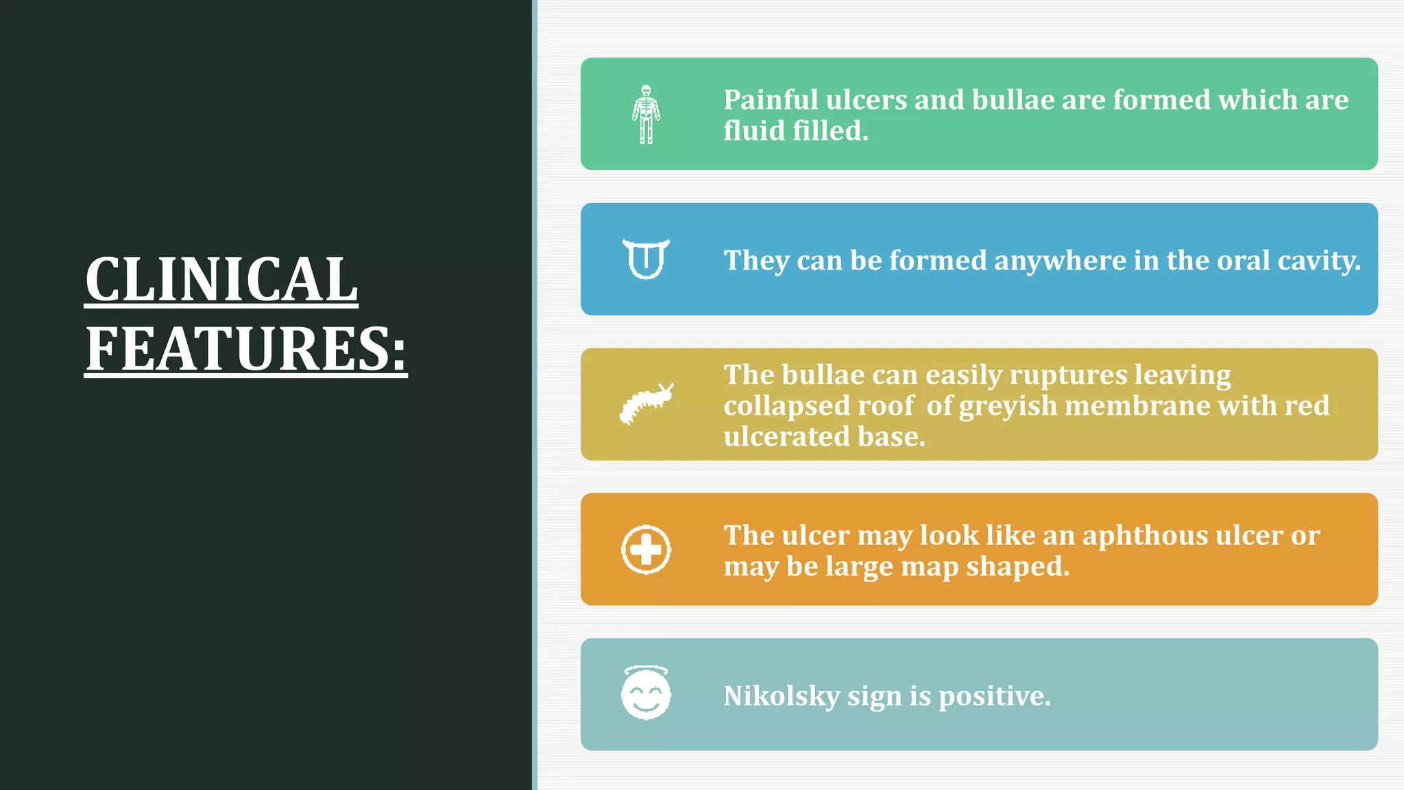
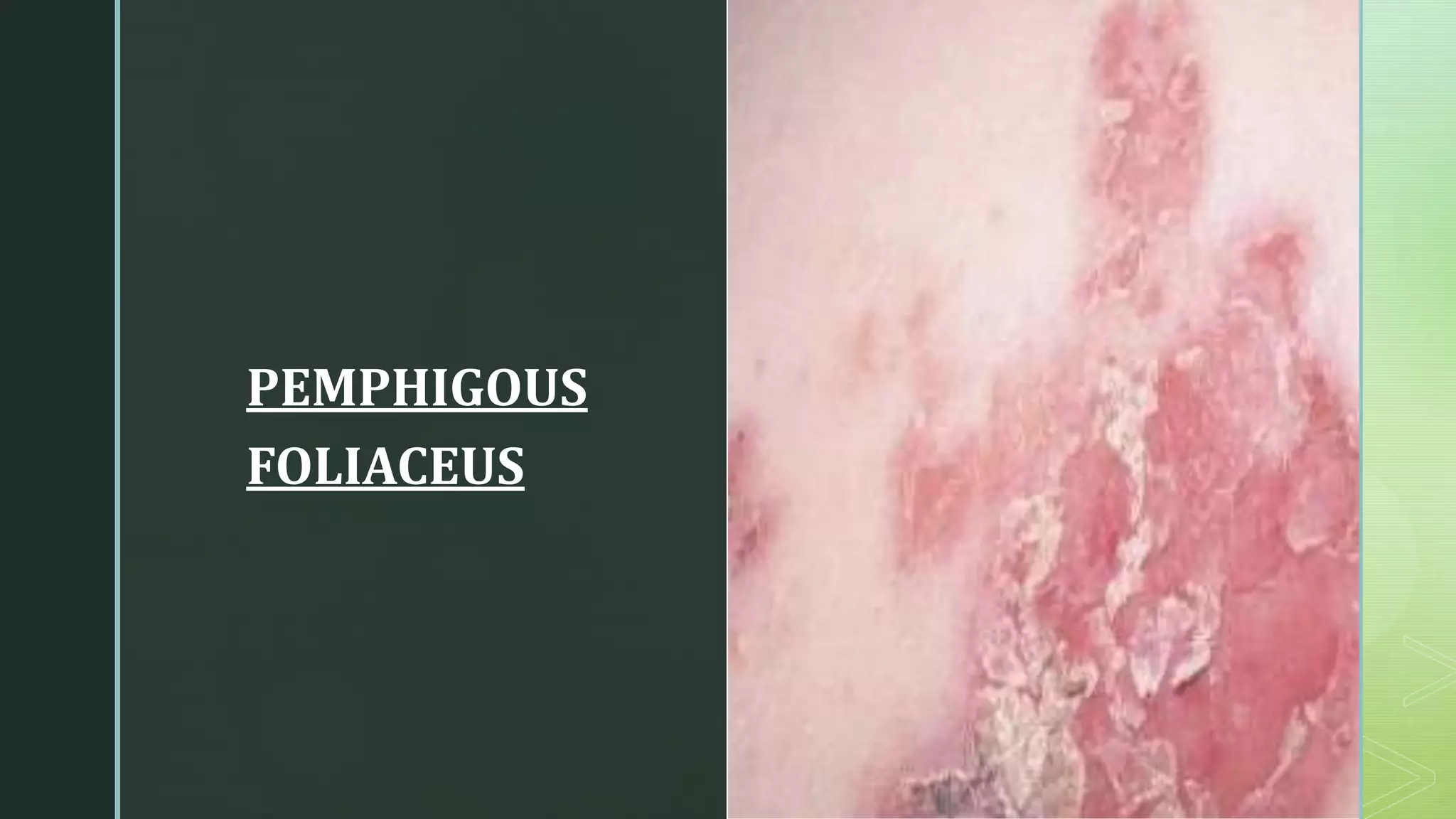
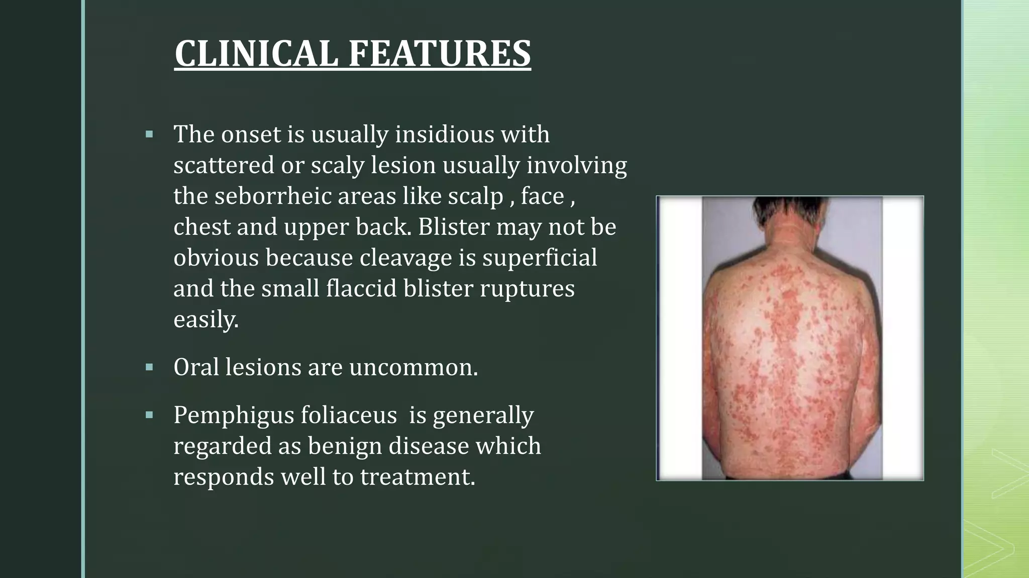
z
VESICULOBULLOUS LESION
Made by: zubaida Abdul qadir
z
CLASSIFICATION
OF
VESICULO BULLOUS
LESION
INTRA EPITHILIAL VESICLES:
Acantholytic Lesions:
•pemphigus vulgaris
•Paraneoplastic Pemphigus
•Deriers disease
Non Acantholytic Lesions:
•Viral infections of oral mucosal
SUB EPITHELIAL VESICLES:
• Erythema multiforme
• pemphigoid
• Dermatitis herpetiform
• Epidermolysis bullous
z
PEMPHIGOUS VULGARIS
 Pemphigus vulgaris is an
uncommon autoimmune
disease causing vesicles and
bullae on skin and mucous
membranes.
 It is usually fatal if untreated.
z
CLINICAL
FEATURES:
Painful ulcers and bullae are formed which are
fluid filled.
They can be formed anywhere in the oral cavity.
The bullae can easily ruptures leaving
collapsed roof of greyish membrane with red
ulcerated base.
The ulcer may look like an aphthous ulcer or
may be large map shaped.
Nikolsky sign is positive.
z
DIAGNOSIS:
Skin Biopsy Microbiology
Direct or indirect
immunofluorescence
ELISA
z
MANAGEMENT
• Systemic steroids like prednisolone in stable
cases.
• prednisolone plus azathioprine, methotrexate and
cyclophosphamide in advanced cases.
z
PEMPHIGOUS
FOLIACEUS
z
CLINICAL FEATURES
 The onset is usually insidious with
scattered or scaly lesion usually involving
the seborrheic areas like scalp , face ,
chest and upper back. Blister may not be
obvious because cleavage is superficial
and the small flaccid blister ruptures
easily.
 Oral lesions are uncommon.
 Pemphigus foliaceus is generally
regarded as benign disease which
responds well to treatment.
z
MANAGEMENT
 Potent topical or intralesional steroids or if control is inadequate
prednisolone 20-40mg/day
 Azathioprine and cyclophosphamide are effective adjuvant to oral
steroids in severe cases.
 Hydroxychloroquine 200mg twice per day is also recommended as
effective adjuvant therapy.
 Intravenous Ig has also been reported as effective in resident cases.
z
BULLOUS
PEMPHIGOID
zBULLOUS PEMPHIGOID
Also called as Para pemphigus or
aging pemphigus.
It is a chronic mucocutaneous bullous
disease that affects older individuals.
Age: above 60 years
Sex: Affects women slightly more
than men's (1.7:1)
Site: gingiva, buccal mucosa, tongue,
floor of the mouth, palate.
z
CLINICAL
FEATURES
Gingival lesion involves generalized edema,
inflammation and desquamation with
localized area of vesicles formation.
The oral lesions usually follow cutaneous
manifestation and being as bullae that soon
ruptures , leaving ulcerated base.
Signs: these vesicles and bullae are thick
walled and may remain intact for some days.
Skin lesions: rash commonly on scalp, limp
nikolsky sign negative.
z
MANAGEMENT
Advise to maintain oral hygiene.
Topical or intralesional steroids.
Systemic steroids: Prednisolone 40-
80mg/day.
Immunosuppression therapy: Azathioprine
50-100mg/day, cyclophosphamide 100-
200mg/day or cyclosporine 5-8mg/day.
Other therapy: Dapsone 50-100 mg/day ,
tetracycline 1.2-2g/day.
z
ERYTHEMA
MULTFORME
z
ERYTHEMA MULTIFORME
 It is a disease of abrupt onset involving the
skin and mucous membrane and have wide
variety of clinical presentation hence called
as ‘multiforme’.
z
CLINICAL FEATURES
age: children and
young adults
sex: males
site: hands, feet,
extensor surface of
elbows and knees.
onset: acute onset
with generalized
symptoms of fever
and malaise.
oral manifestations:
oral lesion start as
bullae on
erythematosus base.
bullae ruptures into
ulcers
lips are usually
involved form thin
haemorrhagic
crusted appearance.
healing occurs at 10-
14 days.
z
MANAGEMENT
SUPPORTIVE TREATMENT:
- liquid diet , electrolytes and nutritional support should start as early
as possible.
-Removal of cause e.g. drugs.
-Rehydration
FOR MILD CASES:
- supportive treatment ( adequate bed rest)
- topical anaesthesia mouth washes
- Soft liquid diet
- Proper rehydration and electrolytes
FOR SEVERE CASES:
- Short course or systemic steroids
- plus immunosuppressants like dapsone.
z
zTHANK YOU

VESICULO BULLOUS LESIONS

  • 1.
  • 2.
    z CLASSIFICATION OF VESICULO BULLOUS LESION INTRA EPITHILIALVESICLES: Acantholytic Lesions: •pemphigus vulgaris •Paraneoplastic Pemphigus •Deriers disease Non Acantholytic Lesions: •Viral infections of oral mucosal SUB EPITHELIAL VESICLES: • Erythema multiforme • pemphigoid • Dermatitis herpetiform • Epidermolysis bullous
  • 3.
    z PEMPHIGOUS VULGARIS  Pemphigusvulgaris is an uncommon autoimmune disease causing vesicles and bullae on skin and mucous membranes.  It is usually fatal if untreated.
  • 4.
    z CLINICAL FEATURES: Painful ulcers andbullae are formed which are fluid filled. They can be formed anywhere in the oral cavity. The bullae can easily ruptures leaving collapsed roof of greyish membrane with red ulcerated base. The ulcer may look like an aphthous ulcer or may be large map shaped. Nikolsky sign is positive.
  • 5.
    z DIAGNOSIS: Skin Biopsy Microbiology Director indirect immunofluorescence ELISA
  • 6.
    z MANAGEMENT • Systemic steroidslike prednisolone in stable cases. • prednisolone plus azathioprine, methotrexate and cyclophosphamide in advanced cases.
  • 7.
  • 8.
    z CLINICAL FEATURES  Theonset is usually insidious with scattered or scaly lesion usually involving the seborrheic areas like scalp , face , chest and upper back. Blister may not be obvious because cleavage is superficial and the small flaccid blister ruptures easily.  Oral lesions are uncommon.  Pemphigus foliaceus is generally regarded as benign disease which responds well to treatment.
  • 9.
    z MANAGEMENT  Potent topicalor intralesional steroids or if control is inadequate prednisolone 20-40mg/day  Azathioprine and cyclophosphamide are effective adjuvant to oral steroids in severe cases.  Hydroxychloroquine 200mg twice per day is also recommended as effective adjuvant therapy.  Intravenous Ig has also been reported as effective in resident cases.
  • 10.
  • 11.
    zBULLOUS PEMPHIGOID Also calledas Para pemphigus or aging pemphigus. It is a chronic mucocutaneous bullous disease that affects older individuals. Age: above 60 years Sex: Affects women slightly more than men's (1.7:1) Site: gingiva, buccal mucosa, tongue, floor of the mouth, palate.
  • 12.
    z CLINICAL FEATURES Gingival lesion involvesgeneralized edema, inflammation and desquamation with localized area of vesicles formation. The oral lesions usually follow cutaneous manifestation and being as bullae that soon ruptures , leaving ulcerated base. Signs: these vesicles and bullae are thick walled and may remain intact for some days. Skin lesions: rash commonly on scalp, limp nikolsky sign negative.
  • 13.
    z MANAGEMENT Advise to maintainoral hygiene. Topical or intralesional steroids. Systemic steroids: Prednisolone 40- 80mg/day. Immunosuppression therapy: Azathioprine 50-100mg/day, cyclophosphamide 100- 200mg/day or cyclosporine 5-8mg/day. Other therapy: Dapsone 50-100 mg/day , tetracycline 1.2-2g/day.
  • 14.
  • 15.
    z ERYTHEMA MULTIFORME  Itis a disease of abrupt onset involving the skin and mucous membrane and have wide variety of clinical presentation hence called as ‘multiforme’.
  • 16.
    z CLINICAL FEATURES age: childrenand young adults sex: males site: hands, feet, extensor surface of elbows and knees. onset: acute onset with generalized symptoms of fever and malaise. oral manifestations: oral lesion start as bullae on erythematosus base. bullae ruptures into ulcers lips are usually involved form thin haemorrhagic crusted appearance. healing occurs at 10- 14 days.
  • 17.
    z MANAGEMENT SUPPORTIVE TREATMENT: - liquiddiet , electrolytes and nutritional support should start as early as possible. -Removal of cause e.g. drugs. -Rehydration FOR MILD CASES: - supportive treatment ( adequate bed rest) - topical anaesthesia mouth washes - Soft liquid diet - Proper rehydration and electrolytes FOR SEVERE CASES: - Short course or systemic steroids - plus immunosuppressants like dapsone.
  • 18.