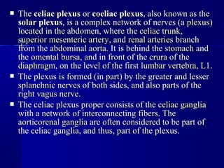
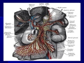
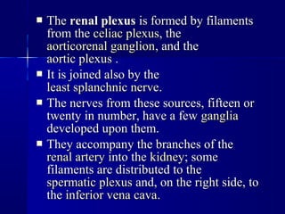
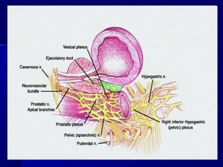
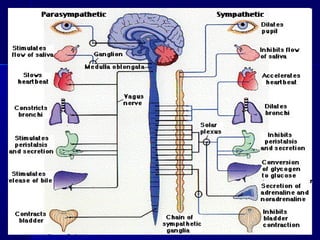
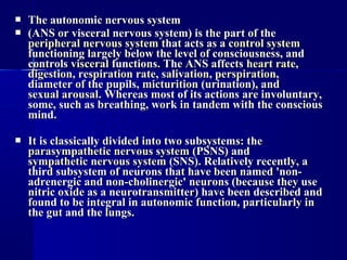
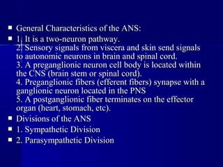
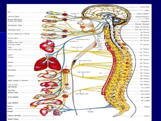
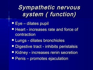
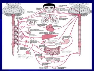
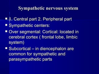
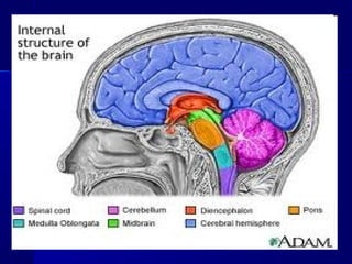
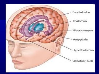
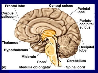
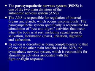
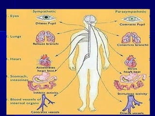
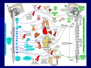
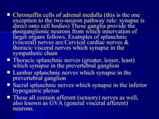
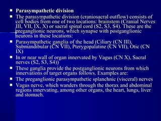
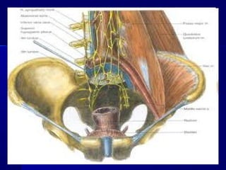
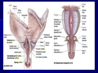
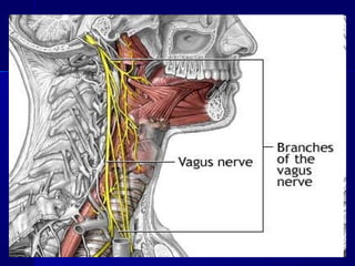
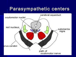
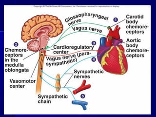
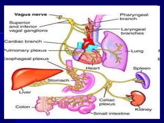
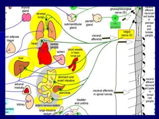
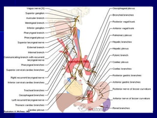
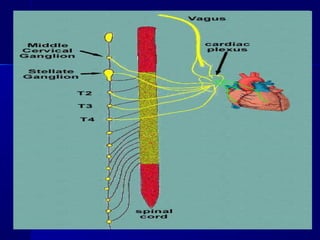
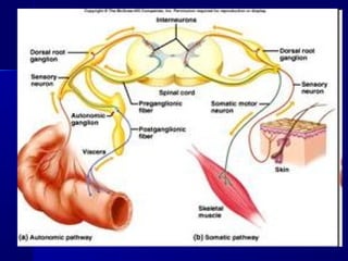
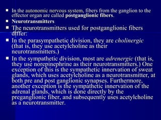
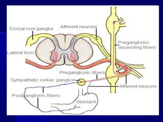
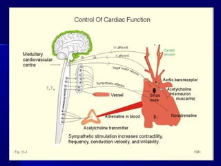
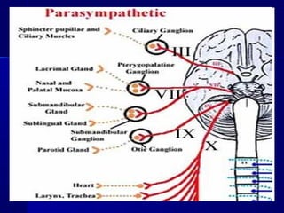
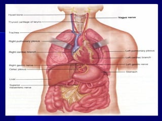
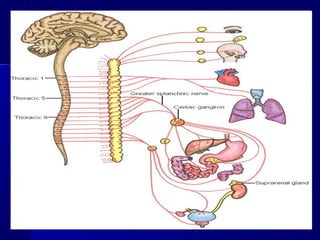
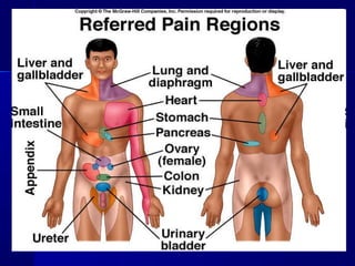
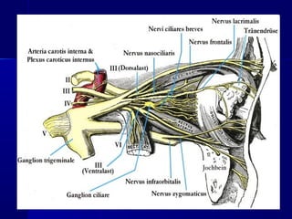
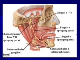
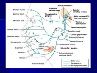
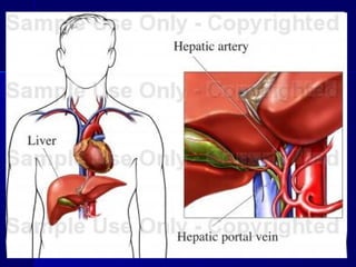
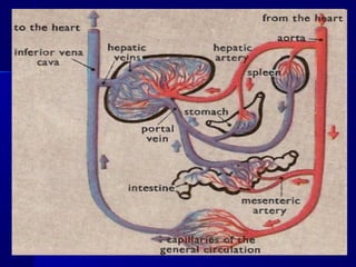
The lecture discusses the anatomy and functions of the autonomic nervous system, specifically the sympathetic and parasympathetic divisions. It describes the central and peripheral structures of these systems, including sympathetic centers in the brain and spinal cord, and ganglia in the periphery. The sympathetic system activates the fight or flight response and increases heart rate and respiration. It diverts blood flow away from the digestive system. The parasympathetic system stimulates rest and digestion functions like salivation and digestion.
The autonomic nervous system regulates the body's internal environment and physical exchanges with the external environment, along with the endocrine system, to maintain homeostasis. It has two divisions - the sympathetic and parasympathetic systems. The central ANS is diffuse in the brain and spinal cord and controls the peripheral ANS neurons and organs. The peripheral ANS includes neurons that release neurotransmitters like acetylcholine and norepinephrine at targets like smooth muscle, cardiac muscle and glands to produce effects like increasing heart rate and relaxing bronchial muscles during the fight or flight response.
The vertebrate brain
The vertebrate brain is the main part of the central nervous system. The brain and the spinal cord make up the central nervous system,
In most of the vertebrates the brain is at the front, in the head. It is protected by the skull and close to the main sense organs.
Brains are extremely complex and the part of human and animal body. The brain controls the other organs of the body, either by activating muscles or by causing secretion of chemicals such as hormones and neurotransmitters.
Muscular action allows rapid and coordinated responses to changes in the environment.
The brain of an adult human weights about 1300–1400 grams .
In vertebrates, the spinal cord by itself can cause reflex responses as well as simple movement such as swimming or walking. However, sophisticated control of behaviour requires a centralized brain.
The structure of all vertebrate brains is basically the same.
At the same time, during the course of evolution, the vertebrate brain has undergone changes, and become more effective.
In so-called 'lower' animals, most or all of the brain structure is inherited, and therefore their behaviour is mostly instinctive.
In mammals, and especially in man, the brain is developed further during life by learning. This has the benefit of helping them fit better into their environment. The capacity to learn is seen best in the cerebral cortex.
Three principles
The brain and nervous system is essentially a system which makes connections. It has input from sense organs and output to muscles. It is connected in several ways with the endocrine system, which makes hormones, and the digestive system and sex system. Hormones work slowly, so those changes are gradual.
The brain is a kind of department store. It has, all inter-connected, departments which do different things. They all help each other gather senses.
Much of what the body does is not conscious. Basically, much of the body runs on automatic (breathing, heart beat, hungry, hair growth) adjusted by the autonomic nervous system. The brain, too, does much of its work without a person noticing it. The unconscious mind refers to the brain activities which are hardly ever noticed.
PHYSIOLOGY OF ANS(AUTONOMIC NERVOUS SYSTEM)
Sympathetic Responses
Parasympathetic Responses
Autonomic Interactions
Control of Autonomic Nervous System Function
The autonomic nervous system (ANS) controls involuntary functions like heartbeat, breathing, and digestion. It has two divisions - the sympathetic and parasympathetic systems.
The sympathetic system prepares the body for "fight or flight" through actions like increasing heart rate. It uses norepinephrine as a neurotransmitter. The parasympathetic system allows the body to "rest and digest" through actions like slowing the heart rate. It uses acetylcholine as a neurotransmitter.
Both systems involve two neurons - a preganglionic neuron from the CNS and a postganglionic neuron. They differ in the lengths of their neurons and the locations of their cell bodies. Together they work to maintain homeostasis through complementary
The document summarizes the anatomy and physiology of the autonomic nervous system. It describes that the ANS has two divisions - the sympathetic and parasympathetic nervous systems. The sympathetic nervous system prepares the body for fight or flight while the parasympathetic system helps with rest and digestion. It also explains that the ANS uses two neurons connected by a ganglion to innervate organs unlike the single neuron pathway of the somatic nervous system. The ANS acts through cholinergic and adrenergic pathways to control functions like heart rate and blood pressure through reflexes.
Nervous system- SYMPATHETIC and PARASYMPATHETICKemberly Lee
The document discusses the autonomic nervous system (ANS), which regulates involuntary body functions. The ANS has two divisions - the sympathetic and parasympathetic nervous systems. The sympathetic system activates the body's fight or flight response and increases functions like heart rate. The parasympathetic system calms the body and increases functions like digestion when the body is at rest.
The document discusses the anatomy and physiology of the autonomic nervous system. It begins by describing the autonomic nervous system's organization based on reflex arcs between receptors, the central nervous system, and effectors. It then discusses the sympathetic and parasympathetic nervous systems in detail, including their anatomy, neurotransmitters, and receptors. Finally, it outlines some common tests used to evaluate autonomic integrity, such as tests of cardiac vagal function like respiratory sinus arrhythmia and tests of sympathetic function like postural stress responses and cold pressor tests.
Autonomic nervous system—arrangement, function, pain,visceral sensebilityRobin Victor
The document provides a history of the study of the autonomic nervous system from ancient Greek philosophers like Aristotle and Descartes to modern scientists like Thomas Willis, Claude Bernard, and Otto Loewi. It discusses the anatomy of the autonomic nervous system including the sympathetic, parasympathetic, and enteric divisions. Key functions and neurotransmitters of the autonomic nervous system like acetylcholine and norepinephrine are also summarized.
The autonomic nervous system and its implications indr anurag giri
The document discusses the autonomic nervous system (ANS), including its anatomy, physiology, and implications for anesthesia. The ANS has sympathetic, parasympathetic, and enteric divisions. It regulates functions like heart rate, digestion, sweating, and pupillary response. During anesthesia and surgery, autonomic reflexes are manipulated through drugs. ANS dysfunction in patients requires special considerations for anesthesia. The ANS also plays a role in intensive care and chronic pain conditions.
The document discusses the evolution of nervous systems across different animal species. It begins with the simplest nervous systems found in cnidarians like hydra, which have a nerve net without central control. It then covers increasing centralization in jellyfish and other species. Bilateral animals showed cephalization with sensory organs and feeding structures concentrated in the head. Invertebrates like worms, insects, and mollusks exhibited more complex centralized nervous systems. Research on invertebrate systems like squid giant axons provided insights. Vertebrate brains evolved from three bulges at the front of the spinal cord. Trends in vertebrate brains included increasing size, compartmentalization, and convolutions. Mammalian brains incorporated
The document provides an overview of the autonomic nervous system (ANS). It discusses that the ANS controls involuntary functions and is divided into the sympathetic and parasympathetic nervous systems. The sympathetic system is activated during fight or flight responses and controls functions like increased heart rate and blood pressure. The parasympathetic system activates rest and digest functions like increased gastrointestinal and salivary gland activity. Key topics covered include the anatomy and pathways of the sympathetic and parasympathetic systems, the receptors they act on, examples of their excitatory and inhibitory effects, and clinical tests of the ANS.
Limbic system and psychiatric disordersKarrar Husain
The limbic system is a set of brain structures located in the medial temporal lobe and midbrain that are involved in emotion, behavior, motivation, long-term memory, and olfaction. It includes structures like the hippocampus, amygdala, hypothalamus, and others. In the 1930s, James Papez proposed a neural circuit involving limbic structures that was important for emotional experiences. Later work expanded understanding of the limbic system's role in additional functions like regulating autonomic and endocrine systems, sleep-wake cycles, sexual behavior, and reward processing.
The autonomic nervous system (ANS) regulates involuntary functions and is divided into the sympathetic and parasympathetic divisions. The sympathetic division, also called the thoracolumbar outflow, supplies smooth muscles, blood vessels, heart, lungs, glands, and gastrointestinal organs. It consists of preganglionic and postganglionic fibers and is arranged in three ganglia groups: paravertebral/sympathetic chain ganglia, prevertebral/collateral ganglia, and terminal/peripheral ganglia. The sympathetic chain ganglia are arranged segmentally along the vertebral column and are divided into cervical, thoracic, lumbar, and sacral ganglia. Preganglionic fibers originate in the
The document summarizes the autonomic nervous system (ANS). It discusses that the ANS acts as an involuntary control system and is divided into the sympathetic, parasympathetic, and enteric nervous systems. The sympathetic "fight or flight" system is activated during stress and increases heart rate while the parasympathetic "rest and digest" system decreases heart rate. Dysregulation of the ANS is associated with psychiatric disorders like depression and anxiety. The ANS plays an important role in mood, stress response, and social behavior according to theories like the polyvagal theory.
The nervous system has two main divisions: the central nervous system (CNS) and the peripheral nervous system (PNS). The autonomic nervous system (ANS) is a division of the PNS and controls involuntary body functions. It has two branches - the sympathetic and parasympathetic systems. The sympathetic system activates the fight or flight response while the parasympathetic system activates the rest and digest response. Both systems involve a two-neuron chain with a preganglionic neuron originating in the CNS and a postganglionic neuron that releases neurotransmitters like acetylcholine and norepinephrine.
The document discusses the autonomic nervous system (ANS), which regulates involuntary body functions. It is divided into the sympathetic and parasympathetic systems. The sympathetic system activates the fight or flight response through increased heart rate and breathing. The parasympathetic system stimulates rest and digestion. Aging causes various changes to the ANS that impair the body's ability to react to stimuli and regulate functions, though memory loss is usually mild. Efficiency of the ANS declines with age, leading to issues like dry eyes and constipation.
2.1 struktur & fisiologi sistem saraf pusatMohd Arif
The document discusses the structure and function of the central nervous system. It describes how the nervous system is divided into the central nervous system, which includes the brain and spinal cord, and the peripheral nervous system. The central nervous system receives sensory information through receptors and interprets it to determine the appropriate motor responses. It provides control over muscles and glands.
The autonomic nervous system regulates involuntary body functions like heart rate, respiration, digestion and more. It has two divisions:
The sympathetic nervous system prepares the body for "fight or flight" through responses like increased heart rate and dilated pupils. It uses norepinephrine as a neurotransmitter.
The parasympathetic nervous system helps the body "rest and digest" with functions like digestion, salivation and pupil constriction. It uses acetylcholine as a neurotransmitter.
Together these two divisions work to maintain homeostasis and control internal organs through a two-neuron pathway, with cell bodies located in the spinal cord or brainstem and ganglia between pre- and postganglionic neurons.
The nervous system is divided into the central nervous system (CNS) and peripheral nervous system (PNS). The CNS consists of the brain and spinal cord. The brain controls functions like awareness, thoughts, movements, sensations, and memory. The PNS includes nerves and ganglia. It has sensory and motor divisions. Neurons transmit signals via electrical and chemical processes at synapses using neurotransmitters. Glial cells support neuron function. The presentation provides detailed information on the structure and function of the nervous system.
The autonomic nervous system regulates involuntary body functions and is divided into the sympathetic and parasympathetic nervous systems. It contains two neurons, with the preganglionic neuron cell body in the CNS and the postganglionic neuron cell body in autonomic ganglia. The sympathetic division is responsible for the fight or flight response and generally opposes the effects of the parasympathetic division, which is responsible for rest and digest functions. The enteric nervous system also helps control the gastrointestinal system independently.
The limbic system is a ring of structures located in the medial temporal lobe that is involved in emotion, motivation, learning, and memory. It includes structures like the hippocampus, amygdala, cingulate gyrus, and hypothalamus that are interconnected. Damage or dysfunction of the limbic system can cause disorders like abnormal emotional states, changes in motivation and drives, and episodic memory problems. The limbic system generates emotions and interprets sensory experiences as pleasant or unpleasant, influencing behavior. It also plays a role in learning, addiction, and various psychological disorders through its effects on neurotransmitters like dopamine and serotonin.
This document provides an overview of the autonomic nervous system (ANS). It discusses the two divisions of the ANS - the sympathetic and parasympathetic divisions. The sympathetic division is involved in the fight or flight response and increases heart rate, breathing, etc. The parasympathetic division is involved in rest and digest functions like digestion. The document details the pathways, ganglia, nerves and effects of both divisions. It notes that the hypothalamus and brainstem have significant control over autonomic function.
The document discusses the autonomic nervous system, which has two divisions - the sympathetic and parasympathetic systems. The sympathetic system prepares the body for fight or flight by increasing heart rate and blood pressure. The parasympathetic system then takes over once the stressor is gone, calming the body down.
The autonomic nervous system regulates involuntary body functions and is divided into the sympathetic and parasympathetic systems. The sympathetic system is associated with the "fight or flight" response and increases heart rate and metabolism. The parasympathetic system is involved in "rest and digest" functions and decreases heart rate and increases digestion. Both systems contain two neurons - a preganglionic neuron originating in the CNS and a postganglionic neuron connecting to the target organ. The sympathetic system uses norepinephrine and the parasympathetic system uses acetylcholine as neurotransmitters. The autonomic nervous system is regulated by regions of the brainstem and limbic system.
The limbic system is a set of brain structures located on the edge of the brainstem that are involved in emotion, behavior, motivation, long-term memory, and olfaction. It includes structures like the hippocampus, amygdala, septum, cingulate gyrus, and others. The hippocampus plays a key role in forming new declarative memories and is susceptible to epileptic activity due to its low seizure threshold. Damage to the hippocampus can cause anterograde amnesia by disrupting the transfer of memories from short-term to long-term storage. The cingulate cortex is connected to structures like the thalamus, striatum, and parahippocampal gyrus and is
The limbic system includes structures involved in emotion processing and memory formation. It contains nuclei such as the hippocampus, amygdala, and septal nuclei, as well as connecting tracts. The hippocampus forms memories and is connected to the amygdala and septal nuclei. The amygdala processes emotions and regulates autonomic functions. Stimulation of the septal nuclei produces pleasurable sensations. Together, these structures form circuits like the circuit of Papez that are important for emotional processing and memory.
The autonomic nervous system regulates involuntary body functions like heart rate and digestion. It is divided into the parasympathetic and sympathetic nervous systems.
The parasympathetic system activates rest and digest functions like increased digestion and decreased heart rate. It originates from the brain stem and sacral spinal cord. The sympathetic system activates fight or flight responses like increased heart and breathing rates and pupil dilation. It originates from the thoracic and lumbar spinal cord.
Horner's syndrome results from damage to the sympathetic nervous system, causing the classic symptoms of ptosis, miosis, and anhidrosis on the affected side. It can be caused by lesions in the neck, chest, or head compressing the sympathetic chain
The nervous system is divided into the central nervous system (CNS) and peripheral nervous system (PNS). The CNS consists of the brain and spinal cord. The brain is made up of the cerebrum, diencephalon, cerebellum, and brain stem. The diencephalon contains the hypothalamus, thalamus, and epithalamus. The PNS has two parts - the somatic nervous system which controls voluntary muscles, and the autonomic nervous system which regulates involuntary functions like digestion. The autonomic system has sympathetic and parasympathetic divisions that work in opposition to mobilize energy or induce calm.
The document summarizes key aspects of the autonomic nervous system, with a focus on the sympathetic and parasympathetic divisions. It describes:
- The autonomic nervous system regulates involuntary body functions and contains the sympathetic and parasympathetic nervous systems.
- The sympathetic nervous system activates the body's fight or flight response through neurons in the spinal cord, increasing heart rate, blood pressure, and diverting blood flow away from the digestive system.
- The parasympathetic nervous system calms the body and activates the rest and digest response through cranial and sacral nerves, lowering heart rate and stimulating digestion.
1. The autonomic nervous system (ANS) controls involuntary functions like digestion, respiration, and heart rate. It is divided into the sympathetic and parasympathetic nervous systems.
2. The ANS is composed of neurons with cell bodies located in the central nervous system that connect to peripheral ganglia. Its pathways involve two neurons, while somatic pathways only involve one neuron.
3. The sympathetic nervous system is responsible for the "fight or flight" response and generally accelerates functions. The parasympathetic nervous system generally acts to slow functions down and is associated with "rest and digest".
The autonomic nervous system (ANS), formerly the vegetative nervous system, is a division of the peripheral nervous system that supplies smooth muscle and glands, and thus influences the function of internal organs. The autonomic nervous system is a control system that acts largely unconsciously and regulates bodily functions, such as the heart rate, digestion, respiratory rate, pupillary response, urination, and sexual arousal.
Autonomic nervous system & neurotransmitter in psychiatryNursing Path
The autonomic nervous system (ANS) regulates involuntary organs and functions. It is divided into the sympathetic and parasympathetic nervous systems. The sympathetic system prepares the body for emergencies through "fight or flight" responses like increased heart rate. The parasympathetic system allows for "rest and digest" functions like decreased heart rate and increased digestion. Neurotransmitters are chemicals that transmit signals between neurons. They allow signals to cross the synaptic cleft and include acetylcholine, norepinephrine, dopamine, serotonin, and GABA.
The document discusses the autonomic nervous system (ANS), which has two divisions - the sympathetic and parasympathetic nervous systems. The sympathetic system prepares the body for "fight or flight" responses and is active during stress. It stimulates organs like the heart. The parasympathetic system calms the body and is active at rest. It stimulates organs like the stomach and intestines. The document outlines the pathways, neurotransmitters, and roles of each division in regulating various organs and body systems.
The nervous system is composed of neurons and glial cells that communicate information throughout the body to cause reactions. Neurons are electrically excitable cells that process and transmit information. They consist of a soma, dendrites, axon, and axon terminals. There are different types of neurons. The brain and spinal cord make up the central nervous system, which controls mental processes and physical actions. The brain is divided into lobes and structures like the limbic system that support functions like emotion and memory. The autonomic nervous system regulates involuntary functions through the sympathetic and parasympathetic nervous systems.
The autonomic nervous system (ANS) innervates the heart, smooth muscles, glands and viscera and is divided into the parasympathetic and sympathetic nervous systems. The parasympathetic system participates in tissue building and the sympathetic system enables responses to stress. Both systems have efferent neurons that travel from the CNS to effector organs via a two-neuron chain, with preganglionic neurons synapsing in ganglia and postganglionic neurons innervating the organs. The sympathetic system originates in the thoracic and lumbar spinal cord and parasympathetic system originates in the cranial and sacral regions. The sympathetic system prepares the body for fight or flight while the parasympathetic
The document discusses the autonomic nervous system and its implications in anaesthesia. It provides an overview of the anatomy and physiology of the sympathetic, parasympathetic, and enteric nervous systems. It describes the effects of these systems on various target organs and the neurotransmitters involved. The document also discusses tests to evaluate autonomic integrity, the implications of anaesthesia on autonomic reflexes, and managing patients with autonomic dysfunction.
The document discusses the autonomic nervous system and its implications in anaesthesia. It provides details on the anatomy and physiology of the sympathetic, parasympathetic, and enteric nervous systems. It describes the effects of these systems on various target organs and the neurotransmitters involved. Tests for evaluating autonomic integrity are discussed. Drugs acting on the autonomic nervous system and their sites of action are also summarized. The implications of autonomic nervous system dysfunction for anaesthesia are outlined.
The document provides information about the autonomic nervous system (ANS). It describes the ANS as having two main divisions - the sympathetic and parasympathetic nervous systems. The sympathetic system prepares the body for "fight or flight" responses, while the parasympathetic system allows for "rest and digest" functions. Key differences between the two divisions are described, including their origins in the spinal cord/brain and targets in the body. The pathways of preganglionic and postganglionic neurons, as well as autonomic ganglia, are outlined. Neurotransmitters and receptors of each division are also detailed.
The brain is one of the largest and most complex organs in the human body. It is made up of more than 100 billion nerves that communicate in trillions of connections called synapses. The brain is made up of many specialized areas that work together: ... The cortex is the outermost layer of brain cells. the brain is how you think.
This document provides an overview of the autonomic nervous system and autonomic drugs. It begins with learning objectives related to the autonomic nervous system and associated drug groups. It then discusses the importance of understanding autonomic drugs for dentists. The remainder of the document details the anatomy and physiology of the parasympathetic and sympathetic divisions of the autonomic nervous system. It explains the functional organization and neurotransmitters of the autonomic nervous system and outlines the four main drug groups that act on the autonomic nervous system.
This document provides an overview of the autonomic nervous system (ANS). It discusses the divisions of the ANS including the sympathetic and parasympathetic nervous systems. Key points covered include the anatomical organization of the ANS from the central nervous system to peripheral ganglia. The roles and effects of the sympathetic and parasympathetic systems on various organs are described. Neurotransmitters, receptors, and reflexes of the ANS are also summarized.
The autonomic nervous system controls internal organs and glands. It has both sympathetic and parasympathetic divisions. The sympathetic division uses norepinephrine and epinephrine to activate the fight or flight response. The parasympathetic uses acetylcholine for rest and digest functions. Both have preganglionic and postganglionic neurons. The autonomic nervous system regulates vital involuntary functions like heart rate, breathing, digestion and pupillary response.
The autonomic nervous system regulates involuntary body functions and is divided into the sympathetic and parasympathetic nervous systems. The sympathetic system activates the body's fight or flight response and increases heart rate and respiration. The parasympathetic system calms the body and increases digestion. Both systems generally have opposing effects on organs with the sympathetic usually stimulating functions and the parasympathetic inhibiting them. Acetylcholine and norepinephrine act as neurotransmitters depending on the type of receptor in the target organ.
The document discusses the nervous system and its main components. It states that any internal or external change that causes a response is called a stimulus. The nervous system includes neurons, which are nerve cells that transmit signals in the form of electrical impulses. There are sensory neurons that receive information, motor neurons that conduct signals to muscles and glands, and interneurons that relay signals between neurons. When an impulse reaches the end of an axon, it releases chemicals that stimulate the next neuron across the synapse. The central nervous system is made up of the brain and spinal cord and coordinates all body activities. The brain is the largest part and is responsible for thinking, interpreting senses, memory, and movement control.
The Indian Dental Academy is the Leader in continuing dental education , training dentists in all aspects of dentistry and
offering a wide range of dental certified courses in different formats.for more details please visit
www.indiandentalacademy.com
a brief description on division of nervous system,SOMATIC NERVOUS SYSTEM and working, SENSORY DIVISION and MOTOR DIVISION, SENSORY RECEPTORS, CATEGORIZATION OF SENSORY RECEPTORS, TYPES OF NERVES, SPINAL NERVES, CRANIAL NERVES, REFLEX ARCS, AUTONOMIC NERVOUS SYSTEM and it's properties and division, PARASYMPATHETIC NERVOUS SYSTEM, SYMPATHETIC NERVOUS SYSTEM
The document discusses the structure and function of the human neural and sensory systems. It describes the central nervous system including the brain, spinal cord, and peripheral nervous system. It explains the divisions of the brain including the forebrain, midbrain, and hindbrain. It also discusses the autonomic nervous system and its sympathetic and parasympathetic divisions. Additionally, it summarizes the structure and function of the eye and ear as sensory organs.
The document discusses the nervous system. It describes the central nervous system as consisting of the brain and spinal cord, which integrate sensory information. The peripheral nervous system includes the afferent and efferent systems, as well as the somatic and autonomic systems. The autonomic system further divides into the sympathetic and parasympathetic divisions, which generally function in complementary, antagonistic ways such as accelerating and braking vital functions. Neurons carry nerve impulses and are supported by neuroglia like Schwann cells.
1. TOPIC OF THE LECTURE:TOPIC OF THE LECTURE:
Anatomy of vegetative part ofAnatomy of vegetative part of
nervous system.nervous system.
The central and peripheralThe central and peripheral
structures of the sympathetic andstructures of the sympathetic and
parasympathetic parts.parasympathetic parts.
Vegetative plexuses.Vegetative plexuses.
2.
3. The autonomic nervous systemThe autonomic nervous system
(ANS or visceral nervous system) is the part of the(ANS or visceral nervous system) is the part of the
peripheral nervous systemperipheral nervous system that acts as athat acts as a control systemcontrol system
functioning largely below the level of consciousness, andfunctioning largely below the level of consciousness, and
controlscontrols visceralvisceral functions. The ANS affectsfunctions. The ANS affects heart rateheart rate,,
digestiondigestion,, respiration raterespiration rate,, salivationsalivation,, perspirationperspiration,,
diameter of the pupils,diameter of the pupils, micturitionmicturition (urination), and(urination), and
sexual arousalsexual arousal. Whereas most of its actions are involuntary,. Whereas most of its actions are involuntary,
some, such as breathing, work in tandem with the conscioussome, such as breathing, work in tandem with the conscious
mind.mind.
It is classically divided into two subsystems: theIt is classically divided into two subsystems: the
parasympathetic nervous systemparasympathetic nervous system (PSNS) and(PSNS) and
sympathetic nervous systemsympathetic nervous system (SNS). Relatively recently, a(SNS). Relatively recently, a
third subsystem of neurons that have been named 'non-third subsystem of neurons that have been named 'non-
adrenergic and non-cholinergic' neurons (because they useadrenergic and non-cholinergic' neurons (because they use
nitric oxide as a neurotransmitter) have been described andnitric oxide as a neurotransmitter) have been described and
found to be integral in autonomic function, particularly infound to be integral in autonomic function, particularly in
the gut and the lungs.the gut and the lungs.
4. General Characteristics of the ANS:General Characteristics of the ANS:
1. It is a two-neuron pathway.1. It is a two-neuron pathway.
2. Sensory signals from viscera and skin send signals2. Sensory signals from viscera and skin send signals
to autonomic neurons in brain and spinal cord.to autonomic neurons in brain and spinal cord.
3. A preganglionic neuron cell body is located within3. A preganglionic neuron cell body is located within
the CNS (brain stem or spinal cord).the CNS (brain stem or spinal cord).
4. Preganglionic fibers (efferent fibers) synapse with a4. Preganglionic fibers (efferent fibers) synapse with a
ganglionic neuron located in the PNSganglionic neuron located in the PNS
5. A postganglionic fiber terminates on the effector5. A postganglionic fiber terminates on the effector
organ (heart, stomach, etc).organ (heart, stomach, etc).
Divisions of the ANSDivisions of the ANS
1. Sympathetic Division1. Sympathetic Division
2. Parasympathetic Division2. Parasympathetic Division
8. The sympathetic nervous system is responsible for up- andThe sympathetic nervous system is responsible for up- and
down-regulating many homeostatic mechanisms in livingdown-regulating many homeostatic mechanisms in living
organisms. Fibers from the SNS innervate tissues in almostorganisms. Fibers from the SNS innervate tissues in almost
every organ system, providing at least some regulatory functionevery organ system, providing at least some regulatory function
to things as diverse asto things as diverse as pupilpupil diameter,diameter, gut motilitygut motility, and, and urinaryurinary
output. It is perhaps best known for mediating the neuronal andoutput. It is perhaps best known for mediating the neuronal and
hormonal stress response commonly known as thehormonal stress response commonly known as the fight-or-fight-or-
flight responseflight response..
This response is also known asThis response is also known as sympatho-adrenal responsesympatho-adrenal response ofof
the body, as thethe body, as the preganglionicpreganglionic sympathetic fibers that end in thesympathetic fibers that end in the
adrenal medulla (but also all other sympathetic fibers) secreteadrenal medulla (but also all other sympathetic fibers) secrete
acetylcholine, which activates the great secretion of adrenalineacetylcholine, which activates the great secretion of adrenaline
(epinephrine) and to a lesser extent noradrenaline(epinephrine) and to a lesser extent noradrenaline
(norepinephrine) from it.(norepinephrine) from it.
Therefore, this response that acts primarily on theTherefore, this response that acts primarily on the
cardiovascular systemcardiovascular system is mediated directly via impulsesis mediated directly via impulses
transmitted through the sympathetic nervous system andtransmitted through the sympathetic nervous system and
indirectly viaindirectly via catecholaminescatecholamines secreted from the adrenalsecreted from the adrenal
medulla.medulla.
9. Sympathetic nervous systemSympathetic nervous system
1. Central part1. Central part 2. Peripheral part2. Peripheral part
Sympathetic centers:Sympathetic centers:
Over segmental: Cortical: located inOver segmental: Cortical: located in
cerebral cortex ( frontal lobecerebral cortex ( frontal lobe,, limbiclimbic
system)system)
Subcortical – in diencephalon areSubcortical – in diencephalon are
common for sympathetic andcommon for sympathetic and
parasympathetic partsparasympathetic parts
10.
11. TheThe limbic systemlimbic system (or Paleomammalian brain)(or Paleomammalian brain)
is a set of brain structures including theis a set of brain structures including the
hippocampushippocampus,, amygdalaamygdala,, anterior thalamic nucleianterior thalamic nuclei
,, septumseptum,, limbic cortexlimbic cortex and fornix, whichand fornix, which
seemingly support a variety of functionsseemingly support a variety of functions
includingincluding emotionemotion,, behaviorbehavior,, long term memorylong term memory,,
andand olfactionolfaction..
The term "limbic" comes from theThe term "limbic" comes from the LatinLatin limbuslimbus,,
for "border" or "edge". Some scientists havefor "border" or "edge". Some scientists have
suggested that the concept of the limbic systemsuggested that the concept of the limbic system
should be abandoned asshould be abandoned as obsoleteobsolete, as it is, as it is
grounded more in transient tradition than ingrounded more in transient tradition than in
facts.facts.
12.
13.
14. Segmental level – in lateral horns of spinal cordSegmental level – in lateral horns of spinal cord
( intermediolateral nucleus)( intermediolateral nucleus)
Nucleus include: tropicNucleus include: tropic,, functionalfunctional,, vasomotorvasomotor,,
visceromotorvisceromotor,, secretorysecretory ссenters.enters.
15. TheThe parasympatheticparasympathetic nervous systemnervous system ((PSNSPSNS) is) is
one of the two main divisions of theone of the two main divisions of the
autonomic nervous systemautonomic nervous system (ANS).(ANS).
The ANS is responsible for regulation of internalThe ANS is responsible for regulation of internal
organs and glands, which occurs unconsciously. Theorgans and glands, which occurs unconsciously. The
parasympathetic system specifically is responsible forparasympathetic system specifically is responsible for
stimulation of "rest-and-digest" activities that occurstimulation of "rest-and-digest" activities that occur
when the body is at rest, including sexual arousal,when the body is at rest, including sexual arousal,
salivationsalivation,, lacrimationlacrimation (tears),(tears), urinationurination,, digestiondigestion
andand defecationdefecation..
Its action is described as being complementary to thatIts action is described as being complementary to that
of one of the other main branches of the ANS, theof one of the other main branches of the ANS, the
sympathetic nervous systemsympathetic nervous system, which is responsible for, which is responsible for
stimulating activities associated with thestimulating activities associated with the
fight-or-flight responsefight-or-flight response..
16.
17.
18. SYMPATHETIC NERVOUSSYMPATHETIC NERVOUS
SYSTEM.SYSTEM.
Diverts blood flow away from the gastro-intestinalDiverts blood flow away from the gastro-intestinal
(GI) tract and(GI) tract and skinskin via vasoconstriction.via vasoconstriction.
Blood flow toBlood flow to skeletal musclesskeletal muscles and theand the lungslungs isis
enhanced (by as much as 1200% in the case ofenhanced (by as much as 1200% in the case of
skeletal muscles). Dilates bronchioles of the lung,skeletal muscles). Dilates bronchioles of the lung,
which allows for greater alveolar oxygen exchange.which allows for greater alveolar oxygen exchange.
IncreasesIncreases heart rateheart rate and theand the contractilitycontractility of cardiacof cardiac
cells (cells (myocytesmyocytes), thereby providing a mechanism for), thereby providing a mechanism for
the enhanced blood flow to skeletal muscles.the enhanced blood flow to skeletal muscles.
Dilates pupils and relaxes the ciliary muscle to theDilates pupils and relaxes the ciliary muscle to the
lens, allowing more light to enter the eye and farlens, allowing more light to enter the eye and far
vision. Provides vasodilation for thevision. Provides vasodilation for the coronary vesselscoronary vessels
of theof the heartheart..
19. Constricts all the intestinalConstricts all the intestinal sphincterssphincters
and the urinary sphincter.and the urinary sphincter.
InhibitsInhibits peristalsisperistalsis..
20. Parasympathetic nervous systemParasympathetic nervous system
Dilates blood vessels leading to the GI tract,Dilates blood vessels leading to the GI tract,
increasing blood flow. This is importantincreasing blood flow. This is important
following the consumption of food, due tofollowing the consumption of food, due to
the greater metabolic demands placed on thethe greater metabolic demands placed on the
body by the gut.body by the gut.
The parasympathetic nervous system canThe parasympathetic nervous system can
also constrict the bronchiolar diameter whenalso constrict the bronchiolar diameter when
the need for oxygen has diminished.the need for oxygen has diminished.
Dedicated cardiac branches of theDedicated cardiac branches of the VagusVagus
and thoracicand thoracic Spinal AccessorySpinal Accessory nerves impartnerves impart
ParasympatheticParasympathetic control of thecontrol of the HeartHeart oror
MyocardiumMyocardium..
21. DuringDuring accommodationaccommodation, the parasympathetic, the parasympathetic
nervous system causes constriction of thenervous system causes constriction of the
pupil and contraction of the ciliary muscle topupil and contraction of the ciliary muscle to
the lens, allowing for closer vision.the lens, allowing for closer vision.
The parasympathetic nervous systemThe parasympathetic nervous system
stimulates salivary gland secretion, andstimulates salivary gland secretion, and
acceleratesaccelerates peristalsisperistalsis, so, in keeping with the, so, in keeping with the
rest and digest functions, appropriate PNSrest and digest functions, appropriate PNS
activity mediates digestion of food andactivity mediates digestion of food and
indirectly, the absorption of nutrients.indirectly, the absorption of nutrients.
Is also involved in erection of genitals, via theIs also involved in erection of genitals, via the
pelvic splanchnic nervespelvic splanchnic nerves 2–4.2–4.
Stimulates sexual arousal.Stimulates sexual arousal.
22.
23.
24. TheThe sympathetic trunkssympathetic trunks ((sympathetic chainsympathetic chain,,
gangliated cordgangliated cord)) are a paired bundle of nerveare a paired bundle of nerve
fibers that run from the base of thefibers that run from the base of the skullskull toto
thethe coccyxcoccyx..
The sympathetic trunk travels in a downwardThe sympathetic trunk travels in a downward
direction from the skull, just lateral to thedirection from the skull, just lateral to the
vertebral bodies. It interacts with thevertebral bodies. It interacts with the
spinal nervesspinal nerves or their ventral rami by way ofor their ventral rami by way of
rami communicantesrami communicantes..
The superior end of it is continued upwardThe superior end of it is continued upward
through thethrough the carotid canalcarotid canal into the skull, andinto the skull, and
forms aforms a plexusplexus on theon the internal carotid arteryinternal carotid artery;;
the inferior part travels in front of thethe inferior part travels in front of the coccyxcoccyx,,
where it converges with the other trunk at awhere it converges with the other trunk at a
structure known as thestructure known as the ganglion imparganglion impar..
25.
26. Along the length of the sympathetic trunkAlong the length of the sympathetic trunk
areare gangliaganglia known asknown as paravertebral gangliaparavertebral ganglia..
FunctionFunction
The sympathetic trunk is a fundamentalThe sympathetic trunk is a fundamental
part of the sympathetic division of thepart of the sympathetic division of the
autonomic nervous systemautonomic nervous system. It allows nerve. It allows nerve
fibers to travel to spinal nerves that arefibers to travel to spinal nerves that are
superior and inferior to the one in whichsuperior and inferior to the one in which
they originated. Also, a number of nerves,they originated. Also, a number of nerves,
such as most of thesuch as most of the splanchnic nervessplanchnic nerves, arise, arise
directly from the trunks.directly from the trunks.
27.
28.
29. The sympathetic division (thoracolumbarThe sympathetic division (thoracolumbar
outflow) consists of cell bodies in theoutflow) consists of cell bodies in the
lateral horn of spinal cordlateral horn of spinal cord (intermediolateral cell(intermediolateral cell
columns) of the spinal cord from T1 to L2.columns) of the spinal cord from T1 to L2.
These cell bodies are GVE (general visceralThese cell bodies are GVE (general visceral
efferent) neurons and are the preganglionicefferent) neurons and are the preganglionic
neurons. There are several locations upon whichneurons. There are several locations upon which
preganglionic neurons can synapse for theirpreganglionic neurons can synapse for their
postganglionic neurons:postganglionic neurons:
Paravertebral gangliaParavertebral ganglia of the sympathetic chainof the sympathetic chain
(these run on either side of the vertebral bodies)(these run on either side of the vertebral bodies)
Prevertebral gangliaPrevertebral ganglia (celiac ganglia, superior(celiac ganglia, superior
mesenteric ganglia, inferior mesenteric ganglia)mesenteric ganglia, inferior mesenteric ganglia)
30.
31.
32.
33. Chromaffin cellsChromaffin cells ofof adrenal medullaadrenal medulla (this is the one(this is the one
exception to the two-neuron pathway rule: synapse isexception to the two-neuron pathway rule: synapse is
direct onto cell bodies) These ganglia provide thedirect onto cell bodies) These ganglia provide the
postganglionic neurons from which innervation ofpostganglionic neurons from which innervation of
target organs follows. Examples of splanchnictarget organs follows. Examples of splanchnic
(visceral) nerves are:Cervical cardiac nerves &(visceral) nerves are:Cervical cardiac nerves &
thoracic visceral nerves which synapse in thethoracic visceral nerves which synapse in the
sympathetic chainsympathetic chain
Thoracic splanchnic nerves (greater, lesser, least)Thoracic splanchnic nerves (greater, lesser, least)
which synapse in the prevertebral ganglionwhich synapse in the prevertebral ganglion
Lumbar splanchnic nerves which synapse in theLumbar splanchnic nerves which synapse in the
prevertebral ganglionprevertebral ganglion
Sacral splanchnic nerves which synapse in the inferiorSacral splanchnic nerves which synapse in the inferior
hypogastric plexushypogastric plexus
These all contain afferent (sensory) nerves as well,These all contain afferent (sensory) nerves as well,
also known as GVA (general visceral afferent)also known as GVA (general visceral afferent)
neurons.neurons.
34. Parasympathetic divisionParasympathetic division
The parasympathetic division (craniosacral outflow) consists ofThe parasympathetic division (craniosacral outflow) consists of
cell bodies from one of two locations:cell bodies from one of two locations: brainstembrainstem (Cranial Nerves(Cranial Nerves
III, VII, IX, X) or sacral spinal cord (S2, S3, S4). These are theIII, VII, IX, X) or sacral spinal cord (S2, S3, S4). These are the
preganglionic neurons, which synapse with postganglionicpreganglionic neurons, which synapse with postganglionic
neurons in these locations:neurons in these locations:
Parasympathetic gangliaParasympathetic ganglia of the head (Ciliary (CN III),of the head (Ciliary (CN III),
Submandibular (CN VII), Pterygopalatine (CN VII), Otic (CNSubmandibular (CN VII), Pterygopalatine (CN VII), Otic (CN
IX)IX)
In or near wall of organ innervated by Vagus (CN X), SacralIn or near wall of organ innervated by Vagus (CN X), Sacral
nerves (S2, S3, S4))nerves (S2, S3, S4))
These ganglia provide the postganglionic neurons from whichThese ganglia provide the postganglionic neurons from which
innervations of target organs follows. Examples are:innervations of target organs follows. Examples are:
The preganglionic parasympathetic splanchnic (visceral) nervesThe preganglionic parasympathetic splanchnic (visceral) nerves
Vagus nerveVagus nerve, which wanders through the thorax and abdominal, which wanders through the thorax and abdominal
regions innervating, among other organs, the heart, lungs, liverregions innervating, among other organs, the heart, lungs, liver
and stomachand stomach..
36. TheThe abdominal aortic plexusabdominal aortic plexus (aortic plexus) is(aortic plexus) is
formed by branches derived, on either side,formed by branches derived, on either side,
from thefrom the celiac plexusceliac plexus andand gangliaganglia, and, and
receives filaments from some of thereceives filaments from some of the
lumbar ganglialumbar ganglia..
It is situated upon the sides and front of theIt is situated upon the sides and front of the
aortaaorta, between the origins of the superior and, between the origins of the superior and
inferior mesentericinferior mesenteric arteries.arteries.
From this plexus arise part of theFrom this plexus arise part of the spermaticspermatic, the, the
inferior mesenteric, and theinferior mesenteric, and the hypogastrichypogastric
plexuses; it also distributes filaments to theplexuses; it also distributes filaments to the
inferior vena cavainferior vena cava..
37. The celiac plexus includes a number ofThe celiac plexus includes a number of
smaller plexuses:smaller plexuses:
Hepatic plexusHepatic plexus
Splenic plexusSplenic plexus
Gastric plexusGastric plexus
Pancreatic plexusPancreatic plexus
Suprarenal plexusSuprarenal plexus
Other plexuses that are derived from theOther plexuses that are derived from the
celiac plexus:celiac plexus:
Renal plexusRenal plexus
Testicular plexusTesticular plexus // ovarian plexusovarian plexus
Superior mesenteric plexusSuperior mesenteric plexus
38.
39. TheThe celiac plexusceliac plexus oror coeliac plexuscoeliac plexus, also known as the, also known as the
solar plexussolar plexus, is a complex network of nerves (a, is a complex network of nerves (a plexusplexus))
located in the abdomen, where thelocated in the abdomen, where the celiac trunkceliac trunk,,
superior mesenteric arterysuperior mesenteric artery, and, and renal arteriesrenal arteries branchbranch
from thefrom the abdominal aortaabdominal aorta. It is behind the. It is behind the stomachstomach andand
thethe omental bursaomental bursa, and in front of the, and in front of the cruracrura of theof the
diaphragmdiaphragm, on the level of the first, on the level of the first lumbar vertebralumbar vertebra, L1., L1.
The plexus is formed (in part) by the greater and lesserThe plexus is formed (in part) by the greater and lesser
splanchnic nervessplanchnic nerves of both sides, and also parts of theof both sides, and also parts of the
rightright vagus nervevagus nerve..
The celiac plexus proper consists of theThe celiac plexus proper consists of the celiac gangliaceliac ganglia
with a network of interconnecting fibers. Thewith a network of interconnecting fibers. The
aorticorenal gangliaaorticorenal ganglia are often considered to be part ofare often considered to be part of
the celiac ganglia, and thus, part of the plexus.the celiac ganglia, and thus, part of the plexus.
40.
41. TheThe renal plexusrenal plexus is formed by filamentsis formed by filaments
from thefrom the celiac plexusceliac plexus, the, the
aorticorenal ganglionaorticorenal ganglion, and the, and the
aortic plexusaortic plexus ..
It is joined also by theIt is joined also by the
least splanchnic nerveleast splanchnic nerve..
The nerves from these sources, fifteen orThe nerves from these sources, fifteen or
twenty in number, have a fewtwenty in number, have a few gangliaganglia
developed upon them.developed upon them.
They accompany the branches of theThey accompany the branches of the
renal arteryrenal artery into theinto the kidneykidney; some; some
filaments are distributed to thefilaments are distributed to the
spermatic plexusspermatic plexus and, on the right side, toand, on the right side, to
thethe inferior vena cavainferior vena cava..
62. AA nerve fibernerve fiber is a threadlike extension of a nerve cellis a threadlike extension of a nerve cell
and consists of anand consists of an axonaxon andand myelin sheathmyelin sheath (if present)(if present)
in thein the nervous systemnervous system. There are nerve fibers in the. There are nerve fibers in the
central nervous systemcentral nervous system andand peripheral nervous systemperipheral nervous system..
A nerve fiber may be myelinated and/or unmyelinated.A nerve fiber may be myelinated and/or unmyelinated.
In the central nervous system (CNS), myelin isIn the central nervous system (CNS), myelin is
produced byproduced by oligodendrogliaoligodendroglia cells.cells.
Schwann cellsSchwann cells form myelin in the peripheral nervousform myelin in the peripheral nervous
system (PNS). Schwann cells can also make a thinsystem (PNS). Schwann cells can also make a thin
covering for an axon which does not consist of myelincovering for an axon which does not consist of myelin
(in the PNS). A peripheral nerve fiber consists of an(in the PNS). A peripheral nerve fiber consists of an
axon, myelin sheath, Schwann cells and itsaxon, myelin sheath, Schwann cells and its
endoneuriumendoneurium. There are no endoneurium and Schwann. There are no endoneurium and Schwann
cells in the central nervous systemcells in the central nervous system..
63. In theIn the autonomic nervous systemautonomic nervous system, fibers from, fibers from
thethe CNSCNS to theto the ganglionganglion are known asare known as
preganglionic fiberspreganglionic fibers..
All preganglionic fibers, whether they are in theAll preganglionic fibers, whether they are in the
sympathetic divisionsympathetic division or in theor in the
parasympathetic divisionparasympathetic division, are, are cholinergiccholinergic (that(that
is, these fibers useis, these fibers use acetylcholineacetylcholine as theiras their
neurotransmitterneurotransmitter).).
Sympathetic preganglionic fibers tend to beSympathetic preganglionic fibers tend to be
shorter than parasympathetic preganglionicshorter than parasympathetic preganglionic
fibers because sympathetic ganglia are oftenfibers because sympathetic ganglia are often
closer to the spinal cord than are thecloser to the spinal cord than are the
parasympathetic ganglia.parasympathetic ganglia.
64.
65. In theIn the autonomic nervous systemautonomic nervous system, fibers from the ganglion to the, fibers from the ganglion to the
effector organ are calledeffector organ are called postganglionic fiberspostganglionic fibers..
NeurotransmittersNeurotransmitters
The neurotransmitters used for postganglionic fibersThe neurotransmitters used for postganglionic fibers
differ:differ:
In the parasympathetic division, they areIn the parasympathetic division, they are cholinergiccholinergic
(that is, they use acetylcholine as their(that is, they use acetylcholine as their
neurotransmitters.)neurotransmitters.)
In the sympathetic division, most areIn the sympathetic division, most are adrenergicadrenergic (that is,(that is,
they use norepinephrine as their neurotransmitters.) Onethey use norepinephrine as their neurotransmitters.) One
exception of this is the sympathetic innervation of sweatexception of this is the sympathetic innervation of sweat
glands, which uses acetylcholine as a neurotransmitter, atglands, which uses acetylcholine as a neurotransmitter, at
both pre and post ganglionic synapses. Furthermore,both pre and post ganglionic synapses. Furthermore,
another exception is the sympathetic innervation of theanother exception is the sympathetic innervation of the
adrenal glands, which is done directly by theadrenal glands, which is done directly by the
preganglionic fiber, and subsequently uses acetylcholinepreganglionic fiber, and subsequently uses acetylcholine
as a neurotransmitter.as a neurotransmitter.
87. BLOOD SUPPLY OFBLOOD SUPPLY OF
LIVER.LIVER. The liver receives a blood supply from two sources. TheThe liver receives a blood supply from two sources. The
first is the hepatic artery which delivers oxygenatedfirst is the hepatic artery which delivers oxygenated
blood from the general circulation. The second is theblood from the general circulation. The second is the
hepatic portal vein delivering deoxygenated blood fromhepatic portal vein delivering deoxygenated blood from
the small intestine containing nutrients.the small intestine containing nutrients.
The blood flows through the liver tissue to the hepaticThe blood flows through the liver tissue to the hepatic
cells where many metabolic functions take place. Thecells where many metabolic functions take place. The
blood drains out of the liver via the hepatic vein.blood drains out of the liver via the hepatic vein.
The liver tissue is not vascularised with a capillaryThe liver tissue is not vascularised with a capillary
network as with most other organs, but consists of bloodnetwork as with most other organs, but consists of blood
filled sinusoids surrounding the hepatic cellsfilled sinusoids surrounding the hepatic cells..