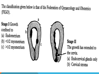
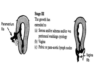
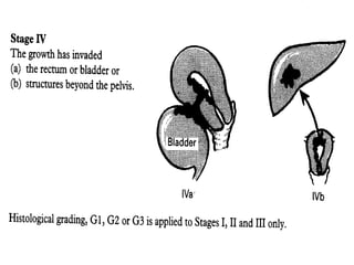
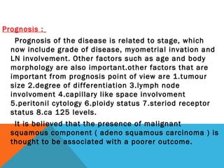
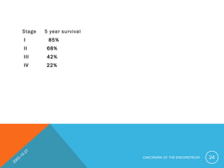
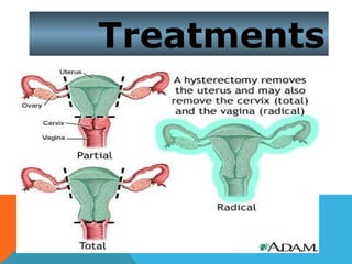
PREM
ALIGNANT
M
ALIGNANT
DISORDERS
OF THE
UTERINE
CORPUS
EMBRYOLOGY OF UTERUS:
The paramesonephric duct develop into main genital
ducts of the female.Three parts can be recognized
in each duct 1.cranial 2.horizental and caudal
vertical part that fuses with its partner from the
opposite side to form uterine canal.The fold ,which
extend from the sides of the fused part fom the
broad ligament of the uterus.The fused
paramesonephric ducts give rise to the corpus and
cervix of uterus.They are sorrounded by a layer of
mesenchyme that forms the muscular coat of uterus
the Myometrium and its peritonail covering, the
perimetrium.
ANOTOMY OF UTERUS
Uterus is the child bearing organ in
females,situated in the in the pelvis between
bladder and rectum.The direction can either be
anteversion or anteflexion.Uterus is divided into
body and cervix.Body is divided into
fundus,anterior ar vesical surface,posterior or
intestinal surface and lateral border.Various
supports of the uterus includes peritonail and
fibromuscular ligaments.Arterial supply is mainly
from uterine artries and partly from ovarian
artries.Lmphatic dranaige follows the track as
upper lymphatics from the fundus and upper part
of the body into aortic and superficial
inguinal.lower to external ilaic.middle to E.iliac.
Most common malignant diseases affecting the uterus
is endometrial carcinoma, which arises from the lining
of the uterus. However, sarcoma also arise from the
stroma of the endometrium or from the myometrium.
Epidemiology :
• The median age of presentation is just over 60 years
of age, however it can occur in their 20s, but the vast
majority of cases occur in women over 45 years of age.
with less than 5 % diagnosed under 40 years of age .
Highest incidence is in white north americans.
Aetiology :
The exact cause is unknown, however risk factors in
postmenapausal and premenapausal women include
the following :
1 – Obesity. 2 – Impaired carbohydrate tolerance.
3 – Nulliparity.
4 – Late menopause.
5 – Unopposed oestrogen therapy.
6 – Functioning ovarian tumors.
7 – Previous pelvic irradiation.
8 – Sequential oral contraceptives with dimethisterone
9 – Family history of carcinoma of breast, ovary or
colon.
10 – Polycystic ovary disease.
11 – Tamoxifin therapy which has weak oestrogen
effects on the endometrium
* Many of the factors are related to an increase in
oestrogen levels.
* In post – menopausal period, the majority of
circulating oestrogen is derived from aromatization of
peripheral androgens. This conversion take place
principally in adipose tissue. Also post – menopausal
women with diabetes have increased oestrogen levels.
* Nulliparity and late menopause are both associated
with increased risk of endometrial cancers, which may
be explained by the prolonged oestrogenic effect on the
endometrium.
* Women who use oral contraceptive or progesterone
have up to a 50 % reduction in the incidence of
endometrial cancer and protection lasts for many years
after the discontinuation of these treatments. Cigarette
smoking has also been associated with the reduced risk
of endometrial cancer.
Screening for endometrial cancer:
Screening stratagies includes
1.Endometrial sampling
2.Vaginal or cervical cytology
3.Progesterone challenge test
4.ultrasonography;Measurement of endometrial
thickness.
Classification :
A Endometrial hyperplasia :
Glandular hyperplasia of the endometrium are benign
conditions that may produce symptoms clinically
indistinguishable from early endometrial carcinoma.
Some of hyperplasias, even though reversible, are
considered premalignant lesions. Divided into:
1/ Hyperplasia without atypia
which is subdivided into either simple ( cystic )
hyperplasia and complex ( adenomatous ) hyperplasia.
2/ Hyperplasia with atypia, these hyperplasia are
generally considered premalignant.
3/ Carcinoma insitu
B Endometrial carcinoma :
Characterized by obvious hyperplasia and anaplasia of
glandular element, with invasion of underlying stroma,
myometrium or vascular spaces.
* Endometrial cancer can spread by 4 possible routs :
1 – Direct extension.
2 – Lymphatic metastasis.
3 – peritoneal implants after transtubal spread.
4 – haematogenous spread.
* Pathologist recognized 3 major histological types of
endometrial carcinoma:
1 – Adenocarcinoma. Mucinous type
2 – Adenocarcinoma with squamous differentiation.
3 – Adenosquamous carcinoma.
2003-10-27
CARCINOMA OF THE ENDOMETRIUM 10
All 3 types have identical presenting symptoms and
signs, patterns of spread, and general clinical behavior.
* Papillary serous and clear cell carcinoma of the
endometrium are other unusual histological subtypes
that carry a poor prognosis even when apparently
confined to the superficial myometrium.
Clinical presentation :
1 – About 75 – 80 % of women with endometrial
carcinoma will present with postmenopausal bleeding.
Sometimes bloody stain postmenopausal vaginal
discharge may be associated with endometrial
carcinoma.
2 – In premenopausal period, most women with
endometrial carcinoma present with intermenstrual
bleeding. Although 1/3 may present with heavy
periods only.
3 - Postmenopausal discharge from pyometra
carries a 50 % risk of associated malignancy.
4 – Pain may occur with pyometra or metastatic
spread . Pain is due to nerve compression on
pelvic side wall.
Diagnosis :
Traditionally, post menopausal bleeding was
investigated by a dilatation and curettage.
Endometrial sampling;
1.Fractional curettage : dilatation and fractional
curettage is the definitive procedure for diagnosis of
endometrial carcinoma. It should be performed with the
patient under anesthesia and by first curetting the
endocervical canal followed by dilatation of the canal
and circumferential curettage of the endometrial cavity.
At curretage care must be observed to avoid perafortion
of uterus, particularly in patients with pyometra.
More recently diagnosis has shifted to outpatient
setting with :
a – Pap smear.
b–Ultrasound determination of endometrial
thickness, also any ovarian pathology may be detected.
In post menopausal woman 5 mm is the cutoff for a
normal unilateral endometrial strip. Color flow imaging
may increase specificity.
c–Out-patient endometrial sampling using
instruments such as a pipelle Sampler and Sharman
currete.
d – Out – patient hysteroscopy.
e- tumour marker ;ca 125 levels
Radiological investigations:
X-ray and MRI may be indicated to see the extent of
2003-10-27
CARCINOMA OF THE ENDOMETRIUM 16
2003-10-27
CARCINOMA OF THE ENDOMETRIUM 17
Staging :
The FIGO classification and staging of endometrial
carcinoma are ( it is surgical staging ) :
Stage I : The carcinoma is confined to the corpus.
Ia : Tumor limited to the endometrium.
Ib : Invasion to less than ½ of the myometrium.
Ic : Invasion more than ½ of the myometrium.
Stage II : The carcinoma has involved the corpus and
the cervix but has not extended outside the uterus.
IIa : Endocervical glandular involvement only.
IIb : Cervical stromal invasion.
Stage III : The carcinoma has extended outside the
uterus but not outside the true pelvis.
Stage IV : The carcinoma has extended outside the
true pelvis or has obviously involved the mucosa of the
bladder or rectum.
2003-10-27
CARCINOMA OF THE ENDOMETRIUM 19
2003-10-27
CARCINOMA OF THE ENDOMETRIUM 20
2003-10-27
CARCINOMA OF THE ENDOMETRIUM 21
* Surgical stage I tumor account for 75 % of all
endometrial carcinoma which explain the relative good
overall prognosis.
Prognosis :
Prognosis of the disease is related to stage, which
now include grade of disease, myometrial invation and
LN involvement. Other factors such as age and body
morphology are also important.other factors that are
important from prognosis point of view are 1.tumour
size 2.degree of differentiation 3.lymph node
involvoment 4.capillary like space involvoment
5.peritonil cytology 6.ploidy status 7.steriod receptor
status 8.ca 125 levels.
It is believed that the presence of malignant
squamous component ( adeno squamous carcinoma ) is
thought to be associated with a poorer outcome.
Stage 5 year survival
I 85%
II 68%
III 42%
IV 22%
2003-10-27
CARCINOMA OF THE ENDOMETRIUM 24
Differential diagnosis :
Clinically the differential diagnosis of endometrial
carcinoma include all the causes of abnormal uterine
bleeding.
* In premeopausal patient the following should be
excluded :
1 – Complication of early pregnancy.
2 – Liomyoma.
3 – Endometrial hyperplasia and polyps.
4 – Cervical polyps.
5 – Various genital or metastatic cancers.
* In the postmenopausal age group, the following
should be considered :
1 – Atrophic vaginitis.
2 – Exogenous oestrogen ( HRT )
3 – Endometrial hyperplasia and polyps.
4 – Various genital neoplasma.
Treatment :
Stage I : - The treatment of choice is total abdominal
hysterectomy and bilateral salpingoopherectomy.
- Radiotherapy is also necessary if invasion of
the myometrium has occurred to more than the inner
half of the myometrium.
Stage II : - If patient is surgically fit, do radical
hysterectomy and bilateral lymphadenectomy with para-
aortic node sampling should be performed.
- If patient unfit surgically then radiotherapy
may be used.
Stage III : If node suggest spread of disease then surgery
with adjuvant radiotherapy.
Stage III & IV : Radiotherapy,chemothrapy,debulking
surgury and hormanal therapy.
Treatments
Radiotherapy is performed and then occasionally
residual disease may be involved by surgical
intervention.
Progesterone :
Some believe that progesterone may be helpful in
preventing recurrence after treatment of early stage
disease.
Follow up;
the objective of post treatment follow up of
endometrial carcinoma are to detect the recurrence
and compications of primary treatment and to manage
tomour related problems.
Compications of therapy;
includes haematuria from radiation
cystitis,diarrhoea,melena from radiation colitis,small
ANY QUERIES;
THANK YOU
Pre Malignant Disorders Of the Uterine Corpus

Pre Malignant Disorders Of the Uterine Corpus

  • 1.
  • 2.
    EMBRYOLOGY OF UTERUS: Theparamesonephric duct develop into main genital ducts of the female.Three parts can be recognized in each duct 1.cranial 2.horizental and caudal vertical part that fuses with its partner from the opposite side to form uterine canal.The fold ,which extend from the sides of the fused part fom the broad ligament of the uterus.The fused paramesonephric ducts give rise to the corpus and cervix of uterus.They are sorrounded by a layer of mesenchyme that forms the muscular coat of uterus the Myometrium and its peritonail covering, the perimetrium.
  • 3.
    ANOTOMY OF UTERUS Uterusis the child bearing organ in females,situated in the in the pelvis between bladder and rectum.The direction can either be anteversion or anteflexion.Uterus is divided into body and cervix.Body is divided into fundus,anterior ar vesical surface,posterior or intestinal surface and lateral border.Various supports of the uterus includes peritonail and fibromuscular ligaments.Arterial supply is mainly from uterine artries and partly from ovarian artries.Lmphatic dranaige follows the track as upper lymphatics from the fundus and upper part of the body into aortic and superficial inguinal.lower to external ilaic.middle to E.iliac.
  • 4.
    Most common malignantdiseases affecting the uterus is endometrial carcinoma, which arises from the lining of the uterus. However, sarcoma also arise from the stroma of the endometrium or from the myometrium. Epidemiology : • The median age of presentation is just over 60 years of age, however it can occur in their 20s, but the vast majority of cases occur in women over 45 years of age. with less than 5 % diagnosed under 40 years of age . Highest incidence is in white north americans.
  • 5.
    Aetiology : The exactcause is unknown, however risk factors in postmenapausal and premenapausal women include the following : 1 – Obesity. 2 – Impaired carbohydrate tolerance. 3 – Nulliparity. 4 – Late menopause. 5 – Unopposed oestrogen therapy. 6 – Functioning ovarian tumors. 7 – Previous pelvic irradiation. 8 – Sequential oral contraceptives with dimethisterone 9 – Family history of carcinoma of breast, ovary or colon. 10 – Polycystic ovary disease. 11 – Tamoxifin therapy which has weak oestrogen effects on the endometrium
  • 6.
    * Many ofthe factors are related to an increase in oestrogen levels. * In post – menopausal period, the majority of circulating oestrogen is derived from aromatization of peripheral androgens. This conversion take place principally in adipose tissue. Also post – menopausal women with diabetes have increased oestrogen levels. * Nulliparity and late menopause are both associated with increased risk of endometrial cancers, which may be explained by the prolonged oestrogenic effect on the endometrium. * Women who use oral contraceptive or progesterone have up to a 50 % reduction in the incidence of endometrial cancer and protection lasts for many years after the discontinuation of these treatments. Cigarette smoking has also been associated with the reduced risk of endometrial cancer.
  • 7.
    Screening for endometrialcancer: Screening stratagies includes 1.Endometrial sampling 2.Vaginal or cervical cytology 3.Progesterone challenge test 4.ultrasonography;Measurement of endometrial thickness.
  • 8.
    Classification : A Endometrialhyperplasia : Glandular hyperplasia of the endometrium are benign conditions that may produce symptoms clinically indistinguishable from early endometrial carcinoma. Some of hyperplasias, even though reversible, are considered premalignant lesions. Divided into: 1/ Hyperplasia without atypia which is subdivided into either simple ( cystic ) hyperplasia and complex ( adenomatous ) hyperplasia. 2/ Hyperplasia with atypia, these hyperplasia are generally considered premalignant. 3/ Carcinoma insitu
  • 9.
    B Endometrial carcinoma: Characterized by obvious hyperplasia and anaplasia of glandular element, with invasion of underlying stroma, myometrium or vascular spaces. * Endometrial cancer can spread by 4 possible routs : 1 – Direct extension. 2 – Lymphatic metastasis. 3 – peritoneal implants after transtubal spread. 4 – haematogenous spread. * Pathologist recognized 3 major histological types of endometrial carcinoma: 1 – Adenocarcinoma. Mucinous type 2 – Adenocarcinoma with squamous differentiation. 3 – Adenosquamous carcinoma.
  • 10.
  • 11.
    All 3 typeshave identical presenting symptoms and signs, patterns of spread, and general clinical behavior. * Papillary serous and clear cell carcinoma of the endometrium are other unusual histological subtypes that carry a poor prognosis even when apparently confined to the superficial myometrium.
  • 12.
    Clinical presentation : 1– About 75 – 80 % of women with endometrial carcinoma will present with postmenopausal bleeding. Sometimes bloody stain postmenopausal vaginal discharge may be associated with endometrial carcinoma. 2 – In premenopausal period, most women with endometrial carcinoma present with intermenstrual bleeding. Although 1/3 may present with heavy periods only.
  • 13.
    3 - Postmenopausaldischarge from pyometra carries a 50 % risk of associated malignancy. 4 – Pain may occur with pyometra or metastatic spread . Pain is due to nerve compression on pelvic side wall.
  • 14.
    Diagnosis : Traditionally, postmenopausal bleeding was investigated by a dilatation and curettage. Endometrial sampling; 1.Fractional curettage : dilatation and fractional curettage is the definitive procedure for diagnosis of endometrial carcinoma. It should be performed with the patient under anesthesia and by first curetting the endocervical canal followed by dilatation of the canal and circumferential curettage of the endometrial cavity. At curretage care must be observed to avoid perafortion of uterus, particularly in patients with pyometra.
  • 15.
    More recently diagnosishas shifted to outpatient setting with : a – Pap smear. b–Ultrasound determination of endometrial thickness, also any ovarian pathology may be detected. In post menopausal woman 5 mm is the cutoff for a normal unilateral endometrial strip. Color flow imaging may increase specificity. c–Out-patient endometrial sampling using instruments such as a pipelle Sampler and Sharman currete. d – Out – patient hysteroscopy. e- tumour marker ;ca 125 levels Radiological investigations: X-ray and MRI may be indicated to see the extent of
  • 16.
  • 17.
  • 18.
    Staging : The FIGOclassification and staging of endometrial carcinoma are ( it is surgical staging ) : Stage I : The carcinoma is confined to the corpus. Ia : Tumor limited to the endometrium. Ib : Invasion to less than ½ of the myometrium. Ic : Invasion more than ½ of the myometrium. Stage II : The carcinoma has involved the corpus and the cervix but has not extended outside the uterus. IIa : Endocervical glandular involvement only. IIb : Cervical stromal invasion. Stage III : The carcinoma has extended outside the uterus but not outside the true pelvis. Stage IV : The carcinoma has extended outside the true pelvis or has obviously involved the mucosa of the bladder or rectum.
  • 19.
  • 20.
  • 21.
  • 22.
    * Surgical stageI tumor account for 75 % of all endometrial carcinoma which explain the relative good overall prognosis.
  • 23.
    Prognosis : Prognosis ofthe disease is related to stage, which now include grade of disease, myometrial invation and LN involvement. Other factors such as age and body morphology are also important.other factors that are important from prognosis point of view are 1.tumour size 2.degree of differentiation 3.lymph node involvoment 4.capillary like space involvoment 5.peritonil cytology 6.ploidy status 7.steriod receptor status 8.ca 125 levels. It is believed that the presence of malignant squamous component ( adeno squamous carcinoma ) is thought to be associated with a poorer outcome.
  • 24.
    Stage 5 yearsurvival I 85% II 68% III 42% IV 22% 2003-10-27 CARCINOMA OF THE ENDOMETRIUM 24
  • 25.
    Differential diagnosis : Clinicallythe differential diagnosis of endometrial carcinoma include all the causes of abnormal uterine bleeding. * In premeopausal patient the following should be excluded : 1 – Complication of early pregnancy. 2 – Liomyoma. 3 – Endometrial hyperplasia and polyps. 4 – Cervical polyps. 5 – Various genital or metastatic cancers. * In the postmenopausal age group, the following should be considered : 1 – Atrophic vaginitis. 2 – Exogenous oestrogen ( HRT ) 3 – Endometrial hyperplasia and polyps. 4 – Various genital neoplasma.
  • 26.
    Treatment : Stage I: - The treatment of choice is total abdominal hysterectomy and bilateral salpingoopherectomy. - Radiotherapy is also necessary if invasion of the myometrium has occurred to more than the inner half of the myometrium. Stage II : - If patient is surgically fit, do radical hysterectomy and bilateral lymphadenectomy with para- aortic node sampling should be performed. - If patient unfit surgically then radiotherapy may be used. Stage III : If node suggest spread of disease then surgery with adjuvant radiotherapy. Stage III & IV : Radiotherapy,chemothrapy,debulking surgury and hormanal therapy.
  • 27.
  • 28.
    Radiotherapy is performedand then occasionally residual disease may be involved by surgical intervention. Progesterone : Some believe that progesterone may be helpful in preventing recurrence after treatment of early stage disease. Follow up; the objective of post treatment follow up of endometrial carcinoma are to detect the recurrence and compications of primary treatment and to manage tomour related problems. Compications of therapy; includes haematuria from radiation cystitis,diarrhoea,melena from radiation colitis,small
  • 29.
  • 30.