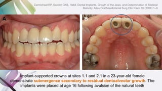
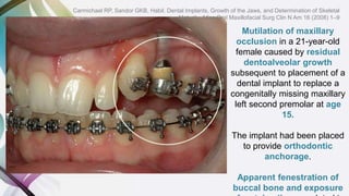
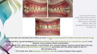
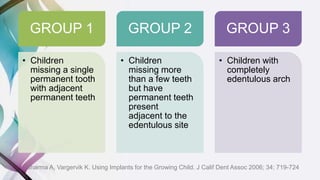
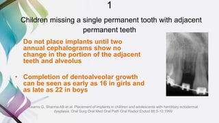
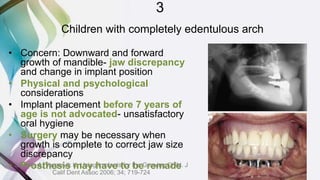
USING IMPLANTS FOR
GROWING PATIENTS
Arun Sharma and Karin Vargervik
J Calif Dent Assoc 2006; 34; 719-724
AAMIR ZAHID GODIL
FIRST YEAR P.G.
DEPARTMENT OF PROSTHODONTICS
M.A.R.D.C.
OUTLINE
• INTRODUCTION
• GROWTH CONSIDERATIONS
– GROWTH OF MAXILLA
– GROWTH OF MANDIBLE
– CONCERNS
• EXPERIENCES OF TREATMENT WITH DENTAL IMPLANTS IN YOUNG INDIVIDUALS:
CASE REPORTS
– BACKGROUND
– CASES
– TREATMENT SUBSTITUTES
– ADVOCATED AGES FOR IMPLANT PLACEMENT
• RECOMMENDED PROTOCOL
– OPTIONS
– GROUP 1: CHILDREN MISSING A SINGLE PERMANENT TOOTH WITH ADJACENT PERMANENT
TEETH
– GROUP 2: CHILDREN MISSING MORE THAN A FEW TEETH BUT HAVE PERMANENT TEETH
PRESENT ADJACENT TO THE EDENTULOUS SITE
– GROUP 3: CHILDREN WITH COMPLETELY EDENTULOUS ARCH
• CONCLUSION AND CRITIQUE
INTRODUCTION
• ‘Hypodontia’ is defined as the congenital missing of <6
permanent teeth, excluding third molars
• ‘Oligodontia’ as the congenital missing of six or more
permanent teeth, excluding third molars
• ‘Anodontia’ as the congenital missing of all deciduous and ⁄ or
permanent teeth
• In the oral habilitation of children with missing teeth a golden
principle is to strive for establishing a good situation from an
aesthetic as well as a functional and psycho-social viewpoint
with minimal replacement of the missing teeth by prosthetic
treatment
GROWTH
CONSIDERATIONS
GROWTH OF MAXILLA
Growth of the maxilla is characterized by remodeling in a
postero-superior direction while simultaneously being
displaced in the opposite antero-inferior direction
GROWTH OF MANDIBLE
Growth of the mandible is characterized by displacement
away from its articulation in the glenoid fossae as the
condyles and rami relocate in a posterosuperior direction
Natural tooth movement occurs as a result of eruption and
of being carried along passively with the maxilla and
mandible, both of which undergo displacement antero-
inferiorly during craniofacial morphogenesis
Transverse development of the maxilla.
(A) Growth at the midpalatal suture is greater posteriorly than anteriorly.
(B) Remodeling allows the dentition to drift horizontally.
A dental implant (red circle) will become displaced palatally (in addition to becoming
submerged) in remodeling regions of the alveolar process
Transverse development of the mandible.
A) Intercanine growth is minimal and ceases early.
B) Mandibular growth is characterized by an opening hinge movement of its
two halves around an axis passing anteroposteriorly through the
symphysis.
CONCERNS
ETIOLOGICAL CONCERNS
• Genetic disorders such as Ectodermal Dysplasia or Down
syndrome
• Hypodontia can also been seen in people with cleft lip and palate
• Hormonal defects: Idiopathic Hypoparathyroidism and
Pseudohypoparathyroidism
• Environmental causes involving exposure to radiation,
anticancer, chemotherapeutic agents, allergy and toxic epidermal
necrolysis after drug
• Infectious causes of hypodontia: rubella, candida
• Trauma: accident or sports injury
TREATMENT
CONCERNS
• Implants inserted into
pediatric patients do not
follow the regular growth
process of the craniofacial
skeleton and are known to
behave similar to
ankylosed teeth, resulting
in both functional and
esthetic disadvantages
(OpHeji et al. 2003)
• Additionally, they can
interfere with the
position and the eruption
of adjacent tooth germs,
thus resulting in potential
severe trauma of the
EXPERIENCES OF TREATMENT
WITH DENTAL IMPLANTS IN
YOUNG INDIVIDUALS: CASE
REPORTS
BACKGROUND• Bjork (1963, 1997):
Implanted pins in the jaws of children for longitudinal cephalometric studies and
reported that those in the path of erupting teeth were displaced and those placed
in resorptive areas were lost. Pins placed in areas of appositional bone growth
became embedded.
• Oesterle (1993):
Compared dental implants to ankylosed primary teeth. They wrote that ankylosis
arrests both dental eruption and alveolar bone formation in the affected area..
The authors proposed that implants placed in the posterior maxilla in children might
become buried to the point that the apical portion may become exposed as the
nasal and antral floor remodel.
They also warned the possibility of loss of implants in the anterior maxilla
because of resorption in the infradental fossa and nasal floor.
• Lederman et al (1993):
In their 7 year follow up with a mean length of 35.5 months, reported a
90% success rate on 42 endosseous dental implants placed in 34
patients aged 9 to 18 years.
There was a positive soft and osseous tissue reaction to the implants, and
most of the failures occurred because of subsequent traumatic
injuries sustained during the healing phase after implant
placement.
The major complication reported was the failure of dental implants to
respond to the vertical growth of adjacent teeth and alveolus due
to ankylosis.
• Brugnolo et al (1996):
Noted the infraocclussion of implants placed in patients aged 13 to
14.5 years, secondary to vertical growth, and prosthesis was
redesigned. Anteroposterior and transverse growth seemed not to
negatively influence the implants position.
• Smith et al (1993):
Implant use in children with ectodermal dysplasia is a treatment of
choice, since its placement in the mandibular anterior region of a
5 year old patient did not affect adjacent tooth buds. Prosthesis
remodeling was performed due to implant submergence.
• Guckes et al (1997):
Described a case of 3-year-old patient with ectodermal dysplasia in
which dental implants located in the mandible and maxilla have
not moved despite growth. During the 5-year follow up, the
prosthesis was remodeled to accommodate eruption of the
maxillary teeth and facial growth.
• Kearns et al (1999):
No evidence of restriction to transverse and sagittal growth due to
implant use in children with ectodermal dysplasia. Prosthesis
remodeling was necessary in some patients secondary to implant
• The youngest child reported was a
French boy who had implants placed
at the age of 1.5 years
Bonin B, Saffarzadeh A, Picard A, Levy P, Romieux G,
Goga D.Early implant treatment of a child with
anhidrotic ectodermal dysplasia. Apropos of a case.
Rev Stomatol Chir Maxillofac.2001;102:313–318
• The first published case of
placing implants in a boy with
hypohidrotic ED and anodontia
of the mandible was treated at
the Institute in Jo¨nko¨ ping
and has been followed for
more than 20 years.
• The inter-implant distance
has not changed and an
overdenture served well
until the patient was 19
years old, when two additional
implants were placed and the
patient was provided with a
mandibular fixed implant-
supported prosthesis.
Guckes AD, Scurria MS, King TS, McCarthy
GR, Brahim JS. Prospective clinical trial
of dental implants in persons with
ectodermal dysplasia. J Prosthet Dent.
2002;88:21–25
• Since there is more vertical growth
in the posterior regions of the
maxilla and mandible during
childhood and adolescence,
implants placed distal to the
canines present more
complications.
Bryant SR. The effects of age, jaw site, and bone condition
on oral implant outcomes. Int J Prosthodont.
1998;11:470–490.
Enzo Rossi and Jens O. Andreasen
Maxillary Bone Growth and Implant Positioning in a
Young Patient: A Case Report
The International Journal of Periodontics & Restorative Dentistry Volume 23, Number 2, 2003; 113-
119
Panoramic radiograph showing the lost maxillary left central
incisor
A 10-year-old
Caucasian male
lost his maxillary
left central incisor
as a result of a
bicycle accident
Surface of the implant at the time
of post cementation
Result after 3 years (13 years old), 3-mm incisal
discrepancy relative to the adjacent teeth because of
maxillary vertical growth and eruption of the natural teeth.
New crown is again fabricated
to create an even incisal line.
Nine years after implantation (age 19),
there is an additional change of 2 mm. Age 21. Now there is a 3-mm distance.
At age 25, 15 years after implantation,
there is a 5-mm discrepancy
Implant-supported crowns at sites 1.1 and 2.1 in a 23-year-old female
demonstrate submergence secondary to residual dentoalveolar growth. The
implants were placed at age 16 following avulsion of the natural teeth
Carmichael RP, Sandor GKB, Habil. Dental Implants, Growth of the Jaws, and Determination of Skeletal
Maturity. Atlas Oral Maxillofacial Surg Clin N Am 16 (2008) 1–9
Mutilation of maxillary
occlusion in a 21-year-old
female caused by residual
dentoalveolar growth
subsequent to placement of a
dental implant to replace a
congenitally missing maxillary
left second premolar at age
15.
The implant had been placed
to provide orthodontic
anchorage.
Apparent fenestration of
buccal bone and exposure
Carmichael RP, Sandor GKB, Habil. Dental Implants, Growth of the Jaws, and Determination of Skeletal
Maturity. Atlas Oral Maxillofacial Surg Clin N Am 16 (2008) 1–9
Mandibular growth has led to a separation of the jaws posteriorly and has advanced the mandible
into an edge-to-edge incisal relationship.
However, no separation of the proximal surfaces of the right and left sides of the bridge (arrows) is
visible, suggesting no transverse growth across the symphysis has occurred. (A) Facial view. (B)
Frontal view of teeth
An 11-year-old boy with
hypohidrotic ectodermal
dysplasia in whom a two-piece
bridge, split at the mandibular
midline, on four anterior mandibular
implants, had been constructed at
age 8
Carmichael RP, Sandor GKB, Habil. Dental Implants, Growth of the Jaws, and Determination of Skeletal
Maturity. Atlas Oral Maxillofacial Surg Clin N Am 16 (2008) 1–9
21-year-old male who had been lost to follow-up since implants replacing all his molars had been placed at age
15.
In the meantime, he had developed a bilateral posterior open bite following from mandibular growth in the
absence of any posterior dental compensation.
(A) Right side view. Note large exophytic, ovoid-shaped, firm, smooth-surfaced, maroon-colored lesion that was
found to be a peripheral giant cell granuloma associated with the dental implant at site 4.6.
(B) Left lateral view.
(C) Frontal view. Note splaying of anterior teeth as a result of absent molar support.
Carmichael RP, Sandor
GKB, Habil. Dental Implants,
Growth of the Jaws, and
Determination of Skeletal
Maturity. Atlas Oral
Maxillofacial Surg Clin N Am
16 (2008) 1–9
Iseri H and Solow B
• No development in the alveolar bone
after integration of the implant with
bone in a growing alveoli
• But the neighboring tissues
continued their 3-dimensional growth
• Therefore artificial teeth upon the
osseointegrated implant remained in
lower positions in occlusion due to
continued eruption of the neighboring
teeth
• Recommendation: no implant therapy
in either anterior or posterior alveolar
segment if there is no intention of
modifying the prosthesis
Iseri H, Solow B. Continued eruption of maxillary incisors
and first molars in girls from 9 to 25 years, studied by
the implant method. Eur J Orthod. 1996;18:245–256
• Emphasized that the implants did not
affect the development of the
neighboring tissues.
Iseri H, Solow B. Average surface remodeling of the
maxillary base and the orbital floor in female subjects
from 8 to 25 years. An implant study. Am J Orthod
Dentofacial Orthop. 1995;107:48–57.
• Measured the width between the
implants at both sides using
posteroanterior radiography and
determined a statistically insignificant
but important increase.
Iseri H, Solow B. Change in the width of the mandibular
body from 6 to 23 years of age: An implant study. Eur J
Orthod. 2000;22:229–238.
• Johnston et al advocated the
application of short fixed partial
dentures to the young patients after
the closure of the permanent teeth,
even if they have no temporary teeth
congenitally
• They stated that the patients should
be subjected to routine controls in
3-month periods for accurate
balancing of the fixed partial
dentures, and monitoring and
correction of the occlusion because of
the rapidly changing relations in jaws
Johnston JF, Phillips RW, Dykema RW. In: Modern Practice in
Crown and Bridge Prosthodontics. Philadelphia,
PA:Saunders; 1980:3–19
• Croll followed the patients with crown
restorations for 8 years and
observed that the treated molars
continued to erupt, and the crown
margins came to a supragingival
position by that time
• That is why he described stainless
steel crowns as intermediate
restorations. He asserted that the
patients could be treated with
noble metal restoration after the
termination of the major effect of
the physical growth
Croll TP. Restorative dentistry for preschool children. Dent Clin
North Am. 1995;39:737–770.
TREATMENT SUBSTITUTES
• Tylman found the application of
fixed prostheses to children and
youth as being contraindicative
due to the fact that the teeth were
not fully erupted, and the pulps
are very large, which may cause
various complications.
• He stated that the age of the
application of prosthesis is 17
years.
Tylman SD. In: Theory and Practice of Crown and Fixed Partial
Prosthodontics (Bridge). 6th ed. Saint Louis, MO: Mosby;1970:13–
• Lederman et al stated that the earliest time when the implants could be
used was 11 years of age for the girls and 13 years of age for the
boys.
Lederman P, Hassell T, Hefti A. Osseointegrated dental implants as alternative therapy to bridge construction or orthodontics in young patients. Seven years
of clinical experience. Pediatr Dent. 1993;15:327–333
ADVOCATED AGES FOR IMPLANT PLACEMENT
Placement of dental implants can
not be recommended before the
age of 6 years, since it is well
established that children can and
should take part in decisions on
elective surgery from the age of
around 5 years
Bradbury ET, Kay SP, Tighe C, Hewison J. Decision-making
by parents and children in paediatric hand surgery. Br J Plast
Surg. 1994;47:324–330
• At the Consensus Conference ‘Oral
Implants in Young Patients’ it was
agreed in a consensus statement that
implants should not be placed until
growth and skeletal development is
completed or nearly completed
Bergendal B, Koch G, Kurol J, Wa¨nndahl G, eds.
Consensus Conference on Ectodermal Dysplasia
with special reference to dental treatment.
Stockholm, Sweden: Fo¨ rlagshuset Gothia AB;
1998.
• This was illustrated in a figure based
on the curve of growth velocity from
infant to adult where the colours
allude to a traffic light
• Anodontia and severe oligodontia
were mentioned as exceptions to
this rule
Bergendal B, Olgart K. Congenitally missing teeth. In: Koch G,
Bergendal T, Kvint S, Johansson UB, eds. Consensus Conference on
Oral Implants in Young Patients. Stockholm, Sweden: Fo¨ rlagshuset
Gothia AB; 1996:16–27.
RECOMMENDED
PROTOCOL
OPTIONS
CONVENTIONAL
DENTURES
FIXED
PROSTHESES
CAST PARTIAL
DENTURES
SPACE
MAINTAINERS/
KIDDY
DENTURES
IMPLANTS WAIT
Maroulakos G, Artopoulou II, Angelopoulou MV, Emmanouil D. Removable partial
dentures vs overdentures in children with ectodermal dysplasia: two case reports.
European Archives of Paediatric Dentistry. 2016 Jun:1-6.
GROUP 1
• Children
missing a single
permanent tooth
with adjacent
permanent teeth
GROUP 2
• Children
missing more
than a few teeth
but have
permanent teeth
present
adjacent to the
edentulous site
GROUP 3
• Children with
completely
edentulous arch
Sharma A, Vargervik K. Using Implants for the Growing Child. J Calif Dent Assoc 2006; 34; 719-724
1
Children missing a single permanent tooth with adjacent
permanent teeth
• Do not place implants until two
annual cephalograms show no
change in the portion of the adjacent
teeth and alveolus
• Completion of dentoalveolar growth
can be seen as early as 16 in girls and
as late as 22 in boys
Kearns G, Sharma AB et al. Placement of implants in children and adolescents with heriditary ectodermal
dysplasia. Oral Surg Oral Med Oral Path Oral Radiol Endod 88:5-10,1999
2
Children missing more than a few teeth but have permanent
teeth present adjacent to the edentulous site
• Most complex to manage
• Initial objective: Orthodontically optimise the
position of the teeth present
• Removable prostheses can be given until
dentoalveolar development is complete
• Consider: psychological and esthetic factors
• Safest approach: wait until dentoalveolar growth is
complete (no change in lateral cephalogram at one
year interval)
• If implants are placed before growth completion,
segmental osteotomy or distraction
osteogenesis can be done for surgical repositioning
• Alternative: Remake the prosthesis using pink
3
Children with completely edentulous arch
• Concern: Downward and forward
growth of mandible- jaw discrepancy
and change in implant position
• Physical and psychological
considerations
• Implant placement before 7 years of
age is not advocated- unsatisfactory
oral hygiene
• Surgery may be necessary when
growth is complete to correct jaw size
discrepancy
• Prosthesis may have to be remadeSharma A, Vargervik K. Using Implants for the Growing Child. J
Calif Dent Assoc 2006; 34; 719-724
CONCLUSION AND CRITIQUE
• NO TREATMENT ALTERNATIVES SUGGESTED FOR
REHABILITATION OF GROWING EDENTULOUS
PATIENTS
• INSUFFICIENT GUIDELINES ON MANAGEMENT OF A
CHILD DURING IMPLANT PLACEMENT- ROLE OF
PAEDIATRIC DENTIST
Thank You

Using implants for growing patients

  • 1.
    USING IMPLANTS FOR GROWINGPATIENTS Arun Sharma and Karin Vargervik J Calif Dent Assoc 2006; 34; 719-724 AAMIR ZAHID GODIL FIRST YEAR P.G. DEPARTMENT OF PROSTHODONTICS M.A.R.D.C.
  • 2.
    OUTLINE • INTRODUCTION • GROWTHCONSIDERATIONS – GROWTH OF MAXILLA – GROWTH OF MANDIBLE – CONCERNS • EXPERIENCES OF TREATMENT WITH DENTAL IMPLANTS IN YOUNG INDIVIDUALS: CASE REPORTS – BACKGROUND – CASES – TREATMENT SUBSTITUTES – ADVOCATED AGES FOR IMPLANT PLACEMENT • RECOMMENDED PROTOCOL – OPTIONS – GROUP 1: CHILDREN MISSING A SINGLE PERMANENT TOOTH WITH ADJACENT PERMANENT TEETH – GROUP 2: CHILDREN MISSING MORE THAN A FEW TEETH BUT HAVE PERMANENT TEETH PRESENT ADJACENT TO THE EDENTULOUS SITE – GROUP 3: CHILDREN WITH COMPLETELY EDENTULOUS ARCH • CONCLUSION AND CRITIQUE
  • 3.
    INTRODUCTION • ‘Hypodontia’ isdefined as the congenital missing of <6 permanent teeth, excluding third molars • ‘Oligodontia’ as the congenital missing of six or more permanent teeth, excluding third molars • ‘Anodontia’ as the congenital missing of all deciduous and ⁄ or permanent teeth • In the oral habilitation of children with missing teeth a golden principle is to strive for establishing a good situation from an aesthetic as well as a functional and psycho-social viewpoint with minimal replacement of the missing teeth by prosthetic treatment
  • 4.
  • 5.
    GROWTH OF MAXILLA Growthof the maxilla is characterized by remodeling in a postero-superior direction while simultaneously being displaced in the opposite antero-inferior direction
  • 6.
    GROWTH OF MANDIBLE Growthof the mandible is characterized by displacement away from its articulation in the glenoid fossae as the condyles and rami relocate in a posterosuperior direction
  • 7.
    Natural tooth movementoccurs as a result of eruption and of being carried along passively with the maxilla and mandible, both of which undergo displacement antero- inferiorly during craniofacial morphogenesis
  • 8.
    Transverse development ofthe maxilla. (A) Growth at the midpalatal suture is greater posteriorly than anteriorly. (B) Remodeling allows the dentition to drift horizontally. A dental implant (red circle) will become displaced palatally (in addition to becoming submerged) in remodeling regions of the alveolar process
  • 9.
    Transverse development ofthe mandible. A) Intercanine growth is minimal and ceases early. B) Mandibular growth is characterized by an opening hinge movement of its two halves around an axis passing anteroposteriorly through the symphysis.
  • 10.
    CONCERNS ETIOLOGICAL CONCERNS • Geneticdisorders such as Ectodermal Dysplasia or Down syndrome • Hypodontia can also been seen in people with cleft lip and palate • Hormonal defects: Idiopathic Hypoparathyroidism and Pseudohypoparathyroidism • Environmental causes involving exposure to radiation, anticancer, chemotherapeutic agents, allergy and toxic epidermal necrolysis after drug • Infectious causes of hypodontia: rubella, candida • Trauma: accident or sports injury TREATMENT CONCERNS • Implants inserted into pediatric patients do not follow the regular growth process of the craniofacial skeleton and are known to behave similar to ankylosed teeth, resulting in both functional and esthetic disadvantages (OpHeji et al. 2003) • Additionally, they can interfere with the position and the eruption of adjacent tooth germs, thus resulting in potential severe trauma of the
  • 11.
    EXPERIENCES OF TREATMENT WITHDENTAL IMPLANTS IN YOUNG INDIVIDUALS: CASE REPORTS
  • 12.
    BACKGROUND• Bjork (1963,1997): Implanted pins in the jaws of children for longitudinal cephalometric studies and reported that those in the path of erupting teeth were displaced and those placed in resorptive areas were lost. Pins placed in areas of appositional bone growth became embedded. • Oesterle (1993): Compared dental implants to ankylosed primary teeth. They wrote that ankylosis arrests both dental eruption and alveolar bone formation in the affected area.. The authors proposed that implants placed in the posterior maxilla in children might become buried to the point that the apical portion may become exposed as the nasal and antral floor remodel. They also warned the possibility of loss of implants in the anterior maxilla because of resorption in the infradental fossa and nasal floor.
  • 13.
    • Lederman etal (1993): In their 7 year follow up with a mean length of 35.5 months, reported a 90% success rate on 42 endosseous dental implants placed in 34 patients aged 9 to 18 years. There was a positive soft and osseous tissue reaction to the implants, and most of the failures occurred because of subsequent traumatic injuries sustained during the healing phase after implant placement. The major complication reported was the failure of dental implants to respond to the vertical growth of adjacent teeth and alveolus due to ankylosis. • Brugnolo et al (1996): Noted the infraocclussion of implants placed in patients aged 13 to 14.5 years, secondary to vertical growth, and prosthesis was redesigned. Anteroposterior and transverse growth seemed not to negatively influence the implants position.
  • 14.
    • Smith etal (1993): Implant use in children with ectodermal dysplasia is a treatment of choice, since its placement in the mandibular anterior region of a 5 year old patient did not affect adjacent tooth buds. Prosthesis remodeling was performed due to implant submergence. • Guckes et al (1997): Described a case of 3-year-old patient with ectodermal dysplasia in which dental implants located in the mandible and maxilla have not moved despite growth. During the 5-year follow up, the prosthesis was remodeled to accommodate eruption of the maxillary teeth and facial growth. • Kearns et al (1999): No evidence of restriction to transverse and sagittal growth due to implant use in children with ectodermal dysplasia. Prosthesis remodeling was necessary in some patients secondary to implant
  • 15.
    • The youngestchild reported was a French boy who had implants placed at the age of 1.5 years Bonin B, Saffarzadeh A, Picard A, Levy P, Romieux G, Goga D.Early implant treatment of a child with anhidrotic ectodermal dysplasia. Apropos of a case. Rev Stomatol Chir Maxillofac.2001;102:313–318 • The first published case of placing implants in a boy with hypohidrotic ED and anodontia of the mandible was treated at the Institute in Jo¨nko¨ ping and has been followed for more than 20 years. • The inter-implant distance has not changed and an overdenture served well until the patient was 19 years old, when two additional implants were placed and the patient was provided with a mandibular fixed implant- supported prosthesis. Guckes AD, Scurria MS, King TS, McCarthy GR, Brahim JS. Prospective clinical trial of dental implants in persons with ectodermal dysplasia. J Prosthet Dent. 2002;88:21–25 • Since there is more vertical growth in the posterior regions of the maxilla and mandible during childhood and adolescence, implants placed distal to the canines present more complications. Bryant SR. The effects of age, jaw site, and bone condition on oral implant outcomes. Int J Prosthodont. 1998;11:470–490.
  • 17.
    Enzo Rossi andJens O. Andreasen Maxillary Bone Growth and Implant Positioning in a Young Patient: A Case Report The International Journal of Periodontics & Restorative Dentistry Volume 23, Number 2, 2003; 113- 119 Panoramic radiograph showing the lost maxillary left central incisor A 10-year-old Caucasian male lost his maxillary left central incisor as a result of a bicycle accident
  • 18.
    Surface of theimplant at the time of post cementation Result after 3 years (13 years old), 3-mm incisal discrepancy relative to the adjacent teeth because of maxillary vertical growth and eruption of the natural teeth. New crown is again fabricated to create an even incisal line. Nine years after implantation (age 19), there is an additional change of 2 mm. Age 21. Now there is a 3-mm distance. At age 25, 15 years after implantation, there is a 5-mm discrepancy
  • 20.
    Implant-supported crowns atsites 1.1 and 2.1 in a 23-year-old female demonstrate submergence secondary to residual dentoalveolar growth. The implants were placed at age 16 following avulsion of the natural teeth Carmichael RP, Sandor GKB, Habil. Dental Implants, Growth of the Jaws, and Determination of Skeletal Maturity. Atlas Oral Maxillofacial Surg Clin N Am 16 (2008) 1–9
  • 21.
    Mutilation of maxillary occlusionin a 21-year-old female caused by residual dentoalveolar growth subsequent to placement of a dental implant to replace a congenitally missing maxillary left second premolar at age 15. The implant had been placed to provide orthodontic anchorage. Apparent fenestration of buccal bone and exposure Carmichael RP, Sandor GKB, Habil. Dental Implants, Growth of the Jaws, and Determination of Skeletal Maturity. Atlas Oral Maxillofacial Surg Clin N Am 16 (2008) 1–9
  • 22.
    Mandibular growth hasled to a separation of the jaws posteriorly and has advanced the mandible into an edge-to-edge incisal relationship. However, no separation of the proximal surfaces of the right and left sides of the bridge (arrows) is visible, suggesting no transverse growth across the symphysis has occurred. (A) Facial view. (B) Frontal view of teeth An 11-year-old boy with hypohidrotic ectodermal dysplasia in whom a two-piece bridge, split at the mandibular midline, on four anterior mandibular implants, had been constructed at age 8 Carmichael RP, Sandor GKB, Habil. Dental Implants, Growth of the Jaws, and Determination of Skeletal Maturity. Atlas Oral Maxillofacial Surg Clin N Am 16 (2008) 1–9
  • 23.
    21-year-old male whohad been lost to follow-up since implants replacing all his molars had been placed at age 15. In the meantime, he had developed a bilateral posterior open bite following from mandibular growth in the absence of any posterior dental compensation. (A) Right side view. Note large exophytic, ovoid-shaped, firm, smooth-surfaced, maroon-colored lesion that was found to be a peripheral giant cell granuloma associated with the dental implant at site 4.6. (B) Left lateral view. (C) Frontal view. Note splaying of anterior teeth as a result of absent molar support. Carmichael RP, Sandor GKB, Habil. Dental Implants, Growth of the Jaws, and Determination of Skeletal Maturity. Atlas Oral Maxillofacial Surg Clin N Am 16 (2008) 1–9
  • 24.
    Iseri H andSolow B • No development in the alveolar bone after integration of the implant with bone in a growing alveoli • But the neighboring tissues continued their 3-dimensional growth • Therefore artificial teeth upon the osseointegrated implant remained in lower positions in occlusion due to continued eruption of the neighboring teeth • Recommendation: no implant therapy in either anterior or posterior alveolar segment if there is no intention of modifying the prosthesis Iseri H, Solow B. Continued eruption of maxillary incisors and first molars in girls from 9 to 25 years, studied by the implant method. Eur J Orthod. 1996;18:245–256 • Emphasized that the implants did not affect the development of the neighboring tissues. Iseri H, Solow B. Average surface remodeling of the maxillary base and the orbital floor in female subjects from 8 to 25 years. An implant study. Am J Orthod Dentofacial Orthop. 1995;107:48–57. • Measured the width between the implants at both sides using posteroanterior radiography and determined a statistically insignificant but important increase. Iseri H, Solow B. Change in the width of the mandibular body from 6 to 23 years of age: An implant study. Eur J Orthod. 2000;22:229–238.
  • 25.
    • Johnston etal advocated the application of short fixed partial dentures to the young patients after the closure of the permanent teeth, even if they have no temporary teeth congenitally • They stated that the patients should be subjected to routine controls in 3-month periods for accurate balancing of the fixed partial dentures, and monitoring and correction of the occlusion because of the rapidly changing relations in jaws Johnston JF, Phillips RW, Dykema RW. In: Modern Practice in Crown and Bridge Prosthodontics. Philadelphia, PA:Saunders; 1980:3–19 • Croll followed the patients with crown restorations for 8 years and observed that the treated molars continued to erupt, and the crown margins came to a supragingival position by that time • That is why he described stainless steel crowns as intermediate restorations. He asserted that the patients could be treated with noble metal restoration after the termination of the major effect of the physical growth Croll TP. Restorative dentistry for preschool children. Dent Clin North Am. 1995;39:737–770. TREATMENT SUBSTITUTES
  • 26.
    • Tylman foundthe application of fixed prostheses to children and youth as being contraindicative due to the fact that the teeth were not fully erupted, and the pulps are very large, which may cause various complications. • He stated that the age of the application of prosthesis is 17 years. Tylman SD. In: Theory and Practice of Crown and Fixed Partial Prosthodontics (Bridge). 6th ed. Saint Louis, MO: Mosby;1970:13– • Lederman et al stated that the earliest time when the implants could be used was 11 years of age for the girls and 13 years of age for the boys. Lederman P, Hassell T, Hefti A. Osseointegrated dental implants as alternative therapy to bridge construction or orthodontics in young patients. Seven years of clinical experience. Pediatr Dent. 1993;15:327–333 ADVOCATED AGES FOR IMPLANT PLACEMENT Placement of dental implants can not be recommended before the age of 6 years, since it is well established that children can and should take part in decisions on elective surgery from the age of around 5 years Bradbury ET, Kay SP, Tighe C, Hewison J. Decision-making by parents and children in paediatric hand surgery. Br J Plast Surg. 1994;47:324–330
  • 27.
    • At theConsensus Conference ‘Oral Implants in Young Patients’ it was agreed in a consensus statement that implants should not be placed until growth and skeletal development is completed or nearly completed Bergendal B, Koch G, Kurol J, Wa¨nndahl G, eds. Consensus Conference on Ectodermal Dysplasia with special reference to dental treatment. Stockholm, Sweden: Fo¨ rlagshuset Gothia AB; 1998. • This was illustrated in a figure based on the curve of growth velocity from infant to adult where the colours allude to a traffic light • Anodontia and severe oligodontia were mentioned as exceptions to this rule Bergendal B, Olgart K. Congenitally missing teeth. In: Koch G, Bergendal T, Kvint S, Johansson UB, eds. Consensus Conference on Oral Implants in Young Patients. Stockholm, Sweden: Fo¨ rlagshuset Gothia AB; 1996:16–27.
  • 28.
  • 29.
  • 30.
    Maroulakos G, ArtopoulouII, Angelopoulou MV, Emmanouil D. Removable partial dentures vs overdentures in children with ectodermal dysplasia: two case reports. European Archives of Paediatric Dentistry. 2016 Jun:1-6.
  • 31.
    GROUP 1 • Children missinga single permanent tooth with adjacent permanent teeth GROUP 2 • Children missing more than a few teeth but have permanent teeth present adjacent to the edentulous site GROUP 3 • Children with completely edentulous arch Sharma A, Vargervik K. Using Implants for the Growing Child. J Calif Dent Assoc 2006; 34; 719-724
  • 32.
    1 Children missing asingle permanent tooth with adjacent permanent teeth • Do not place implants until two annual cephalograms show no change in the portion of the adjacent teeth and alveolus • Completion of dentoalveolar growth can be seen as early as 16 in girls and as late as 22 in boys Kearns G, Sharma AB et al. Placement of implants in children and adolescents with heriditary ectodermal dysplasia. Oral Surg Oral Med Oral Path Oral Radiol Endod 88:5-10,1999
  • 33.
    2 Children missing morethan a few teeth but have permanent teeth present adjacent to the edentulous site • Most complex to manage • Initial objective: Orthodontically optimise the position of the teeth present • Removable prostheses can be given until dentoalveolar development is complete • Consider: psychological and esthetic factors • Safest approach: wait until dentoalveolar growth is complete (no change in lateral cephalogram at one year interval) • If implants are placed before growth completion, segmental osteotomy or distraction osteogenesis can be done for surgical repositioning • Alternative: Remake the prosthesis using pink
  • 34.
    3 Children with completelyedentulous arch • Concern: Downward and forward growth of mandible- jaw discrepancy and change in implant position • Physical and psychological considerations • Implant placement before 7 years of age is not advocated- unsatisfactory oral hygiene • Surgery may be necessary when growth is complete to correct jaw size discrepancy • Prosthesis may have to be remadeSharma A, Vargervik K. Using Implants for the Growing Child. J Calif Dent Assoc 2006; 34; 719-724
  • 35.
    CONCLUSION AND CRITIQUE •NO TREATMENT ALTERNATIVES SUGGESTED FOR REHABILITATION OF GROWING EDENTULOUS PATIENTS • INSUFFICIENT GUIDELINES ON MANAGEMENT OF A CHILD DURING IMPLANT PLACEMENT- ROLE OF PAEDIATRIC DENTIST
  • 36.