SlideShare a Scribd company logo
Prepared by / Yaser Yahya
Basheer.
Supervised by : prof. Maher Fouda.
The growth of the cranium
and facial skeleton
progressat different rates
the face literally emerges
from beneath the
cranium.The upper face,
under the influence of
cranial base inclination,
moves upwards and
forwards; the lower face
moves downwards and
forwards on an 'expanding
V".
The nasomaxillary complexas
it emerges frombeneath the
cranium
Since the maxillary complex is attached to the cranial
base, there is a strong influence of the latter on the
former. Although, there is no sharp line of
demarcation between cranium and maxillary growth
gradients, yet the position of the maxilla is dependent
upon the' growth at sphenooccipital and
sphenoethmoidal synchondroses. Hence, while
discussing the growth of nasomaxillary complex, we
have to look into two aspects.
1. The shift in the position of the maxillary complex,
2. The enlargement of the complex itself.
The maxilla develops entirely by
intramembranous ossification. Sutural
connective tissue proliferations,
ossification, surface apposition,
resorption and translation are the
mechanisms for maxillary growth.
The maxilla is related to the cranium at least partially
by the frontomaxillary suture, the
zygomaticomaxillary suture, zygomaticotemporal
suture and pterygopalatine suture. Weinmann and
Sicher have pointed out that these sutures are all
oblique and more or less parallel with each other
.Thus, growth in these areas would serve to move
the maxilla downward and forward (or the cranium
upward and backward) .
The placement of the various sutures
To analyze the growth of the maxilla better, we must shift
our focus to the functional matrices. It has been noted
that the growth of the eyeball is essential for the
development of the orbital cavity. Experimental
evidence suggests that if there is no primordium for the
eye, there is no orbit. It is clear that this functional
matrix has a direct effect on the contiguous osseous
structures. Also, just as the neurocranial bones are
enclosed within a neurocrania I capsule, the facial
bones are enclosed within the orofacial capsule.
Resultantly the facial bones are passively carried
outward (downward, forward, and laterally) by the
primary expansion of the enclosed orofacial matrices
(orbital, nasal, oral matrices).
In addition there is an essential growth of the sinuses
and spaces themselves, which perform important
functions. The resultant maxillary changes would thus
be secondary, compensatory and mechanically
obligatory. In anteroposterior direction vector, the
forward, passive motion of the maxilla is constantly
being compensated for by the accretions at the
maxillary tuberosity and at the palatal processes of
both the maxillary and the palatine bones.
Specifically mentioning, the vertical growth of the
maxillary complex is due to the continued apposition
of alveolar bone on the free borders of the alveolar
process as the teeth erupt. As the maxilla descends,
continued bony apposition occurs on the orbital floor,
with concomitant resorption on the nasal floor and
apposition of the bone on the inferior palatal surface.
By the alternate process of bone deposition and
resorption, the orbital and nasal floors and the
palatine vault move downward in a parallel fashion.
Transversely, additive growth on the free ends increases
the distance between them. The buccal segments move
downward and outward, as the maxilla itself is moving
downward and forward, following the principle of
expanding "V"
Skeletal Class III malocclusion is one of the most difficult
discrepancies to correct. Skeletal Class III anomalies are
associated with maxillary retrusion, mandibular
protrusion, or both .It has been found that 65% to 67% of
all Class III malocclusions were characterized by maxillary
deficiency .In subjects with maxillary deficiency where the
mandible is not markedly affected, treatment may involve
stimulation and guidance of maxillary growth by
orthopaedic forces. Various types of extraoral appliances,
such as facemasks and reverse pull headgears have been
used to correct maxillary deficiency; however, there are
problems with patient compliance due to their size and
appearance.
Facemask has been successfully used for the correction of
Class III malocclusion since the late 1960s. The main
effects of conventional facemask therapy are maxillary
forward movement and sutural remodeling. Rapid
maxillary expansion (RME) has been recommended for
use in conjunction with facemask because it disrupts
circummaxillary and intermaxillary sutures and
facilitates the orthopedic effect of facemask. However,
it has been reported that circummaxillary sutures may
not be well disarticulated by use of RME alone and
might be better managed by the use of alternate RME
and constriction (Alt-RAMEC).
When using tooth borne appliances in conjunction with
facemask, some dental compensation (maxillary
incisor proclination) is observed. The purpose of
orthopedic treatment should be the achievement of
skeletal changes rather than dental compensation.
Thus, a more rigid anchorage is critical for a pure
orthopedic-forward movement of the maxilla.
Transferring the force to the maxilla with a rigid
anchorage device has been attempted previously.
Facemask treatment with miniplate anchorage has rarely
been discussed. The literature concerning the
facemask treatment anchored with miniplate comes in
the form of case reports, except for one study with a
small study was to evaluate the dentoskeletal and soft
tissue effects of the Delaire-type facemask treatment
anchored with miniplates in maxillary retrusion
patients.
An 11-year-old girl was referred with a complaint
of‘‘small and separated teeth’’ and ‘‘lower jaw
projection.’’ Medical history of the patient was
noncontributory other than her parents were cousins.
Furthermore, her elder brother presented with similar
complaints of maxillary hypoplasia and hypodontia.
Clinical and radiological examination revealed severe
hypodontia and microdontia. Twenty-one of her
permanent teeth were missing, whereas number 11, 21,
36, 46 existed in the dental arch and germs of the
number 15, 37, and 47 could be detected on the
panoramic radiograph.
Furthermore, microdontia existed both in her
primary and permanent dentition. The
maxillary arch was deficient sagittally and
transversally, so that there was an eight mm
negative overjet and a bilateral buccal
crossbite relationship with the lower jaw.
A depression of the midfacial structures included the
maxillary and infraorbital regions with a relative
prominence of the mandible, inadequate projection of
the nasal tip and an old face appearance with an
unesthetic smile constituted general features of the
patient . She also had nasal respiratory problems causing
mouth opening during sleep.
Lateral cephalometric (Figure 4) analysis indicated a Class III skeletal
relationship because of retrusion of the maxilla. Cephalometric
measurements indicated
maxillary depth, 83° (normal, 90°; SD, 21.7);
Nasion perpendicular to A, -7mm;
facial depth, 95° (normal, 87°; SD, 0.8);
SNA, 71°;
SNB angle, 81°;
ANB,-10°; and
convexity, -13 mm.
The patient had a facial axis of 96° (normal, 90°; SD, 21.7),
lower facial height of 41° (normal, 45°; SD, 1.2),
total facial height of 56° (normal, 60°; SD, 1.9),
and mandibular plane to FH of 23° (normal, 25°; SD, 1.1).
Soft tissue analysis indicated decreased nasolabial angle of 95°,
lower lipto E-line of 0 mm and upper lip to E-line of –5 mm.
 Coordination of dental arches both sagittally and
transversally;
 Improvement of facial esthetics by advancing middle
face anteriorly;
 Achievement of normal function both during
mastication and respiration;
 Restoration of the dentition with a removable
denture as she loses her primary teeth;
 Placement of dental implants after the cessation of
skeletal growth.
Three treatment options were considered for maxillary
advancement. The first option was to delay treatment
until growth has ceased and to correct the jaw
relationship by orthognathic surgery. The second option
was to apply rigid external distraction together with
complete Le Fort I osteotomy. The third option was to
try to take advantage of the sutural growth potential by
applying extraoral force with a face mask via rigid
skeletal anchors placed to the maxillary bone.
Because all three treatment options included
surgical intervention, patient and parents
chose the minimally invasive third option.
They were also informed about the fact that if
the advancement with a face mask was
insufficient to correct the discrepancy, the
treatment plan could be switched to the
second option.
A titanium miniplate designed by Erverdi16 (MPI,
Tasarımmed, Istanbul, Turkey) (Figure 5a) was used
as a rigid skeletal anchor to attach the elastic
orthopedic forces to the maxilla. Multipurpose
miniplates were to be placed on both sides of the
apertura piriformis and on the lateral nasal wall of
maxilla. Rapid maxillary expansion was also planned
to correct the transversal maxillary deficiency and to
disturb the circummaxillary sutures. Because the
maxillary dentition was insufficient, it was decided
to place intraosseous titanium screws (two 3 eight
mm IMF screws, Leibinger, Germany) (Figure 5b) on
the palatal bone, near the alveolar crests, to provide
anchorage for the expansion appliance.
After routine surgical preparations,
patient received general anesthesia.
Bilateral mucosal incisions were made on
labial sulcus between lateral incisor and
first cuspid region. Then, mucosal flaps
were carried inferiorly, the muscles and
periosteum were incised and reflected
superomedially, exposing the apertura
piriformis and the lateral nasal wall of
maxilla on both sides. Once an adequate
space was achieved for miniplate
placement, the nasal mucoperiosteum
was elevated. Multipurpose miniplates
were meticulously contoured to the
bilateral lateral nasal wall, and straight
extensions were bent to hook shape
providing retention for face mask elastics
and projected into the oral cavity through
three mm mucoperiosteal incisions made
inferiorly on the attached gingiva.).
Multipurpose miniplate
with intraoral extension
bent to
hook shape placed to the
lateral nasal wall of the
maxilla.
Subsequently, for final stabilization of the bone plates
three, 2.0 mm screws (five mm length) were placed with
a 1.3 mm diameter drill under copious irrigation
Simultaneously, four intraosseous bone screws were
placed in the anterior and posterior palatal region,
close to the alveolar crests, bilaterally .After soft tissue
healing, orthopedic forces were applied.
IMF screws placed in anterior
and posterior palatal region.
Impressions and stone casts were obtained with th IMF screws in place.
The screws were blocked out with wax on the stone model, and an acrylic
plate was prepared with an expansion screw in the midline. Appliance
adaptation was checked intraorally and then connected to the IMF screw
heads using cold curing methyl methacrylate–free acrylic resin (Ufi Gel
hard, Voco GmbH, Cuxhaven, Germany). One of the parents was asked to
activate the screw a quarter turn once a day (Figure 8).,
Intraosseous screw–supported
maxillary expansion appliance.
An elastic force of approximately 150 g was applied
bilaterally to the miniplate extensions after the
adaptation of face mask (Leone spa, Firenze, Italy). After
being sure of the stability, the force was increased
gradually to 350 g. The direction of the force was
adjusted approximately 308 to the occlusal plane, and
the patient was asked to wear the face mask full time
except during meals.
The application of the orthopedic forces via elastics directly
to the anterior part of the maxillary bone by using miniplate
anchorage resulted in a remarkable improvement in the
middle face (Figure 9). Together with the maxillary bone
advancement, significant enhancement in the soft tissue
profile revealed improved facial esthetics (Figure 10). The
maxilla was expanded from the median palatal suture, and
seven mm of expansion was achieved across the buccal
segments. Coordination of the dental arches both in the
sagittal and transversal planes created improved
physiological functions.
Pre- and posttreatment cephalometric radiographs were
superimposed18 to demonstrate the skeletalchange (Figure
12). The maxilla was displaced eight mm anteriorly, the
depth was increased by 9º, and Nasion perpendicular to A
increased by eight mm. SNA increased by 7º and SNB
decreased by 3º. The mandible rotated posteriorly, the facial
axis decreased by 2º, and the facial depth decreased by 18.
Lower facial height remained unchanged, and total facial
height increased by 1º. The palatal plane angle rotated 2ºin
acounterclockwise direction. PNS and ANS descended
three and one mm, respectively. The soft tissue changes
showed the nasal tip advanced five mm and the nasolabial
angle increased 7º. The treatment results were achieved in
12 months.
Pretreatment and posttreatment superimpositions (a) on Basion-Nasion
at CC point demonstrating the overall skeletal change, (b) on Basion-
Nasion at Nasion showing an eight mm maxillary advancement, and (c)
on ANS-PNS at PNS indicating the dramatic sagittal growth of the palatal
bone as a consequence of the treatment.
In Class III malocclusion patients with mild to
moderate maxillary hypoplasia, the protraction
facemask has been used to stimulate sutural growth
at the circum-maxillary suture sites in growing
patients. To transmit the orthopedic force from the
protraction facemask to the maxilla, intraoral
devices such as a labiolingual arch, quad helix, and
rapid maxillaryexpansion (RME) have been used.
However, the use of the upper dentition as anchorage
cannot avoid unwanted side effects such as labioversion
of the upper incisors, extrusion of the upper molars,
counterclockwise rotation of the upper occlusal plane,
and eventual clockwise rotation of the mandible.3–6
Therefore, labial inclined maxillary incisors and/or a
vertical facial growth pattern would be contraindications
for facemask therapy with tooth-borne anchorage.
To allow the direct transmission of orthopedic force to
the circum-maxillary sutures, intentionally ankylosed
primary canines, osseointegrated implants, and
orthodontic miniscrews have been used as skeletal
anchorage for protraction facemasks.7–11 Since surgical
miniplates are a reliable means for applying orthodontic
and orthopedic forces,12 Kircelli et al.,13 Cha et al.,14 and
Kircelli and Pektas15 introduced the protraction
facemask with miniplate anchorage (FM/MP) therapy to
treat Class III malocclusion with maxillary hypoplasia
and hypodontia.
Comparison of pretreatment (left) and posttreatment (right) in patient with
Class III malocclusion. (a) Facial photographs. (b) Intraoral photographs. (c)
Lateral cephalograms. (d) Superimposition (solid line: pretreatment; dotted
line: posttreatment).
The protocol of FM/MP is as follows:
(1) After an approximate 1–2 cm horizontal vestibular
incision is made just below the zygomatic buttress
area under local anesthesia, the zygomatic buttress is
exposed with a subperiosteal flap.
(2) Curvilinear type surgical miniplates (Martin,
Tuttlinger, Germany) are bent according to the
anatomical shape of the zygomatic buttress.
(3) The distal end hole of the miniplate should be cut to
make a hook for elastics.
(4) After the miniplates are placed into the zygomatic
buttress areas, three self-tapping type screws are used per
side to fix the miniplates .
(5) The distal end of the miniplate should be exposed
through the attached gingiva between the upper canine and
first premolar to control the vector of elastic traction.
(6) Four weeks after placement of the miniplates, their
mobility is checked and the orthopedic force (500 g per side,
30u downward and forward from the occlusal plane) is
applied for 12 to 14 hours per day.
(7) It is recommended to overcorrect the malocclusion into
positive overjet and a slight Class II canine and molar
relationship.
Cleft patients often develop Class III malocclusion
with maxillary hypoplasia and vertical facial growth
pattern due to the combined effects of the congenital
deformity itself and the scar tissues after surgical
repair.16 These are contraindications for conventional
facemask therapy. However, little research has been
done on the use of FM/MP in cleft patients.
Therefore, the purpose of this case report is to
present three cleftpatients who were treated with
FM/MP and to demonstrate the effect of FM/MP on
maxillary hypoplasia in cleft patients.
CASE 1:
Skeletal Class III malocclusion with cleft palate (CP) and anterior open bite.
Comparison of pretreatment (left) and posttreatment (right) in patient with cleft palate
(case 1). (a) Facial photographs. (b) Intraoral photographs. (c) Lateral cephalograms. (d)
Superimposition (solid line: pretreatment; dotted line: posttreatment).
The patient was a 12 year 1 month old girl with CP only.
She presented with concave facial profile, anterior
crossbite (29 mm overjet), and anterior open bite (22
mm overbite). Cephalometric analysis showed skeletal
Class III malocclusion with maxillary hypoplasia
(ANB, -5.4; A to N perp, -3.4 mm), steep mandibular
plane angle (FMA, 32.7u), and a skeletal age after the
pubertal growth spurt according to the cervical vertebrae
maturation index (CVMI, stage 4). Her condition was
one of the contraindications for conventional facemask
therapy.
Although growth observation and reassessment after 2
years were proposed, her parents wanted to receive the
FM/MP therapy. The possibility of orthognathic surgery
after pubertal growth was explained.
FM/MP therapy was started 4 weeks after placement
of the miniplates according to the protocol.
During protraction, the fixed appliances were placed
to align the dentition.
After 16 months of FM/MP therapy, there was significant
forward movement of the point A (A to N perp, 5.6 mm). The
ANB angle was changed from - 5.4° to 2.9°, and a Class II
canine and molar relationship, normal overbite, and overjet
were obtained. A slight counterclockwise rotation of the
occlusal plane angle (-1.8) was interpreted to mean that there
was almost no side effect such as extrusion of the upper
molars. Although the FMA was increased 4.3, the anterior
open bite was corrected by downward and forward
movement of the maxilla. Slight labial tipping of the upper
incisors (U1 to SN, 2.0°) occurred after correction of anterior
crossbite and open bite.
Skeletal Class III malocclusion with unilateral cleft lip
and alveolus (UCLA) and vertical facial growth pattern.
The patient was a 12 year 1 month old boy with UCLA on
the left side. Although he presented with a straight facial
profile, he had an anterior crossbite (22.5 mm overjet),
upper anterior crowding, and peg laterals on the cleft
side. Although the anteroposterior skeletal relationship
(ANB, 1.4°) was within normal range and the upper and
lower incisors were lingually inclined (U1 to SN, 95.1°;
IMPA, 86.9°), a vertical facial growth pattern (FMA,
33.2°) existed. His skeletal age was before his pubertal
growth spurt according to the CVMI (stage 3).
Conventional facemask protraction with a toothborne
anchorage device was not appropriate because the
patient had a vertical facial growth pattern. Therefore,
the FM/MP was used to avoid unwanted side effects.
Initially, the fixed orthodontic appliance was placed to
correct the anterior crowding in the upper arch. The
FM/MP therapy was started 4 weeks after placement of
the miniplates according to the protocol.
After 24 months of FM/MP therapy, there was a 3.1- mm
forward movement of point A (A to N perp). ANB angle
was changed from 1.4° to 3.5 °, and a Class II canine and
molar relationship was obtained. The finding that there
was a negligible counterclockwise rotation of the
mandibular plane (0.4 °) and occlusal plane angle (20.9°)
indicated that there were almost no side effects such as
extrusion of the upper molars and bite opening. Labial
tipping of the upper incisors (U1 to SN, 4.9 °) occurred
due to alignment.
Skeletal Class III malocclusion with unilateral cleft lip
and palate (UCLP) and vertical facial growth pattern
Comparison of pretreatment (left) and posttreatment (right) in patient with unilateral cleft
lip and palate (case 3). (a) Facial photographs. (b) Intraoral photographs. (c) Lateral
cephalograms. (d) Superimposition (solid line: pretreatment; dotted line: posttreatment).
The patient was a 7 year 2 month old boy with a UCLP on
the right side. Although he presented with a straight
facial profile, he had an anterior crossbite (22.7 mm
overjet). Although the anteroposterior skeletal
relationship (ANB, 1.6°) was within normal range and the
upper incisors were lingually inclined (U1 to SN, 93.9°), a
vertical facial growth pattern existed (FMA, 34.6°). His
skeletal age was before his pubertal growth spurt
according to CVMI (stage 2).
Treatment Plan
FM/MP was planned to maximize protraction
of the maxilla and to avoid unwanted side
effects.
Treatment Progress
FM/MP therapy was started 4 weeks after
placement of the miniplates according to the
protocol.
Similar to Case 2, there was a 3.0-mm forward movement
of point A (A to N perp) after 13 months of protraction
facemask therapy. The ANB angle was changed from 1.6° to
3.1 °, and Class II canine and molar relationships were
obtained. Although there was a slight counterclockwise
rotation of the mandibular plane (20.9 °) and occlusal
plane angle (22.5 °), there was no bite opening in the
anterior teeth. Some labial tipping of the upper incisors
(U1 to SN, 2.7 °) occurred after correction of the anterior
crossbite.
An 11-year five-month-old Chinese female was seen in
the Department of Orthodontics School of Stomatology,
Wuhan University, Wuhan, Hubei Province, China. Extraoral
examination revealed a concave facial profile characterized
by a maxillary retrusion with hypoplasia of the infraorbital
region. Intraoral examination revealed an anterior
crossbite with a reverse overjet of 23 mm (Figure 1). There
was no anteroposterior centric relation discrepancy on
closure. The transverse width was within normal limits with a
dental crossbite in the premolar region.
Pretreatment
extra-oral and
intra-oral
photographs.
The maxillary midline was centered in the face and the
mandibular midline was off two mm to the patient’s left
side. The maxillary incisors were within normal limit (1
to palatal plane = 110°), and the mandibular incisors were
slightly retroclined (l to mandibular plane = 89°). The
molar relationship was half-step Class III. There was
crowding in both the maxillary and mandibular arches
with a blocked-out maxillary right canine.
Temporomandibular joint function was normal. There
was no pain on palpation, no clicking, popping,
or crepitus noise, and a normal range of motion.
Cephalometric analysis indicated a mild skeletal Class III
pattern due to a retrusive maxilla (SNA=76°, SNB =
78.2°, ANB =22.2°). The occlusal plane was tilted 7° to
SN and the mandibular plane was 36.8° to SN .
Pretreatment lateral cephalometric radiograph.
The patient was concerned about her dental and facial
esthetics. Two treatment plans were discussed with the
patient and her parents. The first option was to delay
treatment until growth was completed and then use
orthodontic treatment in combination with orthognathic
surgery to advance the maxilla. The second option was to use
a facemask combined with orthodontic treatment to correct
the anterior crossbite and improve facial esthetics. The
patient chose to proceed with the latter because of a desire to
improve her dentofacial appearance. The patient was
informed that although facemask treatment may be able to
correct the anterior crossbite,
it did not eliminate the possibility that orthognathic
surgery may eventually be needed to correct the jaw
discrepancy. Because of the severe arch length deficiency
in the maxillary arch, the use of an onplant as anchorage
for maxillary protraction was suggested. The patient
agreed to the placement of an onplant and treatment
with maxillary protraction for approximately 12 months
followed by fixed orthodontic appliances to eliminate
crowding of the dentition.
A 7.7-mm hexagonal onplant (Nobel Biocare, Gotenberg,
Sweden) was surgically placed on the flat part of the palatal
bone near the maxillary molar region. An incision was made in
the palatal mucosa from the premolar area toward the midline.
The tissue was tunneled under in full-thickness fashion, past
the midline to the eventual implantation site. The onplant was
then slipped under the soft tissue and brought into position,
and the incision was sutured. The onplant was placed as close
to the midline as possible but not on the midsagittal maxillary
suture, so as not to disturb lateral growth with the surgical
operation. A vacuformed stent was worn for 10 days to place
pressure on the onplant. This step was crucial to minimize the
movement of onplant during osseointegration, which took
approximately three to four months.
Once integration was achieved, the onplant was
exposed, its cover screw removed, and an open-tray
impression was taken. A simulated implant was used in
pouring the working cast. A transpalatal arch was
attached to the onplant and soldered to a silver cast
splint, which was connected to all the maxillary teeth .
Placement of onplant and cast splint as anchorage
for maxillary protraction.
Maxillary protraction was started
four months after placement of the
onplant. A Petit facemask (Ormco
Corporation, Glendora, Calif) was
fitted with elastics that delivered
approximately 400 g of force on each
side (Figure 3B). The force was
directed from the canine area, 30°
from the occlusal plane, to
counteract the anticlockwise rotation
of the palatal plane. Patient was
instructed to wear the facemask for 12
hours per day. Traction was
continued for 12 months until
sufficient clinical movement of the
maxilla had been achieved to
improve the midface esthetics.
Protraction facemask with
elastics pulling 30°forward
and downward from the
occlusal plane.
At the end of the protraction period, the onplant was
removed using a surgical elevator. All surgical
procedures were carried out under local anesthesia. The
peri-onplant soft tissue healed uneventfully within two
weeks. The patient’s acceptance regarding surgical
effects was positive, and postoperative pain and
discomfort symptoms were negligible.
The application of an anteriorly directed force from a
facemask to an osseointegrated onplant placed in the
palatal bone resulted in a significant improvement in
midface esthetics .This was noted by an increase in
fullness of the infraorbital region and the correction of
the skeletal discrepancy between the maxillary and
mandibular jaw relationship (ANB from -2.2° to 3.7 °,
Wits from -6.1 mm to -1.0 mm). Pre- and post-treatment
cephalometric radiographs were superimposed on the
anterior cranial base structures at Sella as described by
Bjork and Skieller22 to demonstrate the skeletal changes
.
Twelve-month progress extra-oral and intra-oral photographs
showing improvement in midface profile and overjet.
To verify whether the onplant was
stable during application of
orthopedic force, superimposition
was also performed on the lingual
contour of the maxilla along ANS
PNS to detect any movement of the
onplant.
The results showed no movement of the onplant
during both the six-month and 12-month period of
protraction. The onplant was then used as an
internal reference point to measure spatial
movement of the maxilla, which was found to have
been displaced 2.9 mm horizontally and 2.9 mm
vertically during the 12-month period of protraction.
This movement is more than the average horizontal
movement of two mm reported by other
investigators using toothborne appliances as
anchorage for a facemask alone or in combination
with expansion techniques.
It has been shown that transverse expansion of the
maxilla may result in an anterior movement of point A,
and the whole maxillary complex may be movable up to
seven to eight years of age.6,23 Because the maxilla
articulates with nine other bones of the craniofacial
complex, palatal expansion may disarticulate the maxilla
and initiate cellular response in the sutures, allowing a
more positive reaction to protraction forces.24 It is
therefore of interest to note that in the present case
report, no transverse expansion was necessary to obtain
a similar amount of displacement of the maxilla.
Superimposition of the pre- and post-treatment
cephalometric radiographs on the lingual contour of the
maxilla revealed no mesial movement of the maxillary
molars. This is in contrast to maxillary protraction with
tooth-borne appliances that usually results in mesial
movement of maxillary molars and the loss of arch length.1–6
The maxillary palatal plane angle was tipped 2.48 in an
anticlockwise direction despite the 308 downward and
forward pull of the facemask to counteract such a rotational
effect. This is acceptable when protraction is carried out
along the occlusal plane below the center of resistance of the
maxilla.
Long-term studies with protraction facemask
have shown that this effect was transient with
the palatal plane and returns to normal
inclination four years after completion of
maxillary protraction.
The occlusal plane angle was increased 11.8°. Extrusion of
the maxillary molars was not observed. This differs from
facemask treatment with tooth-borne appliances where a
change in the occlusal plane is a combination of downward
movement of the posterior nasal spine and extrusion of
maxillary molars. The 3°increase in the SN-mandibular
plane angle and an overall increase in the lower face height
by 3.6 mm are similar to those reported by other
investigatorsand is caused by the downward movement of
the maxilla and downward and backward rotation of the
mandible. These transient changes are favorable for dental
correction in brachyfacial individuals with deep overbite.
Long-term studies showed a return of the mandibular
plane angle to pretreatment level four years after
protraction facemask treatment.
Long-term stability of early facemask treatment depends on
the ability of treatment changes to compensate for
subsequent growth, which is usually unfavorable.
Elimination of dental changes such as extrusion and mesial
movements of upper molars by using onplant as anchorage
may be helpful in improving the stability of facemask
treatment. Another advantage of using onplant as compared
with implants as anchorage is that onplant can be placed
anywhere on the anterior part of the hard palate. In contrast,
placement of implants in this area creates a potential risk of
damage to the roots of adjacent teeth because of the length
of the implant fixtures available. Placement of an implant on
the anterior part of the hard palate would not only have a risk
of root damage of anterior teeth but also the risk of
penetration of the nasal floor.
Clinicians should be aware that complications such as
soft tissue irritation can develop after placement of an
onplant. This irritation usually subsides over a two-
month period Other possible problems that could arise
include the failure of integration and loosening of the
onplant because of torsional force.
Pretreatment
photographs.
The patient, an 8-year-old girl, came to the Kangnung
National University Orthodontic Clinic in Gangneung,
South Korea, with a chief concern of “my bite is not
right.” Clinically, she had a concave facial profile, and
acute nasolabial angle, and a protrusive mandible.
Intraorally, she had an anterior crossbite and a low anterior
tongue posture. The maxillary right first deciduous
molar and left second deciduous molar had exfoliated
prematurely, and midarch crowding was noted on the
dental casts and panoramic radiograph. The
cephalometric radiograph and tracing showed a skeletal
Class III malocclusion with maxillary deficiency,
mandibular prognathism (ANB, –2.2), and a normal
mandibular plane angle (FMA, 23). The maxillary
incisors were proclined (U1 to FH, 109), and the
mandibular incisors were retroclined (IMPA, 86),
compensating for the skeletal malocclusion (Figs 2 and 3,
Table). There was no family history of mandibular
prognathism.
In determining our treatment objectives, we askedthe
patient whether she was willing to undergo a surgical
operation. She was willing if necessary. For that
reason, our treatment consisted of phase 1 orthopedic
treatment to protract the maxilla with a skeletal
anchorage system. Surgical miniplates were used as
anchorage instead of the conventional tooth-borne
appliances to prevent possible mesial movement of the
posterior dentition.
The objective of this early phase of treatment was to
induce harmonious growth of the maxilla with
improvement in facial esthetics. Overcorrection of the
maxilla to an overjet of 3 to 4 mm was desirable to
anticipate excessive growth of the mandible during the
pubertal growth spurt. The patient was followed for a
period of time to determine whether the malocclusion
could be camouflaged by orthodontic tooth
movement.
The phase 2 treatment was initiated at 11 years of age for
18 months to correct the remaining crowding, overjet,
and overbite problems. The patient was placed in
retention for 27 months after fixed appliance therapy
to determine the stability of treatment without
orthognathic surgery.
Based on the objectives, 2 treatment options were proposed.
The first option was an early phase of orthopedic treatment
to induce harmonious skeletal growth and improve facial
esthetics followed by phase 2 treatment to correct the
remaining crowding, overjet, and overbite problems. This
option would not eliminate the necessity for orthognathic
surgery. The patient would be followed to determine the
stability of treatment. The second option would be to wait
until all growth was completed and determine whether the
malocclusion could be camouflaged by orthodontic
treatment or a combination of surgical and orthodontic
treatment.
Phase 1 treatment was started at age 8 years 4 months with a
maxillary removable appliance to regain space lost from the
early loss of the deciduous molars (Fig 4). After 6 months
of observation, a surgical miniplate was placed. Local
infiltration anesthesia was administered to the maxillary
left and right buccalvestibular areas after surgical
disinfection. A vestibular incision around the canine area
was performed. After an atraumatic subperiosteal
dissection to the infrazygomatic crest, a curvilinear
miniplate was adapted, bent to the zygomatic buttress’s
bony surface, and fixated with 3 self-tapping miniscrews
per side (Fig 5, A).
Phase 1 treatment included a maxillary
removable appliance to regain space lost by
early loss of the deciduous molars.
From our experience, at least 3 to 4 screws should be
placed to resist the maxillary protraction force of about
300 to 400 cN per side. Screw placement should be in a
posteriorsuperior direction to prevent damage to the
premolar tooth follicles (Fig 5, B). The end of the
miniplate entered the oral cavity between the canine
and first premolar area in the keratinized attached
gingiva to prevent gingival irritation. The oral portion
of the miniplate was modified into a hook for elastic
traction.
A, After atraumatic subperiosteal dissection to the infrazygomatic crest, a
curvilinear miniplate was adapted, bent to the zygomatic buttress bony surface,
and fixated with 3 self-tapping miniscrews; B, screws should be placed in a
posterior-superior direction to prevent damage to the premolar tooth follicles.
Cha et al 103 American
Maxillary protraction was started 2 weeks after
placement of the miniplates, with a force of 300cN per
side applied 12 to 14 hours per day (Fig 6). Within 10
months of treatment, a three quarters premolar width
Class II molar relationship was established. Thereafter,
the patient’s wearing of protraction headgear was
limited to nighttime only as a retainer for 10 months.
The plates were removed after the facemask treatment.
A mucoperiosteal incision and a subperiosteal
dissection were performed to expose the miniplate.
The monocortical screws were removed first, and the
miniplate was then detached because often new bone
is deposited next to the plate. The surgical site was
then closed and sutured.
Maxillary
protraction was
started 2 weeks
after
placement of the
miniplates.
Progress records taken at age 10 years 7 months
showed favorable growth between the maxilla and
the mandible, and the malocclusion could be
camouflaged by orthodontic treatment. The
patient was treated with fixed appliances for 18
months to establish a good molar relationship and
correct the midline discrepancy. A maxillary
circumferential retainer and a mandibular lingual
fixed retainer were placed after appliance removal.
The patient was instructed to wear the retainer at
night for 10 to 12 hours.
Figures 7 through 9 show the results 14 months after
protraction headgear treatment. The malocclusion was
overcorrected to a Class II molar relationship to
compensate for future excessive mandibular growth.
Superimposition of pretreatment and posttreatment
cephalometric tracings showed 8.1 mm of forward
movement of A-point (A-point to NtFH) and 3.3 of
counterclockwise tipping of the palatal plane (Fig 10). The
ANB angle changed from –2.2 to 1 6.7. The SNO, or angle
between the anterior cranial base and orbitale, changed
from 63 to 70. Labial tipping of the maxillary incisors and
lingual tipping of the mandibular incisors, which are
typically observed after tooth-borne protraction, were not
seen with the miniplates.
Progress
photographs
after 14
months of
protraction
headgear
treatment.
Progress
radiograph
s and
cephalome
tric tracing
after
14months
of
protraction
headgear
treatment.
Cha et al
105
American
Figures 11 through 14 show the results after phase 2 fixed
appliance treatment at age 12 years 6 months. The
ANB angle was reduced from 6.7 to 3.9, indicating
normalization of the jaw relationship after
overcorrection in the phase 1 treatment. Class I canine
and molar relationships were obtained, and overjet
and overbite were returned to normal after phase 2
treatment. Superimposition of the postprotraction and
posttreatment radiographs showed differential
forward growth of the maxilla and the mandible, and
compensation of the incisors to the skeletal
growth (Fig 15).
Intraoral photographs near the end of phase 2
fixed appliance treatment.
Posttreatment radiograph and tracing after phase 2
fixed appliance treatment.
Superimposition of postprotraction and
posttreatment cephalometric tracings.
Figures 16 and 17 show the patient at age 14 years 9 months,
27 months after the removal of the orthodontic appliances.
During the retention period, the maxilla and the mandible
showed relatively harmonious growth, maintaining an ANB
difference of 3. The angle of convexity was reduced from 7.4
to 5.4. Superimposition of the posttreatment and
postretention cephalometric tracings (Fig 18) showed
continuous dental compensation to the skeletal
discrepancy was observed with proclination of the
maxillary incisors and slight retroclination of the
mandibular incisors. Despite the mild lip protrusion and
0.5 mm of midline deviation, the patient was pleased with
the final results without orthognathic surgery
Postretention
photographs
27 months
after
appliance
removal.
Postretention radiograph and cephalometric tracing 27 months
after appliance removal.
Superimposition of posttreatment and postretention
cephalometric tracings.
A 12-year-old girl presented with a skeletal Class III
malocclusion, a concave facial profile due to a combination
of a retruded maxilla and protruded mandible and a 3-mm
functional deviation of the mandibular midline to the right
. Intraoral examination revealed a 1-mm reverse overjet,
Class III molar relationship and congenital absence of the
mandibular right and left second premolars .
Cephalometric analysis showed an ANB angle of -0.6°, an angle of convexity of -
0.8°, a decrease in maxillary length (79.6 mm) and an increase in mandibular
length (114.1 mm). Vertically, the patient presented with an increase in the
mandibular plane angle (MP/FH=38.6°). Dentally, the incisors were
compensated to the skeletal malocclusion. The maxillary incisors to the
Frankfurt plane (U1/FH) angle was 112° and the mandibular incisors to the
mandibular plane (IMPA) angle was 83° (Table). There was no family history of
Class III malocclusion.
Based on the above diagnosis, the treatment
objective was to normalize the skeletal
discrepancy, correct the reverse overjet, establish a
functional occlusion, prepare the patient for
prosthetic replacement of the congenital missing
mandibular premolars, and improve facial
esthetics.
The treatment plan for this patient included
protraction of the maxilla, comprehensive
orthodontic treatment with a fixed appliance, and
eventually prosthetic treatment for the congenitally
absent mandibular right and left second premolars.
After due consideration of the subject’s age and the
patency of the circum-maxillary sutures, the
patient was informed that maxillary protraction in
conjunction with a tooth-borne anchorage device
such as a maxillary expansion appliance might
result in significant dental side effects rather than
skeletal advancement of the maxilla.
The patient consented to the placement of
surgical miniplates as skeletal anchorage to
minimize forward movement of the maxillary
molars, maxillary incisors and extrusion of
the molars. It was also decided that the
mandibular second deciduous molars be
retained during the maxillary protraction
phase of treatment to prevent alveolar
atrophy. Thus, the implants were to be placed
so as to replace the missing second premolar
after all growth was complete.
An alternative treatment plan offered to the
patient included removal of two lower
retained second deciduous molars and
closure of the space by losing anchorage
from behind (with/without extraction of
premolars in the upper arch). This option
could reduce the complexity and duration of
the treatment, and eliminate implant
replacement for the congenitally missing
premolars.
After local infiltration anesthesia and surgical disinfection,
a vestibular incision around the canine area was
performed. After performing an atraumatic subperiosteal
dissection of the infrazygomatic crest area, curvilinear-
type surgical miniplates (OsteoMed Corp., Addison [TX],
USA) were fixated with three self-tapping miniscrews on
both sides of the infrazygomatic crest area. The end of the
miniplate was exposed between the canine and the first
premolar area and modified as a hook .
Protraction headgear
therapy using surgical
miniplates
Maxillary protraction was started 3 weeks later with a force of
300 cN to each side, and applied 12 to 14 hours per day. After
7.5 months of protraction therapy, an improvement of facial
esthetics and occlusion was noted .The functional deviation
of the mandible was eliminated. The dental midline was
coincident after correction of the anterior occlusal
interferences.
Analysis of the post-treatment cephalometric radiograph
showed that the position of the maxilla (SNA) had
improved from 75.7° to 77.8°. The position of the
mandible (SNB) had decreased from 76.2° to 73.6°. The
angle of convexity was improved from -0.8° to 8.5°. The
mandibular plane angle increased from 38.6° to 40.9°,
and the maxillary incisors proclined from 112.0° to 113.7°
.Superimposition of the pre- and post-facemask
treatment radiographs showed a forward and downward
movement of the maxilla, which is usually only obtained
with protraction headgear therapy in a much younger
age-group. Yet there was relatively little dental tooth
movement
Comprehensive orthodontic treatment with a fixed appliance
was initiated immediately after completion of the facemask
treatment. The protraction headgear was used at night for 2
months during this second phase of treatment, so as to
maintain the forward position of the maxilla. After 12 months
of fixed appliance treatment, the facial esthetics had
improved and a functional occlusion was established
Facial and intraoral
photographs of the
patient (at 14
years and 2 months)
after second-phase
treatment
Moreover, the overbite and overjet were improved and the midlines were
coincident. The spaces for the congenitally absent mandibular second
premolars were maintained and patient was referred to the
Department of Prosthetics for placement of implants. Superimposition
of the pre– and post–second-phase treatment cephalometric
radiographs revealed that the skeletal change during this
comprehensive protraction facemask therapy was stable
Facial and intraoral
photographs of the patients
(at 16 years and 8 months)
after 30 months of retention
and
superimpositions of
cephalometric tracings after
second-phase
treatment and after 30 months
of retention
Pre-treatment
Post- treatment
Location of the miniplate relative to the
zygomaticomaxillary suture. Note that this figure is
only for visualization purposes as this is a skull of an
adult, and the anatomic contour of the zygomatic
process of the maxilla changes with age
Three girls (aged 10 to 11 years) presenting with a severe
skeletal Class III relationship with a maxillary deficiency
and concave soft tissue profile were treated according to
the same treatment plan (Figs 1A, 2A, 3A). Two of them
had an anterior crossbite without anterior shift of the
mandible (cases 2 and 3). One had an edge-to-edge
incisor occlusion in centric relation, with a forward
posture into maximum intercuspation (case 1)
Pretreatment cephalometric evaluation of the 3 cases
showed a skeletal Class III relationship with hypoplasia
of the maxilla combined with a normal or increased
mandibular size and normal or slightly decreased vertical
dimensions (Table 1). The patients’ upper incisors were
proclined or retroclined, and the lower incisors were
normal or proclined.
The main treatment objective was to
achieve a reduction of the facial
concavity, maximize skeletal maxillary
changes, and minimize dentoalveolar
movement.
The 3 patients were treated exclusively by
intermaxillary traction between miniplates
placed in the maxilla and in the mandible,
in combination with a bite plane to jump the
crossbite.
The skeletal deformity of these patients was judged
too severe to consider treatment by dentoalveolar
compensation alone, and the degree of maxillary
hypoplasia and age of the patients were not
favorable for facemask therapy. Orthognathic
surgery after growth completion was offered to the
patients. However, to avoid retaining such severe
facial deformity until adulthood, each of the 3
patients and their parents preferred to try
orthopedic traction from skeletal anchorage, even
though they had been informed about the possible
need for future orthognathic surgery.
Four orthodontic miniplates (Bollard; Tita-Link,
Brussels, Belgium) were inserted into the
infrazygomatic crests and between the canine and
lateral incisor (cases 1 and 2) or between the canine
and first premolar (case 3) in the mandible, on both
the right and left sides (Fig 3C). Surgery was
performed with patients under general anesthesia
(cases 1 and 2) or local anesthesia (case 3). The
miniplates were fixed to the bone with 2 or 3 titanium
screws (2.3 mm in diameter and 5 mm in length) after
predrilling with a 1.6-mm-diameter bur, as previously
described.
Three weeks after surgery, maxillomandibular elastics
were attached between the upper and lower miniplates
on each side, applying a force of 100 g per side (Fig 3D).
The patients were asked to replace the elastics once a
day and to wear them 24 hours per day. After 1 month
(case 1) or 2 months (cases 2 and 3), a removable bite
plane was placed to eliminate the occlusal interference
in the incisor region (Fig 3D). At this time, the elastic
force was increased to 200 g per side.
After 7 months (cases 1 and 2) or 12 months (case 3) of
orthopedic traction, the bite plane was removed. The
traction was maintained full time for a total period of
12 months (cases 1 and 2) or 16 months (case 3). No
local infections were observed around any of the
miniplates. They remained stable throughout
treatment. During the follow-up period after the active
treatment, the patients wore the elastics at night for
retention.
The anterior crossbite was corrected in each patient
(Figs 1B, 2B, 3B). Their soft tissue profiles considerably
improved, with anterior displacement of the whole
midfacen(infraorbital ridge, nose, and upper lip),
reducing the paranasal concavity. Almost no anterior
displacement of the lower lip and chin was observed at
the end of the traction,nleading to an improvement of
the relationship between the upper and lower lip. The
tip of the nose moved slightly upward.
Cephalometric evaluation between the beginning of
treatment and end of treatment showed
marked increase of ANB, Wits, and facial convexity
(G′-Sn-Pg′) values in all 3 cases(Table 1). No
rotation of the mandible was observed in cases 1
and 3, whereas a slightclockwise rotation was seen
in case 2; there was a slight counterclockwise
rotation of the maxilla in all patients. No major
changes occurred in the upper incisor inclination,
whereas the lower incisors were proclined. During
the follow-up period (from end of treatment to 11
to 38 months later), the Class III correction was
maintained.
Figure 5 shows the cone-beam computed
tomography scans from case 1 superimposed
on the anterior cranial base. The post-
treatment scan is a semitransparent
overlay.9 The maxilla and the infraorbital
border moved forward, whereas the
horizontal growth of the mandiblewas
restricted.
A and B, Visualization of
treatment changes as shown
by 3-dimensional surface
models from cone-beam
computed tomography scans
registered on anterior cranial
base. Initial models are
shown in red, and end-of-
treatment models are shown
as semitransparent mesh.
Skeletal Class III malocclusion is one of the most
difficult discrepancies to correct. Skeletal Class III
anomalies are associated with maxillary retrusion,
mandibular protrusion, or both .It has been found that
65% to 67% of all Class III malocclusions were
characterized by maxillary deficiency .In subjects with
maxillary deficiency where the mandible is not
markedly affected, treatment may involve stimulation
and guidance of maxillary growth by orthopaedic
forces. Various types of extraoral appliances, such as
facemasks and reverse pull headgears have been used
to correct maxillary deficiency; however, there are
problems with patient compliance due to their size and
appearance.
Dental implants, miniplates, and modified fixation
screws provide bone anchorage in orthodontic
treatment.Miniscrews (mini-implants) have also
become popular because they are easier to both
insert and remove . In this case report, two
miniplates were inserted in connected to a
removable appliance in the upper jaw by use of
elastics in order to correct maxillary deficiency.the
anterior part of the mandible in the canine areas
andconnected to a removable appliance in the
upper jaw by use of elastics in order to correct
maxillary deficiency.
The patient was an 11-year-old boy who was referred for
treatment of maxillary deficiency. He had no medical
problems, and there were no signs of
temporomandibular joint dysfunction. The patient had
a skeletal Class III malocclusion and maxillary
deficiency. His parents had no Class III characteristics.
The facial photographs showed a Class III appearance
with a concave profile because of maxillary deficiency.
The pretreatment intraoral photographs and dental
casts showed Class III relationship of the central
incisors and anterior crossbite. The patient had a Class
III molar relationship on the right and Class I on the
left side (Figures 1 and 2). Cephalometric analysis
confirmed the Class III skeletal pattern (Table 1)
(Figure 3).
The treatment objectives for this patient were to
(1) correct the deficient maxillary arch, ideally by
forward positioning of the maxilla;
(2) obtain an ideal overjet and overbite;
(3) correct the anterior crossbites
Extraoral appliances, such as protraction facemask, Class
III functional appliance, any modified maxillary
protraction devices, and orthognathic surgery, were
considered as alternative treatments for the correction
of this Class III malocclusion. However, the patient
refused the use of extraoral appliances and major
surgery. Therefore, in this case, it was decided to use
miniplates to protract the maxillaby application of
Class III elastics.
Plates for Orthodontic Anchorage were placed under
local anaesthesia in the canine areas of the
mandible by a maxillofacial surgeon. The ideal
position for miniplates insertion was evaluated by
using a panoramic radiograph in order to avoid
damage to the roots of the adjacent teeth and
mental foramen.
A tightly fitting and well-retained upper removable
appliance was fabricated with two Adams clasps on
the upper first permanent molars. Each of the
Adams clasps had a loop which was used for
retaining the elastics. A labial bow was also used
on the anterior teeth for retention. A maxillary
posterior bite plate was used to disclude the upper
and lower jaws.
Removable appliance
in the upper jaw.
Orthodontic latex elastics (3/16 heavy size—Unitek
Elastics) were connected from the hooks of the
miniplates to the Adams clasps of the removable
appliance to generate approximately 500 g of anterior
retraction. The patient was instructed to wear the
appliance full-time except for eating, contact sports,
and tooth brushing; he was also told to change the
elastics every day. In order to retain these elastics, the
Adams clasps on the molars were bent to form loops
After 10 months of active treatment a positive overjet
and Class I buccal segments were achieved and the
anterior crossbite was corrected (Figures 5 and 6). The
posttreatment cephalometric radiograph tracing
showed a favourable increase of 5.1◦ and 4.4◦ in the
SNA and ANB angles, The pre- and posttreatment
cephalometric problem caused by extraoral appliances
is that they can cause skin abrasions on the chin
especially in hot climates. Therefore patients may
simply refrain from wearing the appliance, and the
lack of cooperation might lead to an unsatisfactory
result. respectively
One of the disadvantages of extraoral appliances
is that, when extraoral force is applied against
the chin, it is difficult to avoid tipping the
lower incisors lingually. In other words, use of a
chin cup can lead to lingual tipping of the
lower incisors as a result of the pressure of the
chin cup component on the lower lip and
dentition .In most cases, lingual tipping is an
undesirable side effect and can cause crowding
.In a case report miniscrews have been used for
treatment of maxillary deficiency.
One of the limitations of miniscrew is their
loosening, which can be distressing for the
clinician and the patient. In order to overcome this
problem, wider diameter and deeper insertion of
miniscrews must be used. De Clerck et al. used the
miniplates to protract the maxilla however; the
design of current case report is different from that
study. In a recent study bone-anchored maxillary
protraction (BAMP) with miniplates was used in
patients with Class III malocclusion, and
significant improvements of over jet and molar
relationship were recorded .
In this case report, minor surgery and miniplates
were used to overcome these various problems. As
undertaken in this case, applying a force to the
teeth in order to correct the skeletal discrepancy
will inevitably result in toothmovement; therefore,
a full coverage upper removable appliance was
used to cover all the maxillary dentition. The
treatment process lasted for 10 months. However,
since the patient was only 11 years old and still had
considerable residual growth, treatment was
continued by fixed appliance.
The forces generated by elastics may be divided into two
components. One force component is in a horizontal
direction, moving the maxilla forwards, which is
favourable in maxillary deficiency cases. The second
component is in a vertical direction, moving the
posterior maxillary dentition downwards. This might
lead to unfavourable tooth movements in high angle
cases, but it is not a problem in patients with a lowor
average face height. Maxillary posterior bite plate can
overcome this problem in high angle cases by
decreasing facial height.
This case report demonstrates a different method of
using miniplates to treat an 11-year-old boy with a
skeletal Class III malocclusion and maxillary
deficiency. This treatment was found to be an
acceptable alternative to the use of extraoral
appliances such as facemasks and major surgery.
An 11-year seven-month-old Chinese boy in late
mixed dentition was seen in the Department of
Orthodontics, School of Stomatology, Peking
University, Beijing, China. Extraoral examination
revealed that the maxilla was retrognathic and the
mandible was slightly prognathic. Intraoral
examination revealed an anterior and posterior
crossbite with a reverse overjet of 4 mm (Figs. 1
and 2). The molars were in class III relationship on
both sides. There was no crowding in both
maxillary and mandibular arches.
Temporomandibular joint function was normal.
CASE REPORT
Cephalometric analysis indicated a moderate
skeletal class III malocclusion due to both
maxillary retrusion and mandibular protrusion.
The mandibular plane was tilted 30.72° to S-N
plane.
The patient and parents were concerned about
dentofacial appearance. Treatment option was to use a
facemask combined with fixed appliance to correct the
anterior and posterior crossbite and improve facial
esthetics. The patient and parents were informed that
the treatment plan did not eliminate the possibility of
orthognathic surgery later. The unfavorable growth of
the jaws during or after treatment might necessitate a
surgical treatment plan. The use of miniplate implant
as anchorage for maxillary protraction was suggested
and the patient and parents agreed.
Miniplate implant placement
Titanium miniplates were implanted by an experienced
oral surgeon. After mouth rinsing for 3 minutes with
0.2% chlorhexidine gluconate, under local anesthesia, a
mucoperiosteal incision was made at the labial vestibule
between the upper lateral incisors and canines on both
sides. The mucoperiosteal flap was then elevated, and
the surface of the cortical bone at the apical region of
lateral incisors and canines was exposed.
An appropriate length of I-shaped miniplate (plate
thickness 1 mm, 3 holes) was selected and fixed in
position with self-tapping screws (diameter 2 mm, length
7 mm), with the head exposed to the oral cavity from the
incised wound. Care was taken toadjust the angle between
the head and the body of the plates so that the head
portion would not apply pressure on the attached gingiva.
The incisions were finally closed and sutured with
absorbable thread around the miniplate. This patient
showed mild facial swelling for a week after the operation.
It was necessary to take antibiotics and brush carefully.
Miniplates were placed on the lateral nasal
wall of maxilla.
A month was allowed for healing before application of
force to the miniplates. After the month, the clinical
evaluation for the patient included an assessment of
platemobility and infection. If nothing was abnormal,
maxillary protraction was started. First, thread a
segment of brass wire through the hole in the head of
miniplate. Second, a ganoid composite resin ball
(diameter 2−3 mm) was made at top of brass wire. The
ball was used for protraction hook.
A protraction force of 450 g per side at first, 500−600 g
per side after one month with an anteroinferior force
vector 30° to the occlusal plane, was applied from the
composite ball in anchor miniplates to the facemask
by using elastic modules. The patient was instructed to
wear facemask at least 10 hours a day and prolong the
wearing time as much as possible. Traction was
continued for 6 months until enough forward
movement of the maxilla had been achieved to
improve the midface esthetics. After maxillary
protraction, the miniplate implant was removed under
local anesthesia, then fixed appliance was bonded.
The application of protraction force from a facemask to a
miniplate implant placed in the anterior maxilla
resulted in a significant improvement in facial
esthetics (Fig. 4) and the maxillo-mandibular jaw
relation (Figs. 5 and 6). SNA angel was changed from
79.89° to 82.53°, SNB angle from 84.16° to 81.67°, ANB
angle from −4.27° to 0.86°, Wits from −10.18 mm to
−2.66 mm, A-NP distance increased by 5.48 mm,
mandibular plane angle increased by 2°, while the
change of U1/SN was not significant.
Post-treatment extraoral frontal (A) and lateral (B)
view photographs.
Post-treatment intraoral photographs. A: Occlusal view of
maxillary arch; B: Normal overjet; C: Occusal view of
mandibular arch; D: Class Ⅰ molar relationship on right
side; E: Normal overbite; F: Class Ⅰ molar relationship on
left side.
Pretreatment (A) and post-treatment (B) lateral cephalometric
radiograph.
THANX

More Related Content

What's hot

Management of impacted canine (2)
Management of impacted canine (2)Management of impacted canine (2)
Management of impacted canine (2)
Indian dental academy
 
Molar distalisation
Molar distalisationMolar distalisation
Molar distalisation
Tony Pious
 
Biomechanics of molar distalization /certified fixed orthodontic courses by I...
Biomechanics of molar distalization /certified fixed orthodontic courses by I...Biomechanics of molar distalization /certified fixed orthodontic courses by I...
Biomechanics of molar distalization /certified fixed orthodontic courses by I...
Indian dental academy
 
Mc namara analysis
Mc namara  analysisMc namara  analysis
Mc namara analysis
stanly stan
 
Bionator
BionatorBionator
Tweeds analysis & wits appraisal / dental crown & bridge courses
Tweeds analysis & wits appraisal / dental crown & bridge coursesTweeds analysis & wits appraisal / dental crown & bridge courses
Tweeds analysis & wits appraisal / dental crown & bridge courses
Indian dental academy
 
Basic concepts of functional appliances ashok
Basic concepts of functional appliances ashokBasic concepts of functional appliances ashok
Basic concepts of functional appliances ashok
Ashok Kumar
 
Management of open-bite in orthodontics
Management of open-bite in orthodonticsManagement of open-bite in orthodontics
Management of open-bite in orthodontics
Indian dental academy
 
Management of cleft lip and palate 2. /certified fixed orthodontic courses ...
Management of cleft lip and palate 2.   /certified fixed orthodontic courses ...Management of cleft lip and palate 2.   /certified fixed orthodontic courses ...
Management of cleft lip and palate 2. /certified fixed orthodontic courses ...
Indian dental academy
 
Bjork& jarabak cephalometric analysis
Bjork& jarabak cephalometric analysisBjork& jarabak cephalometric analysis
Bjork& jarabak cephalometric analysis
Indian dental academy
 
Schwarz analysis (mothi krishna)
Schwarz analysis (mothi krishna)Schwarz analysis (mothi krishna)
Schwarz analysis (mothi krishna)
Mothi Krishna
 
Extrusion by reverse curves archwires by Dr Maher Fouda
Extrusion by reverse curves archwires by Dr Maher FoudaExtrusion by reverse curves archwires by Dr Maher Fouda
Extrusion by reverse curves archwires by Dr Maher Fouda
Maher Fouda
 
Mangement of openbite in orthodontics
Mangement of openbite in orthodonticsMangement of openbite in orthodontics
Mangement of openbite in orthodontics
Ravikanth lakkakula
 
Mode of action of functional appliances /certified fixed orthodontic courses ...
Mode of action of functional appliances /certified fixed orthodontic courses ...Mode of action of functional appliances /certified fixed orthodontic courses ...
Mode of action of functional appliances /certified fixed orthodontic courses ...
Indian dental academy
 
Quadilateral analysis
Quadilateral analysisQuadilateral analysis
Quadilateral analysis
Tony Pious
 
Headgear
HeadgearHeadgear
Retention of open bite
Retention of open bite Retention of open bite
Retention of open bite
Maher Fouda
 
Vertical maxillary excess
Vertical maxillary excessVertical maxillary excess
Vertical maxillary excess
Indian dental academy
 
Open bite
Open bite Open bite
Open bite
Maher Fouda
 

What's hot (20)

Management of impacted canine (2)
Management of impacted canine (2)Management of impacted canine (2)
Management of impacted canine (2)
 
Molar distalisation
Molar distalisationMolar distalisation
Molar distalisation
 
Biomechanics of molar distalization /certified fixed orthodontic courses by I...
Biomechanics of molar distalization /certified fixed orthodontic courses by I...Biomechanics of molar distalization /certified fixed orthodontic courses by I...
Biomechanics of molar distalization /certified fixed orthodontic courses by I...
 
Mc namara analysis
Mc namara  analysisMc namara  analysis
Mc namara analysis
 
Bionator
BionatorBionator
Bionator
 
Tweeds analysis & wits appraisal / dental crown & bridge courses
Tweeds analysis & wits appraisal / dental crown & bridge coursesTweeds analysis & wits appraisal / dental crown & bridge courses
Tweeds analysis & wits appraisal / dental crown & bridge courses
 
Basic concepts of functional appliances ashok
Basic concepts of functional appliances ashokBasic concepts of functional appliances ashok
Basic concepts of functional appliances ashok
 
Management of open-bite in orthodontics
Management of open-bite in orthodonticsManagement of open-bite in orthodontics
Management of open-bite in orthodontics
 
Management of cleft lip and palate 2. /certified fixed orthodontic courses ...
Management of cleft lip and palate 2.   /certified fixed orthodontic courses ...Management of cleft lip and palate 2.   /certified fixed orthodontic courses ...
Management of cleft lip and palate 2. /certified fixed orthodontic courses ...
 
Bjork& jarabak cephalometric analysis
Bjork& jarabak cephalometric analysisBjork& jarabak cephalometric analysis
Bjork& jarabak cephalometric analysis
 
Schwarz analysis (mothi krishna)
Schwarz analysis (mothi krishna)Schwarz analysis (mothi krishna)
Schwarz analysis (mothi krishna)
 
Extrusion by reverse curves archwires by Dr Maher Fouda
Extrusion by reverse curves archwires by Dr Maher FoudaExtrusion by reverse curves archwires by Dr Maher Fouda
Extrusion by reverse curves archwires by Dr Maher Fouda
 
Mangement of openbite in orthodontics
Mangement of openbite in orthodonticsMangement of openbite in orthodontics
Mangement of openbite in orthodontics
 
Mode of action of functional appliances /certified fixed orthodontic courses ...
Mode of action of functional appliances /certified fixed orthodontic courses ...Mode of action of functional appliances /certified fixed orthodontic courses ...
Mode of action of functional appliances /certified fixed orthodontic courses ...
 
Quadilateral analysis
Quadilateral analysisQuadilateral analysis
Quadilateral analysis
 
Headgear
HeadgearHeadgear
Headgear
 
non compliance class 2 correcters
non compliance class 2 correctersnon compliance class 2 correcters
non compliance class 2 correcters
 
Retention of open bite
Retention of open bite Retention of open bite
Retention of open bite
 
Vertical maxillary excess
Vertical maxillary excessVertical maxillary excess
Vertical maxillary excess
 
Open bite
Open bite Open bite
Open bite
 

Similar to Treatment of maxillary deficiency by miniplates last

Maxillary protraction /certified fixed orthodontic courses by Indian dental a...
Maxillary protraction /certified fixed orthodontic courses by Indian dental a...Maxillary protraction /certified fixed orthodontic courses by Indian dental a...
Maxillary protraction /certified fixed orthodontic courses by Indian dental a...
Indian dental academy
 
Expansion appliances /certified fixed orthodontic courses by Indian dental ac...
Expansion appliances /certified fixed orthodontic courses by Indian dental ac...Expansion appliances /certified fixed orthodontic courses by Indian dental ac...
Expansion appliances /certified fixed orthodontic courses by Indian dental ac...
Indian dental academy
 
POST NATAL GROWTH AND DEVELOPMENT OF MAXILLA AND MANDIBLE
POST NATAL GROWTH AND DEVELOPMENT OF MAXILLA AND MANDIBLEPOST NATAL GROWTH AND DEVELOPMENT OF MAXILLA AND MANDIBLE
POST NATAL GROWTH AND DEVELOPMENT OF MAXILLA AND MANDIBLE
Shehnaz Jahangir
 
Expansion appliances
Expansion appliancesExpansion appliances
Expansion appliances
Indian dental academy
 
Growth of the nasomaxillary /certified fixed orthodontic courses by Indian de...
Growth of the nasomaxillary /certified fixed orthodontic courses by Indian de...Growth of the nasomaxillary /certified fixed orthodontic courses by Indian de...
Growth of the nasomaxillary /certified fixed orthodontic courses by Indian de...
Indian dental academy
 
Long face syndrome /certified fixed orthodontic courses by Indian dental ac...
Long face syndrome   /certified fixed orthodontic courses by Indian dental ac...Long face syndrome   /certified fixed orthodontic courses by Indian dental ac...
Long face syndrome /certified fixed orthodontic courses by Indian dental ac...
Indian dental academy
 
Protraction face mask /certified fixed orthodontic courses by Indian dental a...
Protraction face mask /certified fixed orthodontic courses by Indian dental a...Protraction face mask /certified fixed orthodontic courses by Indian dental a...
Protraction face mask /certified fixed orthodontic courses by Indian dental a...
Indian dental academy
 
Trajectories and rotations 6
Trajectories and rotations   6Trajectories and rotations   6
Trajectories and rotations 6
bejoy unni
 
Growth & Development of Maxilla
Growth & Development of MaxillaGrowth & Development of Maxilla
Growth & Development of Maxilla
Saibel Farishta
 
A magnetic resonance imaging studyof the temporomandibular joint and the disc...
A magnetic resonance imaging studyof the temporomandibular joint and the disc...A magnetic resonance imaging studyof the temporomandibular joint and the disc...
A magnetic resonance imaging studyof the temporomandibular joint and the disc...
Abu-Hussein Muhamad
 
A magnetic resonance imaging studyof the temporomandibular joint and the disc...
A magnetic resonance imaging studyof the temporomandibular joint and the disc...A magnetic resonance imaging studyof the temporomandibular joint and the disc...
A magnetic resonance imaging studyof the temporomandibular joint and the disc...
iosrjce
 
model planing mock up for orthognathic surgery
 model planing mock up for orthognathic surgery  model planing mock up for orthognathic surgery
model planing mock up for orthognathic surgery
bilal falahi
 
Growth rotations
Growth rotationsGrowth rotations
Growth rotations
B NITIN KUMAR
 
Rapid maxillary expansion in growing patients.
Rapid maxillary expansion in growing patients.Rapid maxillary expansion in growing patients.
Rapid maxillary expansion in growing patients.
Dr. Carlos Joel Sequeira.
 
ghthj.pptx
ghthj.pptxghthj.pptx
ghthj.pptx
SPradhan10
 
Growth rotations /certified fixed orthodontic courses by Indian dental aca...
Growth rotations    /certified fixed orthodontic courses by Indian dental aca...Growth rotations    /certified fixed orthodontic courses by Indian dental aca...
Growth rotations /certified fixed orthodontic courses by Indian dental aca...
Indian dental academy
 
Rapid maxillary expansion in orthodontics
Rapid maxillary expansion in orthodonticsRapid maxillary expansion in orthodontics
Rapid maxillary expansion in orthodontics
Indian dental academy
 
bio mechanical consideration to orthopedic force.docx
bio mechanical consideration to orthopedic force.docxbio mechanical consideration to orthopedic force.docx
bio mechanical consideration to orthopedic force.docx
Dr.Mohammed Alruby
 
Postero anterior cephalometric _ mansoura university _ Egypt
Postero anterior cephalometric _  mansoura university _ EgyptPostero anterior cephalometric _  mansoura university _ Egypt
Postero anterior cephalometric _ mansoura university _ Egypt
ameen qulah
 

Similar to Treatment of maxillary deficiency by miniplates last (20)

Maxillary protraction /certified fixed orthodontic courses by Indian dental a...
Maxillary protraction /certified fixed orthodontic courses by Indian dental a...Maxillary protraction /certified fixed orthodontic courses by Indian dental a...
Maxillary protraction /certified fixed orthodontic courses by Indian dental a...
 
Expansion appliances /certified fixed orthodontic courses by Indian dental ac...
Expansion appliances /certified fixed orthodontic courses by Indian dental ac...Expansion appliances /certified fixed orthodontic courses by Indian dental ac...
Expansion appliances /certified fixed orthodontic courses by Indian dental ac...
 
POST NATAL GROWTH AND DEVELOPMENT OF MAXILLA AND MANDIBLE
POST NATAL GROWTH AND DEVELOPMENT OF MAXILLA AND MANDIBLEPOST NATAL GROWTH AND DEVELOPMENT OF MAXILLA AND MANDIBLE
POST NATAL GROWTH AND DEVELOPMENT OF MAXILLA AND MANDIBLE
 
Expansion appliances
Expansion appliancesExpansion appliances
Expansion appliances
 
Growth of the nasomaxillary /certified fixed orthodontic courses by Indian de...
Growth of the nasomaxillary /certified fixed orthodontic courses by Indian de...Growth of the nasomaxillary /certified fixed orthodontic courses by Indian de...
Growth of the nasomaxillary /certified fixed orthodontic courses by Indian de...
 
Long face syndrome /certified fixed orthodontic courses by Indian dental ac...
Long face syndrome   /certified fixed orthodontic courses by Indian dental ac...Long face syndrome   /certified fixed orthodontic courses by Indian dental ac...
Long face syndrome /certified fixed orthodontic courses by Indian dental ac...
 
Protraction face mask /certified fixed orthodontic courses by Indian dental a...
Protraction face mask /certified fixed orthodontic courses by Indian dental a...Protraction face mask /certified fixed orthodontic courses by Indian dental a...
Protraction face mask /certified fixed orthodontic courses by Indian dental a...
 
Protraction face mask /certified fixed orthodontic courses by Indian dental a...
Protraction face mask /certified fixed orthodontic courses by Indian dental a...Protraction face mask /certified fixed orthodontic courses by Indian dental a...
Protraction face mask /certified fixed orthodontic courses by Indian dental a...
 
Trajectories and rotations 6
Trajectories and rotations   6Trajectories and rotations   6
Trajectories and rotations 6
 
Growth & Development of Maxilla
Growth & Development of MaxillaGrowth & Development of Maxilla
Growth & Development of Maxilla
 
A magnetic resonance imaging studyof the temporomandibular joint and the disc...
A magnetic resonance imaging studyof the temporomandibular joint and the disc...A magnetic resonance imaging studyof the temporomandibular joint and the disc...
A magnetic resonance imaging studyof the temporomandibular joint and the disc...
 
A magnetic resonance imaging studyof the temporomandibular joint and the disc...
A magnetic resonance imaging studyof the temporomandibular joint and the disc...A magnetic resonance imaging studyof the temporomandibular joint and the disc...
A magnetic resonance imaging studyof the temporomandibular joint and the disc...
 
model planing mock up for orthognathic surgery
 model planing mock up for orthognathic surgery  model planing mock up for orthognathic surgery
model planing mock up for orthognathic surgery
 
Growth rotations
Growth rotationsGrowth rotations
Growth rotations
 
Rapid maxillary expansion in growing patients.
Rapid maxillary expansion in growing patients.Rapid maxillary expansion in growing patients.
Rapid maxillary expansion in growing patients.
 
ghthj.pptx
ghthj.pptxghthj.pptx
ghthj.pptx
 
Growth rotations /certified fixed orthodontic courses by Indian dental aca...
Growth rotations    /certified fixed orthodontic courses by Indian dental aca...Growth rotations    /certified fixed orthodontic courses by Indian dental aca...
Growth rotations /certified fixed orthodontic courses by Indian dental aca...
 
Rapid maxillary expansion in orthodontics
Rapid maxillary expansion in orthodonticsRapid maxillary expansion in orthodontics
Rapid maxillary expansion in orthodontics
 
bio mechanical consideration to orthopedic force.docx
bio mechanical consideration to orthopedic force.docxbio mechanical consideration to orthopedic force.docx
bio mechanical consideration to orthopedic force.docx
 
Postero anterior cephalometric _ mansoura university _ Egypt
Postero anterior cephalometric _  mansoura university _ EgyptPostero anterior cephalometric _  mansoura university _ Egypt
Postero anterior cephalometric _ mansoura university _ Egypt
 

More from Maher Fouda

selection of preformed archwires during the alignment stage of preadjusted or...
selection of preformed archwires during the alignment stage of preadjusted or...selection of preformed archwires during the alignment stage of preadjusted or...
selection of preformed archwires during the alignment stage of preadjusted or...
Maher Fouda
 
orthodontic initial alignmen.pptx
orthodontic initial alignmen.pptxorthodontic initial alignmen.pptx
orthodontic initial alignmen.pptx
Maher Fouda
 
selection of preformed archwires during the alignment stage of preadjusted or...
selection of preformed archwires during the alignment stage of preadjusted or...selection of preformed archwires during the alignment stage of preadjusted or...
selection of preformed archwires during the alignment stage of preadjusted or...
Maher Fouda
 
selection of preformed archwires during the alignment stage of preadjusted or...
selection of preformed archwires during the alignment stage of preadjusted or...selection of preformed archwires during the alignment stage of preadjusted or...
selection of preformed archwires during the alignment stage of preadjusted or...
Maher Fouda
 
orthodontic alignment of teeth part 3
orthodontic alignment  of teeth part 3orthodontic alignment  of teeth part 3
orthodontic alignment of teeth part 3
Maher Fouda
 
Moment-to-Force Ratios and Controlling RootNew Microsoft PowerPoint Presentat...
Moment-to-Force Ratios and Controlling RootNew Microsoft PowerPoint Presentat...Moment-to-Force Ratios and Controlling RootNew Microsoft PowerPoint Presentat...
Moment-to-Force Ratios and Controlling RootNew Microsoft PowerPoint Presentat...
Maher Fouda
 
orthodontic bracket prescription 1
orthodontic bracket prescription 1 orthodontic bracket prescription 1
orthodontic bracket prescription 1
Maher Fouda
 
orthodontic controlled space closure
orthodontic controlled space closureorthodontic controlled space closure
orthodontic controlled space closure
Maher Fouda
 
Retention after orthodontic therapy
Retention after orthodontic therapy    Retention after orthodontic therapy
Retention after orthodontic therapy
Maher Fouda
 
orthodontic Bracket variations
orthodontic Bracket variations orthodontic Bracket variations
orthodontic Bracket variations
Maher Fouda
 
Andrew’s six keys of normal occlusion
Andrew’s six keys of normal occlusion Andrew’s six keys of normal occlusion
Andrew’s six keys of normal occlusion
Maher Fouda
 
MBT wire sequence during orthodontic alignment and leveling
MBT wire sequence  during  orthodontic alignment and levelingMBT wire sequence  during  orthodontic alignment and leveling
MBT wire sequence during orthodontic alignment and leveling
Maher Fouda
 
orthodontic arch form
orthodontic arch form  orthodontic arch form
orthodontic arch form
Maher Fouda
 
Orthodontic alignment phase of pre-adjusted fixed appliance ...
Orthodontic alignment phase of pre-adjusted fixed appliance                  ...Orthodontic alignment phase of pre-adjusted fixed appliance                  ...
Orthodontic alignment phase of pre-adjusted fixed appliance ...
Maher Fouda
 
Orthodontic alignment phase of pre-adjusted fixed appliance ...
    Orthodontic alignment phase of pre-adjusted fixed appliance              ...    Orthodontic alignment phase of pre-adjusted fixed appliance              ...
Orthodontic alignment phase of pre-adjusted fixed appliance ...
Maher Fouda
 
Hazards of swallowing orthodontic appliances
Hazards of swallowing  orthodontic appliancesHazards of swallowing  orthodontic appliances
Hazards of swallowing orthodontic appliances
Maher Fouda
 
Functional appliances
Functional appliances Functional appliances
Functional appliances
Maher Fouda
 
orthodontic deep bite
orthodontic deep biteorthodontic deep bite
orthodontic deep bite
Maher Fouda
 
Biology of orthodontic tooth movement
Biology of  orthodontic tooth movement Biology of  orthodontic tooth movement
Biology of orthodontic tooth movement
Maher Fouda
 
Classll etiology and management
Classll  etiology and management  Classll  etiology and management
Classll etiology and management
Maher Fouda
 

More from Maher Fouda (20)

selection of preformed archwires during the alignment stage of preadjusted or...
selection of preformed archwires during the alignment stage of preadjusted or...selection of preformed archwires during the alignment stage of preadjusted or...
selection of preformed archwires during the alignment stage of preadjusted or...
 
orthodontic initial alignmen.pptx
orthodontic initial alignmen.pptxorthodontic initial alignmen.pptx
orthodontic initial alignmen.pptx
 
selection of preformed archwires during the alignment stage of preadjusted or...
selection of preformed archwires during the alignment stage of preadjusted or...selection of preformed archwires during the alignment stage of preadjusted or...
selection of preformed archwires during the alignment stage of preadjusted or...
 
selection of preformed archwires during the alignment stage of preadjusted or...
selection of preformed archwires during the alignment stage of preadjusted or...selection of preformed archwires during the alignment stage of preadjusted or...
selection of preformed archwires during the alignment stage of preadjusted or...
 
orthodontic alignment of teeth part 3
orthodontic alignment  of teeth part 3orthodontic alignment  of teeth part 3
orthodontic alignment of teeth part 3
 
Moment-to-Force Ratios and Controlling RootNew Microsoft PowerPoint Presentat...
Moment-to-Force Ratios and Controlling RootNew Microsoft PowerPoint Presentat...Moment-to-Force Ratios and Controlling RootNew Microsoft PowerPoint Presentat...
Moment-to-Force Ratios and Controlling RootNew Microsoft PowerPoint Presentat...
 
orthodontic bracket prescription 1
orthodontic bracket prescription 1 orthodontic bracket prescription 1
orthodontic bracket prescription 1
 
orthodontic controlled space closure
orthodontic controlled space closureorthodontic controlled space closure
orthodontic controlled space closure
 
Retention after orthodontic therapy
Retention after orthodontic therapy    Retention after orthodontic therapy
Retention after orthodontic therapy
 
orthodontic Bracket variations
orthodontic Bracket variations orthodontic Bracket variations
orthodontic Bracket variations
 
Andrew’s six keys of normal occlusion
Andrew’s six keys of normal occlusion Andrew’s six keys of normal occlusion
Andrew’s six keys of normal occlusion
 
MBT wire sequence during orthodontic alignment and leveling
MBT wire sequence  during  orthodontic alignment and levelingMBT wire sequence  during  orthodontic alignment and leveling
MBT wire sequence during orthodontic alignment and leveling
 
orthodontic arch form
orthodontic arch form  orthodontic arch form
orthodontic arch form
 
Orthodontic alignment phase of pre-adjusted fixed appliance ...
Orthodontic alignment phase of pre-adjusted fixed appliance                  ...Orthodontic alignment phase of pre-adjusted fixed appliance                  ...
Orthodontic alignment phase of pre-adjusted fixed appliance ...
 
Orthodontic alignment phase of pre-adjusted fixed appliance ...
    Orthodontic alignment phase of pre-adjusted fixed appliance              ...    Orthodontic alignment phase of pre-adjusted fixed appliance              ...
Orthodontic alignment phase of pre-adjusted fixed appliance ...
 
Hazards of swallowing orthodontic appliances
Hazards of swallowing  orthodontic appliancesHazards of swallowing  orthodontic appliances
Hazards of swallowing orthodontic appliances
 
Functional appliances
Functional appliances Functional appliances
Functional appliances
 
orthodontic deep bite
orthodontic deep biteorthodontic deep bite
orthodontic deep bite
 
Biology of orthodontic tooth movement
Biology of  orthodontic tooth movement Biology of  orthodontic tooth movement
Biology of orthodontic tooth movement
 
Classll etiology and management
Classll  etiology and management  Classll  etiology and management
Classll etiology and management
 

Recently uploaded

The Normal Electrocardiogram - Part I of II
The Normal Electrocardiogram - Part I of IIThe Normal Electrocardiogram - Part I of II
The Normal Electrocardiogram - Part I of II
MedicoseAcademics
 
New Directions in Targeted Therapeutic Approaches for Older Adults With Mantl...
New Directions in Targeted Therapeutic Approaches for Older Adults With Mantl...New Directions in Targeted Therapeutic Approaches for Older Adults With Mantl...
New Directions in Targeted Therapeutic Approaches for Older Adults With Mantl...
i3 Health
 
heat stroke and heat exhaustion in children
heat stroke and heat exhaustion in childrenheat stroke and heat exhaustion in children
heat stroke and heat exhaustion in children
SumeraAhmad5
 
Ophthalmology Clinical Tests for OSCE exam
Ophthalmology Clinical Tests for OSCE examOphthalmology Clinical Tests for OSCE exam
Ophthalmology Clinical Tests for OSCE exam
KafrELShiekh University
 
Are There Any Natural Remedies To Treat Syphilis.pdf
Are There Any Natural Remedies To Treat Syphilis.pdfAre There Any Natural Remedies To Treat Syphilis.pdf
Are There Any Natural Remedies To Treat Syphilis.pdf
Little Cross Family Clinic
 
Ozempic: Preoperative Management of Patients on GLP-1 Receptor Agonists
Ozempic: Preoperative Management of Patients on GLP-1 Receptor Agonists  Ozempic: Preoperative Management of Patients on GLP-1 Receptor Agonists
Ozempic: Preoperative Management of Patients on GLP-1 Receptor Agonists
Saeid Safari
 
ARTIFICIAL INTELLIGENCE IN HEALTHCARE.pdf
ARTIFICIAL INTELLIGENCE IN  HEALTHCARE.pdfARTIFICIAL INTELLIGENCE IN  HEALTHCARE.pdf
ARTIFICIAL INTELLIGENCE IN HEALTHCARE.pdf
Anujkumaranit
 
How to Give Better Lectures: Some Tips for Doctors
How to Give Better Lectures: Some Tips for DoctorsHow to Give Better Lectures: Some Tips for Doctors
How to Give Better Lectures: Some Tips for Doctors
LanceCatedral
 
KDIGO 2024 guidelines for diabetologists
KDIGO 2024 guidelines for diabetologistsKDIGO 2024 guidelines for diabetologists
KDIGO 2024 guidelines for diabetologists
د.محمود نجيب
 
Report Back from SGO 2024: What’s the Latest in Cervical Cancer?
Report Back from SGO 2024: What’s the Latest in Cervical Cancer?Report Back from SGO 2024: What’s the Latest in Cervical Cancer?
Report Back from SGO 2024: What’s the Latest in Cervical Cancer?
bkling
 
Superficial & Deep Fascia of the NECK.pptx
Superficial & Deep Fascia of the NECK.pptxSuperficial & Deep Fascia of the NECK.pptx
Superficial & Deep Fascia of the NECK.pptx
Dr. Rabia Inam Gandapore
 
Evaluation of antidepressant activity of clitoris ternatea in animals
Evaluation of antidepressant activity of clitoris ternatea in animalsEvaluation of antidepressant activity of clitoris ternatea in animals
Evaluation of antidepressant activity of clitoris ternatea in animals
Shweta
 
Flu Vaccine Alert in Bangalore Karnataka
Flu Vaccine Alert in Bangalore KarnatakaFlu Vaccine Alert in Bangalore Karnataka
Flu Vaccine Alert in Bangalore Karnataka
addon Scans
 
Surat @ℂall @Girls ꧁❤8527049040❤꧂@ℂall @Girls Service Vip Top Model Safe
Surat @ℂall @Girls ꧁❤8527049040❤꧂@ℂall @Girls Service Vip Top Model SafeSurat @ℂall @Girls ꧁❤8527049040❤꧂@ℂall @Girls Service Vip Top Model Safe
Surat @ℂall @Girls ꧁❤8527049040❤꧂@ℂall @Girls Service Vip Top Model Safe
Savita Shen $i11
 
Non-respiratory Functions of the Lungs.pdf
Non-respiratory Functions of the Lungs.pdfNon-respiratory Functions of the Lungs.pdf
Non-respiratory Functions of the Lungs.pdf
MedicoseAcademics
 
How STIs Influence the Development of Pelvic Inflammatory Disease.pptx
How STIs Influence the Development of Pelvic Inflammatory Disease.pptxHow STIs Influence the Development of Pelvic Inflammatory Disease.pptx
How STIs Influence the Development of Pelvic Inflammatory Disease.pptx
FFragrant
 
Couples presenting to the infertility clinic- Do they really have infertility...
Couples presenting to the infertility clinic- Do they really have infertility...Couples presenting to the infertility clinic- Do they really have infertility...
Couples presenting to the infertility clinic- Do they really have infertility...
Sujoy Dasgupta
 
basicmodesofventilation2022-220313203758.pdf
basicmodesofventilation2022-220313203758.pdfbasicmodesofventilation2022-220313203758.pdf
basicmodesofventilation2022-220313203758.pdf
aljamhori teaching hospital
 
New Drug Discovery and Development .....
New Drug Discovery and Development .....New Drug Discovery and Development .....
New Drug Discovery and Development .....
NEHA GUPTA
 
Cervical & Brachial Plexus By Dr. RIG.pptx
Cervical & Brachial Plexus By Dr. RIG.pptxCervical & Brachial Plexus By Dr. RIG.pptx
Cervical & Brachial Plexus By Dr. RIG.pptx
Dr. Rabia Inam Gandapore
 

Recently uploaded (20)

The Normal Electrocardiogram - Part I of II
The Normal Electrocardiogram - Part I of IIThe Normal Electrocardiogram - Part I of II
The Normal Electrocardiogram - Part I of II
 
New Directions in Targeted Therapeutic Approaches for Older Adults With Mantl...
New Directions in Targeted Therapeutic Approaches for Older Adults With Mantl...New Directions in Targeted Therapeutic Approaches for Older Adults With Mantl...
New Directions in Targeted Therapeutic Approaches for Older Adults With Mantl...
 
heat stroke and heat exhaustion in children
heat stroke and heat exhaustion in childrenheat stroke and heat exhaustion in children
heat stroke and heat exhaustion in children
 
Ophthalmology Clinical Tests for OSCE exam
Ophthalmology Clinical Tests for OSCE examOphthalmology Clinical Tests for OSCE exam
Ophthalmology Clinical Tests for OSCE exam
 
Are There Any Natural Remedies To Treat Syphilis.pdf
Are There Any Natural Remedies To Treat Syphilis.pdfAre There Any Natural Remedies To Treat Syphilis.pdf
Are There Any Natural Remedies To Treat Syphilis.pdf
 
Ozempic: Preoperative Management of Patients on GLP-1 Receptor Agonists
Ozempic: Preoperative Management of Patients on GLP-1 Receptor Agonists  Ozempic: Preoperative Management of Patients on GLP-1 Receptor Agonists
Ozempic: Preoperative Management of Patients on GLP-1 Receptor Agonists
 
ARTIFICIAL INTELLIGENCE IN HEALTHCARE.pdf
ARTIFICIAL INTELLIGENCE IN  HEALTHCARE.pdfARTIFICIAL INTELLIGENCE IN  HEALTHCARE.pdf
ARTIFICIAL INTELLIGENCE IN HEALTHCARE.pdf
 
How to Give Better Lectures: Some Tips for Doctors
How to Give Better Lectures: Some Tips for DoctorsHow to Give Better Lectures: Some Tips for Doctors
How to Give Better Lectures: Some Tips for Doctors
 
KDIGO 2024 guidelines for diabetologists
KDIGO 2024 guidelines for diabetologistsKDIGO 2024 guidelines for diabetologists
KDIGO 2024 guidelines for diabetologists
 
Report Back from SGO 2024: What’s the Latest in Cervical Cancer?
Report Back from SGO 2024: What’s the Latest in Cervical Cancer?Report Back from SGO 2024: What’s the Latest in Cervical Cancer?
Report Back from SGO 2024: What’s the Latest in Cervical Cancer?
 
Superficial & Deep Fascia of the NECK.pptx
Superficial & Deep Fascia of the NECK.pptxSuperficial & Deep Fascia of the NECK.pptx
Superficial & Deep Fascia of the NECK.pptx
 
Evaluation of antidepressant activity of clitoris ternatea in animals
Evaluation of antidepressant activity of clitoris ternatea in animalsEvaluation of antidepressant activity of clitoris ternatea in animals
Evaluation of antidepressant activity of clitoris ternatea in animals
 
Flu Vaccine Alert in Bangalore Karnataka
Flu Vaccine Alert in Bangalore KarnatakaFlu Vaccine Alert in Bangalore Karnataka
Flu Vaccine Alert in Bangalore Karnataka
 
Surat @ℂall @Girls ꧁❤8527049040❤꧂@ℂall @Girls Service Vip Top Model Safe
Surat @ℂall @Girls ꧁❤8527049040❤꧂@ℂall @Girls Service Vip Top Model SafeSurat @ℂall @Girls ꧁❤8527049040❤꧂@ℂall @Girls Service Vip Top Model Safe
Surat @ℂall @Girls ꧁❤8527049040❤꧂@ℂall @Girls Service Vip Top Model Safe
 
Non-respiratory Functions of the Lungs.pdf
Non-respiratory Functions of the Lungs.pdfNon-respiratory Functions of the Lungs.pdf
Non-respiratory Functions of the Lungs.pdf
 
How STIs Influence the Development of Pelvic Inflammatory Disease.pptx
How STIs Influence the Development of Pelvic Inflammatory Disease.pptxHow STIs Influence the Development of Pelvic Inflammatory Disease.pptx
How STIs Influence the Development of Pelvic Inflammatory Disease.pptx
 
Couples presenting to the infertility clinic- Do they really have infertility...
Couples presenting to the infertility clinic- Do they really have infertility...Couples presenting to the infertility clinic- Do they really have infertility...
Couples presenting to the infertility clinic- Do they really have infertility...
 
basicmodesofventilation2022-220313203758.pdf
basicmodesofventilation2022-220313203758.pdfbasicmodesofventilation2022-220313203758.pdf
basicmodesofventilation2022-220313203758.pdf
 
New Drug Discovery and Development .....
New Drug Discovery and Development .....New Drug Discovery and Development .....
New Drug Discovery and Development .....
 
Cervical & Brachial Plexus By Dr. RIG.pptx
Cervical & Brachial Plexus By Dr. RIG.pptxCervical & Brachial Plexus By Dr. RIG.pptx
Cervical & Brachial Plexus By Dr. RIG.pptx
 

Treatment of maxillary deficiency by miniplates last

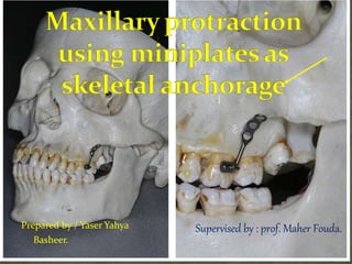
  • 1. Prepared by / Yaser Yahya Basheer. Supervised by : prof. Maher Fouda.
  • 2. The growth of the cranium and facial skeleton progressat different rates the face literally emerges from beneath the cranium.The upper face, under the influence of cranial base inclination, moves upwards and forwards; the lower face moves downwards and forwards on an 'expanding V". The nasomaxillary complexas it emerges frombeneath the cranium
  • 3. Since the maxillary complex is attached to the cranial base, there is a strong influence of the latter on the former. Although, there is no sharp line of demarcation between cranium and maxillary growth gradients, yet the position of the maxilla is dependent upon the' growth at sphenooccipital and sphenoethmoidal synchondroses. Hence, while discussing the growth of nasomaxillary complex, we have to look into two aspects. 1. The shift in the position of the maxillary complex, 2. The enlargement of the complex itself.
  • 4. The maxilla develops entirely by intramembranous ossification. Sutural connective tissue proliferations, ossification, surface apposition, resorption and translation are the mechanisms for maxillary growth.
  • 5. The maxilla is related to the cranium at least partially by the frontomaxillary suture, the zygomaticomaxillary suture, zygomaticotemporal suture and pterygopalatine suture. Weinmann and Sicher have pointed out that these sutures are all oblique and more or less parallel with each other .Thus, growth in these areas would serve to move the maxilla downward and forward (or the cranium upward and backward) .
  • 6. The placement of the various sutures
  • 7. To analyze the growth of the maxilla better, we must shift our focus to the functional matrices. It has been noted that the growth of the eyeball is essential for the development of the orbital cavity. Experimental evidence suggests that if there is no primordium for the eye, there is no orbit. It is clear that this functional matrix has a direct effect on the contiguous osseous structures. Also, just as the neurocranial bones are enclosed within a neurocrania I capsule, the facial bones are enclosed within the orofacial capsule. Resultantly the facial bones are passively carried outward (downward, forward, and laterally) by the primary expansion of the enclosed orofacial matrices (orbital, nasal, oral matrices).
  • 8. In addition there is an essential growth of the sinuses and spaces themselves, which perform important functions. The resultant maxillary changes would thus be secondary, compensatory and mechanically obligatory. In anteroposterior direction vector, the forward, passive motion of the maxilla is constantly being compensated for by the accretions at the maxillary tuberosity and at the palatal processes of both the maxillary and the palatine bones.
  • 9. Specifically mentioning, the vertical growth of the maxillary complex is due to the continued apposition of alveolar bone on the free borders of the alveolar process as the teeth erupt. As the maxilla descends, continued bony apposition occurs on the orbital floor, with concomitant resorption on the nasal floor and apposition of the bone on the inferior palatal surface. By the alternate process of bone deposition and resorption, the orbital and nasal floors and the palatine vault move downward in a parallel fashion.
  • 10. Transversely, additive growth on the free ends increases the distance between them. The buccal segments move downward and outward, as the maxilla itself is moving downward and forward, following the principle of expanding "V"
  • 11.
  • 12. Skeletal Class III malocclusion is one of the most difficult discrepancies to correct. Skeletal Class III anomalies are associated with maxillary retrusion, mandibular protrusion, or both .It has been found that 65% to 67% of all Class III malocclusions were characterized by maxillary deficiency .In subjects with maxillary deficiency where the mandible is not markedly affected, treatment may involve stimulation and guidance of maxillary growth by orthopaedic forces. Various types of extraoral appliances, such as facemasks and reverse pull headgears have been used to correct maxillary deficiency; however, there are problems with patient compliance due to their size and appearance.
  • 13. Facemask has been successfully used for the correction of Class III malocclusion since the late 1960s. The main effects of conventional facemask therapy are maxillary forward movement and sutural remodeling. Rapid maxillary expansion (RME) has been recommended for use in conjunction with facemask because it disrupts circummaxillary and intermaxillary sutures and facilitates the orthopedic effect of facemask. However, it has been reported that circummaxillary sutures may not be well disarticulated by use of RME alone and might be better managed by the use of alternate RME and constriction (Alt-RAMEC).
  • 14. When using tooth borne appliances in conjunction with facemask, some dental compensation (maxillary incisor proclination) is observed. The purpose of orthopedic treatment should be the achievement of skeletal changes rather than dental compensation. Thus, a more rigid anchorage is critical for a pure orthopedic-forward movement of the maxilla. Transferring the force to the maxilla with a rigid anchorage device has been attempted previously.
  • 15. Facemask treatment with miniplate anchorage has rarely been discussed. The literature concerning the facemask treatment anchored with miniplate comes in the form of case reports, except for one study with a small study was to evaluate the dentoskeletal and soft tissue effects of the Delaire-type facemask treatment anchored with miniplates in maxillary retrusion patients.
  • 16.
  • 17. An 11-year-old girl was referred with a complaint of‘‘small and separated teeth’’ and ‘‘lower jaw projection.’’ Medical history of the patient was noncontributory other than her parents were cousins. Furthermore, her elder brother presented with similar complaints of maxillary hypoplasia and hypodontia. Clinical and radiological examination revealed severe hypodontia and microdontia. Twenty-one of her permanent teeth were missing, whereas number 11, 21, 36, 46 existed in the dental arch and germs of the number 15, 37, and 47 could be detected on the panoramic radiograph.
  • 18.
  • 19. Furthermore, microdontia existed both in her primary and permanent dentition. The maxillary arch was deficient sagittally and transversally, so that there was an eight mm negative overjet and a bilateral buccal crossbite relationship with the lower jaw.
  • 20.
  • 21. A depression of the midfacial structures included the maxillary and infraorbital regions with a relative prominence of the mandible, inadequate projection of the nasal tip and an old face appearance with an unesthetic smile constituted general features of the patient . She also had nasal respiratory problems causing mouth opening during sleep.
  • 22. Lateral cephalometric (Figure 4) analysis indicated a Class III skeletal relationship because of retrusion of the maxilla. Cephalometric measurements indicated maxillary depth, 83° (normal, 90°; SD, 21.7); Nasion perpendicular to A, -7mm; facial depth, 95° (normal, 87°; SD, 0.8); SNA, 71°; SNB angle, 81°; ANB,-10°; and convexity, -13 mm. The patient had a facial axis of 96° (normal, 90°; SD, 21.7), lower facial height of 41° (normal, 45°; SD, 1.2), total facial height of 56° (normal, 60°; SD, 1.9), and mandibular plane to FH of 23° (normal, 25°; SD, 1.1). Soft tissue analysis indicated decreased nasolabial angle of 95°, lower lipto E-line of 0 mm and upper lip to E-line of –5 mm.
  • 23.
  • 24.  Coordination of dental arches both sagittally and transversally;  Improvement of facial esthetics by advancing middle face anteriorly;  Achievement of normal function both during mastication and respiration;  Restoration of the dentition with a removable denture as she loses her primary teeth;  Placement of dental implants after the cessation of skeletal growth.
  • 25. Three treatment options were considered for maxillary advancement. The first option was to delay treatment until growth has ceased and to correct the jaw relationship by orthognathic surgery. The second option was to apply rigid external distraction together with complete Le Fort I osteotomy. The third option was to try to take advantage of the sutural growth potential by applying extraoral force with a face mask via rigid skeletal anchors placed to the maxillary bone.
  • 26. Because all three treatment options included surgical intervention, patient and parents chose the minimally invasive third option. They were also informed about the fact that if the advancement with a face mask was insufficient to correct the discrepancy, the treatment plan could be switched to the second option.
  • 27. A titanium miniplate designed by Erverdi16 (MPI, Tasarımmed, Istanbul, Turkey) (Figure 5a) was used as a rigid skeletal anchor to attach the elastic orthopedic forces to the maxilla. Multipurpose miniplates were to be placed on both sides of the apertura piriformis and on the lateral nasal wall of maxilla. Rapid maxillary expansion was also planned to correct the transversal maxillary deficiency and to disturb the circummaxillary sutures. Because the maxillary dentition was insufficient, it was decided to place intraosseous titanium screws (two 3 eight mm IMF screws, Leibinger, Germany) (Figure 5b) on the palatal bone, near the alveolar crests, to provide anchorage for the expansion appliance.
  • 28.
  • 29. After routine surgical preparations, patient received general anesthesia. Bilateral mucosal incisions were made on labial sulcus between lateral incisor and first cuspid region. Then, mucosal flaps were carried inferiorly, the muscles and periosteum were incised and reflected superomedially, exposing the apertura piriformis and the lateral nasal wall of maxilla on both sides. Once an adequate space was achieved for miniplate placement, the nasal mucoperiosteum was elevated. Multipurpose miniplates were meticulously contoured to the bilateral lateral nasal wall, and straight extensions were bent to hook shape providing retention for face mask elastics and projected into the oral cavity through three mm mucoperiosteal incisions made inferiorly on the attached gingiva.). Multipurpose miniplate with intraoral extension bent to hook shape placed to the lateral nasal wall of the maxilla.
  • 30. Subsequently, for final stabilization of the bone plates three, 2.0 mm screws (five mm length) were placed with a 1.3 mm diameter drill under copious irrigation Simultaneously, four intraosseous bone screws were placed in the anterior and posterior palatal region, close to the alveolar crests, bilaterally .After soft tissue healing, orthopedic forces were applied. IMF screws placed in anterior and posterior palatal region.
  • 31. Impressions and stone casts were obtained with th IMF screws in place. The screws were blocked out with wax on the stone model, and an acrylic plate was prepared with an expansion screw in the midline. Appliance adaptation was checked intraorally and then connected to the IMF screw heads using cold curing methyl methacrylate–free acrylic resin (Ufi Gel hard, Voco GmbH, Cuxhaven, Germany). One of the parents was asked to activate the screw a quarter turn once a day (Figure 8)., Intraosseous screw–supported maxillary expansion appliance.
  • 32. An elastic force of approximately 150 g was applied bilaterally to the miniplate extensions after the adaptation of face mask (Leone spa, Firenze, Italy). After being sure of the stability, the force was increased gradually to 350 g. The direction of the force was adjusted approximately 308 to the occlusal plane, and the patient was asked to wear the face mask full time except during meals.
  • 33. The application of the orthopedic forces via elastics directly to the anterior part of the maxillary bone by using miniplate anchorage resulted in a remarkable improvement in the middle face (Figure 9). Together with the maxillary bone advancement, significant enhancement in the soft tissue profile revealed improved facial esthetics (Figure 10). The maxilla was expanded from the median palatal suture, and seven mm of expansion was achieved across the buccal segments. Coordination of the dental arches both in the sagittal and transversal planes created improved physiological functions.
  • 34.
  • 35.
  • 36. Pre- and posttreatment cephalometric radiographs were superimposed18 to demonstrate the skeletalchange (Figure 12). The maxilla was displaced eight mm anteriorly, the depth was increased by 9º, and Nasion perpendicular to A increased by eight mm. SNA increased by 7º and SNB decreased by 3º. The mandible rotated posteriorly, the facial axis decreased by 2º, and the facial depth decreased by 18. Lower facial height remained unchanged, and total facial height increased by 1º. The palatal plane angle rotated 2ºin acounterclockwise direction. PNS and ANS descended three and one mm, respectively. The soft tissue changes showed the nasal tip advanced five mm and the nasolabial angle increased 7º. The treatment results were achieved in 12 months.
  • 37. Pretreatment and posttreatment superimpositions (a) on Basion-Nasion at CC point demonstrating the overall skeletal change, (b) on Basion- Nasion at Nasion showing an eight mm maxillary advancement, and (c) on ANS-PNS at PNS indicating the dramatic sagittal growth of the palatal bone as a consequence of the treatment.
  • 38.
  • 39. In Class III malocclusion patients with mild to moderate maxillary hypoplasia, the protraction facemask has been used to stimulate sutural growth at the circum-maxillary suture sites in growing patients. To transmit the orthopedic force from the protraction facemask to the maxilla, intraoral devices such as a labiolingual arch, quad helix, and rapid maxillaryexpansion (RME) have been used.
  • 40. However, the use of the upper dentition as anchorage cannot avoid unwanted side effects such as labioversion of the upper incisors, extrusion of the upper molars, counterclockwise rotation of the upper occlusal plane, and eventual clockwise rotation of the mandible.3–6 Therefore, labial inclined maxillary incisors and/or a vertical facial growth pattern would be contraindications for facemask therapy with tooth-borne anchorage.
  • 41. To allow the direct transmission of orthopedic force to the circum-maxillary sutures, intentionally ankylosed primary canines, osseointegrated implants, and orthodontic miniscrews have been used as skeletal anchorage for protraction facemasks.7–11 Since surgical miniplates are a reliable means for applying orthodontic and orthopedic forces,12 Kircelli et al.,13 Cha et al.,14 and Kircelli and Pektas15 introduced the protraction facemask with miniplate anchorage (FM/MP) therapy to treat Class III malocclusion with maxillary hypoplasia and hypodontia.
  • 42. Comparison of pretreatment (left) and posttreatment (right) in patient with Class III malocclusion. (a) Facial photographs. (b) Intraoral photographs. (c) Lateral cephalograms. (d) Superimposition (solid line: pretreatment; dotted line: posttreatment).
  • 43. The protocol of FM/MP is as follows: (1) After an approximate 1–2 cm horizontal vestibular incision is made just below the zygomatic buttress area under local anesthesia, the zygomatic buttress is exposed with a subperiosteal flap. (2) Curvilinear type surgical miniplates (Martin, Tuttlinger, Germany) are bent according to the anatomical shape of the zygomatic buttress. (3) The distal end hole of the miniplate should be cut to make a hook for elastics.
  • 44. (4) After the miniplates are placed into the zygomatic buttress areas, three self-tapping type screws are used per side to fix the miniplates . (5) The distal end of the miniplate should be exposed through the attached gingiva between the upper canine and first premolar to control the vector of elastic traction. (6) Four weeks after placement of the miniplates, their mobility is checked and the orthopedic force (500 g per side, 30u downward and forward from the occlusal plane) is applied for 12 to 14 hours per day. (7) It is recommended to overcorrect the malocclusion into positive overjet and a slight Class II canine and molar relationship.
  • 45.
  • 46. Cleft patients often develop Class III malocclusion with maxillary hypoplasia and vertical facial growth pattern due to the combined effects of the congenital deformity itself and the scar tissues after surgical repair.16 These are contraindications for conventional facemask therapy. However, little research has been done on the use of FM/MP in cleft patients. Therefore, the purpose of this case report is to present three cleftpatients who were treated with FM/MP and to demonstrate the effect of FM/MP on maxillary hypoplasia in cleft patients.
  • 47. CASE 1: Skeletal Class III malocclusion with cleft palate (CP) and anterior open bite. Comparison of pretreatment (left) and posttreatment (right) in patient with cleft palate (case 1). (a) Facial photographs. (b) Intraoral photographs. (c) Lateral cephalograms. (d) Superimposition (solid line: pretreatment; dotted line: posttreatment).
  • 48. The patient was a 12 year 1 month old girl with CP only. She presented with concave facial profile, anterior crossbite (29 mm overjet), and anterior open bite (22 mm overbite). Cephalometric analysis showed skeletal Class III malocclusion with maxillary hypoplasia (ANB, -5.4; A to N perp, -3.4 mm), steep mandibular plane angle (FMA, 32.7u), and a skeletal age after the pubertal growth spurt according to the cervical vertebrae maturation index (CVMI, stage 4). Her condition was one of the contraindications for conventional facemask therapy.
  • 49. Although growth observation and reassessment after 2 years were proposed, her parents wanted to receive the FM/MP therapy. The possibility of orthognathic surgery after pubertal growth was explained.
  • 50. FM/MP therapy was started 4 weeks after placement of the miniplates according to the protocol. During protraction, the fixed appliances were placed to align the dentition.
  • 51. After 16 months of FM/MP therapy, there was significant forward movement of the point A (A to N perp, 5.6 mm). The ANB angle was changed from - 5.4° to 2.9°, and a Class II canine and molar relationship, normal overbite, and overjet were obtained. A slight counterclockwise rotation of the occlusal plane angle (-1.8) was interpreted to mean that there was almost no side effect such as extrusion of the upper molars. Although the FMA was increased 4.3, the anterior open bite was corrected by downward and forward movement of the maxilla. Slight labial tipping of the upper incisors (U1 to SN, 2.0°) occurred after correction of anterior crossbite and open bite.
  • 52. Skeletal Class III malocclusion with unilateral cleft lip and alveolus (UCLA) and vertical facial growth pattern.
  • 53. The patient was a 12 year 1 month old boy with UCLA on the left side. Although he presented with a straight facial profile, he had an anterior crossbite (22.5 mm overjet), upper anterior crowding, and peg laterals on the cleft side. Although the anteroposterior skeletal relationship (ANB, 1.4°) was within normal range and the upper and lower incisors were lingually inclined (U1 to SN, 95.1°; IMPA, 86.9°), a vertical facial growth pattern (FMA, 33.2°) existed. His skeletal age was before his pubertal growth spurt according to the CVMI (stage 3).
  • 54. Conventional facemask protraction with a toothborne anchorage device was not appropriate because the patient had a vertical facial growth pattern. Therefore, the FM/MP was used to avoid unwanted side effects.
  • 55. Initially, the fixed orthodontic appliance was placed to correct the anterior crowding in the upper arch. The FM/MP therapy was started 4 weeks after placement of the miniplates according to the protocol.
  • 56. After 24 months of FM/MP therapy, there was a 3.1- mm forward movement of point A (A to N perp). ANB angle was changed from 1.4° to 3.5 °, and a Class II canine and molar relationship was obtained. The finding that there was a negligible counterclockwise rotation of the mandibular plane (0.4 °) and occlusal plane angle (20.9°) indicated that there were almost no side effects such as extrusion of the upper molars and bite opening. Labial tipping of the upper incisors (U1 to SN, 4.9 °) occurred due to alignment.
  • 57. Skeletal Class III malocclusion with unilateral cleft lip and palate (UCLP) and vertical facial growth pattern Comparison of pretreatment (left) and posttreatment (right) in patient with unilateral cleft lip and palate (case 3). (a) Facial photographs. (b) Intraoral photographs. (c) Lateral cephalograms. (d) Superimposition (solid line: pretreatment; dotted line: posttreatment).
  • 58. The patient was a 7 year 2 month old boy with a UCLP on the right side. Although he presented with a straight facial profile, he had an anterior crossbite (22.7 mm overjet). Although the anteroposterior skeletal relationship (ANB, 1.6°) was within normal range and the upper incisors were lingually inclined (U1 to SN, 93.9°), a vertical facial growth pattern existed (FMA, 34.6°). His skeletal age was before his pubertal growth spurt according to CVMI (stage 2).
  • 59. Treatment Plan FM/MP was planned to maximize protraction of the maxilla and to avoid unwanted side effects. Treatment Progress FM/MP therapy was started 4 weeks after placement of the miniplates according to the protocol.
  • 60. Similar to Case 2, there was a 3.0-mm forward movement of point A (A to N perp) after 13 months of protraction facemask therapy. The ANB angle was changed from 1.6° to 3.1 °, and Class II canine and molar relationships were obtained. Although there was a slight counterclockwise rotation of the mandibular plane (20.9 °) and occlusal plane angle (22.5 °), there was no bite opening in the anterior teeth. Some labial tipping of the upper incisors (U1 to SN, 2.7 °) occurred after correction of the anterior crossbite.
  • 61.
  • 62. An 11-year five-month-old Chinese female was seen in the Department of Orthodontics School of Stomatology, Wuhan University, Wuhan, Hubei Province, China. Extraoral examination revealed a concave facial profile characterized by a maxillary retrusion with hypoplasia of the infraorbital region. Intraoral examination revealed an anterior crossbite with a reverse overjet of 23 mm (Figure 1). There was no anteroposterior centric relation discrepancy on closure. The transverse width was within normal limits with a dental crossbite in the premolar region.
  • 64. The maxillary midline was centered in the face and the mandibular midline was off two mm to the patient’s left side. The maxillary incisors were within normal limit (1 to palatal plane = 110°), and the mandibular incisors were slightly retroclined (l to mandibular plane = 89°). The molar relationship was half-step Class III. There was crowding in both the maxillary and mandibular arches with a blocked-out maxillary right canine. Temporomandibular joint function was normal. There was no pain on palpation, no clicking, popping, or crepitus noise, and a normal range of motion.
  • 65. Cephalometric analysis indicated a mild skeletal Class III pattern due to a retrusive maxilla (SNA=76°, SNB = 78.2°, ANB =22.2°). The occlusal plane was tilted 7° to SN and the mandibular plane was 36.8° to SN . Pretreatment lateral cephalometric radiograph.
  • 66. The patient was concerned about her dental and facial esthetics. Two treatment plans were discussed with the patient and her parents. The first option was to delay treatment until growth was completed and then use orthodontic treatment in combination with orthognathic surgery to advance the maxilla. The second option was to use a facemask combined with orthodontic treatment to correct the anterior crossbite and improve facial esthetics. The patient chose to proceed with the latter because of a desire to improve her dentofacial appearance. The patient was informed that although facemask treatment may be able to correct the anterior crossbite,
  • 67. it did not eliminate the possibility that orthognathic surgery may eventually be needed to correct the jaw discrepancy. Because of the severe arch length deficiency in the maxillary arch, the use of an onplant as anchorage for maxillary protraction was suggested. The patient agreed to the placement of an onplant and treatment with maxillary protraction for approximately 12 months followed by fixed orthodontic appliances to eliminate crowding of the dentition.
  • 68. A 7.7-mm hexagonal onplant (Nobel Biocare, Gotenberg, Sweden) was surgically placed on the flat part of the palatal bone near the maxillary molar region. An incision was made in the palatal mucosa from the premolar area toward the midline. The tissue was tunneled under in full-thickness fashion, past the midline to the eventual implantation site. The onplant was then slipped under the soft tissue and brought into position, and the incision was sutured. The onplant was placed as close to the midline as possible but not on the midsagittal maxillary suture, so as not to disturb lateral growth with the surgical operation. A vacuformed stent was worn for 10 days to place pressure on the onplant. This step was crucial to minimize the movement of onplant during osseointegration, which took approximately three to four months.
  • 69. Once integration was achieved, the onplant was exposed, its cover screw removed, and an open-tray impression was taken. A simulated implant was used in pouring the working cast. A transpalatal arch was attached to the onplant and soldered to a silver cast splint, which was connected to all the maxillary teeth . Placement of onplant and cast splint as anchorage for maxillary protraction.
  • 70. Maxillary protraction was started four months after placement of the onplant. A Petit facemask (Ormco Corporation, Glendora, Calif) was fitted with elastics that delivered approximately 400 g of force on each side (Figure 3B). The force was directed from the canine area, 30° from the occlusal plane, to counteract the anticlockwise rotation of the palatal plane. Patient was instructed to wear the facemask for 12 hours per day. Traction was continued for 12 months until sufficient clinical movement of the maxilla had been achieved to improve the midface esthetics. Protraction facemask with elastics pulling 30°forward and downward from the occlusal plane.
  • 71. At the end of the protraction period, the onplant was removed using a surgical elevator. All surgical procedures were carried out under local anesthesia. The peri-onplant soft tissue healed uneventfully within two weeks. The patient’s acceptance regarding surgical effects was positive, and postoperative pain and discomfort symptoms were negligible.
  • 72. The application of an anteriorly directed force from a facemask to an osseointegrated onplant placed in the palatal bone resulted in a significant improvement in midface esthetics .This was noted by an increase in fullness of the infraorbital region and the correction of the skeletal discrepancy between the maxillary and mandibular jaw relationship (ANB from -2.2° to 3.7 °, Wits from -6.1 mm to -1.0 mm). Pre- and post-treatment cephalometric radiographs were superimposed on the anterior cranial base structures at Sella as described by Bjork and Skieller22 to demonstrate the skeletal changes .
  • 73. Twelve-month progress extra-oral and intra-oral photographs showing improvement in midface profile and overjet.
  • 74.
  • 75.
  • 76. To verify whether the onplant was stable during application of orthopedic force, superimposition was also performed on the lingual contour of the maxilla along ANS PNS to detect any movement of the onplant.
  • 77. The results showed no movement of the onplant during both the six-month and 12-month period of protraction. The onplant was then used as an internal reference point to measure spatial movement of the maxilla, which was found to have been displaced 2.9 mm horizontally and 2.9 mm vertically during the 12-month period of protraction. This movement is more than the average horizontal movement of two mm reported by other investigators using toothborne appliances as anchorage for a facemask alone or in combination with expansion techniques.
  • 78. It has been shown that transverse expansion of the maxilla may result in an anterior movement of point A, and the whole maxillary complex may be movable up to seven to eight years of age.6,23 Because the maxilla articulates with nine other bones of the craniofacial complex, palatal expansion may disarticulate the maxilla and initiate cellular response in the sutures, allowing a more positive reaction to protraction forces.24 It is therefore of interest to note that in the present case report, no transverse expansion was necessary to obtain a similar amount of displacement of the maxilla.
  • 79. Superimposition of the pre- and post-treatment cephalometric radiographs on the lingual contour of the maxilla revealed no mesial movement of the maxillary molars. This is in contrast to maxillary protraction with tooth-borne appliances that usually results in mesial movement of maxillary molars and the loss of arch length.1–6 The maxillary palatal plane angle was tipped 2.48 in an anticlockwise direction despite the 308 downward and forward pull of the facemask to counteract such a rotational effect. This is acceptable when protraction is carried out along the occlusal plane below the center of resistance of the maxilla.
  • 80. Long-term studies with protraction facemask have shown that this effect was transient with the palatal plane and returns to normal inclination four years after completion of maxillary protraction.
  • 81. The occlusal plane angle was increased 11.8°. Extrusion of the maxillary molars was not observed. This differs from facemask treatment with tooth-borne appliances where a change in the occlusal plane is a combination of downward movement of the posterior nasal spine and extrusion of maxillary molars. The 3°increase in the SN-mandibular plane angle and an overall increase in the lower face height by 3.6 mm are similar to those reported by other investigatorsand is caused by the downward movement of the maxilla and downward and backward rotation of the mandible. These transient changes are favorable for dental correction in brachyfacial individuals with deep overbite. Long-term studies showed a return of the mandibular plane angle to pretreatment level four years after protraction facemask treatment.
  • 82. Long-term stability of early facemask treatment depends on the ability of treatment changes to compensate for subsequent growth, which is usually unfavorable. Elimination of dental changes such as extrusion and mesial movements of upper molars by using onplant as anchorage may be helpful in improving the stability of facemask treatment. Another advantage of using onplant as compared with implants as anchorage is that onplant can be placed anywhere on the anterior part of the hard palate. In contrast, placement of implants in this area creates a potential risk of damage to the roots of adjacent teeth because of the length of the implant fixtures available. Placement of an implant on the anterior part of the hard palate would not only have a risk of root damage of anterior teeth but also the risk of penetration of the nasal floor.
  • 83. Clinicians should be aware that complications such as soft tissue irritation can develop after placement of an onplant. This irritation usually subsides over a two- month period Other possible problems that could arise include the failure of integration and loosening of the onplant because of torsional force.
  • 84.
  • 86. The patient, an 8-year-old girl, came to the Kangnung National University Orthodontic Clinic in Gangneung, South Korea, with a chief concern of “my bite is not right.” Clinically, she had a concave facial profile, and acute nasolabial angle, and a protrusive mandible.
  • 87. Intraorally, she had an anterior crossbite and a low anterior tongue posture. The maxillary right first deciduous molar and left second deciduous molar had exfoliated prematurely, and midarch crowding was noted on the dental casts and panoramic radiograph. The cephalometric radiograph and tracing showed a skeletal Class III malocclusion with maxillary deficiency, mandibular prognathism (ANB, –2.2), and a normal mandibular plane angle (FMA, 23). The maxillary incisors were proclined (U1 to FH, 109), and the mandibular incisors were retroclined (IMPA, 86), compensating for the skeletal malocclusion (Figs 2 and 3, Table). There was no family history of mandibular prognathism.
  • 88.
  • 89.
  • 90.
  • 91. In determining our treatment objectives, we askedthe patient whether she was willing to undergo a surgical operation. She was willing if necessary. For that reason, our treatment consisted of phase 1 orthopedic treatment to protract the maxilla with a skeletal anchorage system. Surgical miniplates were used as anchorage instead of the conventional tooth-borne appliances to prevent possible mesial movement of the posterior dentition.
  • 92. The objective of this early phase of treatment was to induce harmonious growth of the maxilla with improvement in facial esthetics. Overcorrection of the maxilla to an overjet of 3 to 4 mm was desirable to anticipate excessive growth of the mandible during the pubertal growth spurt. The patient was followed for a period of time to determine whether the malocclusion could be camouflaged by orthodontic tooth movement.
  • 93. The phase 2 treatment was initiated at 11 years of age for 18 months to correct the remaining crowding, overjet, and overbite problems. The patient was placed in retention for 27 months after fixed appliance therapy to determine the stability of treatment without orthognathic surgery.
  • 94. Based on the objectives, 2 treatment options were proposed. The first option was an early phase of orthopedic treatment to induce harmonious skeletal growth and improve facial esthetics followed by phase 2 treatment to correct the remaining crowding, overjet, and overbite problems. This option would not eliminate the necessity for orthognathic surgery. The patient would be followed to determine the stability of treatment. The second option would be to wait until all growth was completed and determine whether the malocclusion could be camouflaged by orthodontic treatment or a combination of surgical and orthodontic treatment.
  • 95. Phase 1 treatment was started at age 8 years 4 months with a maxillary removable appliance to regain space lost from the early loss of the deciduous molars (Fig 4). After 6 months of observation, a surgical miniplate was placed. Local infiltration anesthesia was administered to the maxillary left and right buccalvestibular areas after surgical disinfection. A vestibular incision around the canine area was performed. After an atraumatic subperiosteal dissection to the infrazygomatic crest, a curvilinear miniplate was adapted, bent to the zygomatic buttress’s bony surface, and fixated with 3 self-tapping miniscrews per side (Fig 5, A).
  • 96. Phase 1 treatment included a maxillary removable appliance to regain space lost by early loss of the deciduous molars.
  • 97. From our experience, at least 3 to 4 screws should be placed to resist the maxillary protraction force of about 300 to 400 cN per side. Screw placement should be in a posteriorsuperior direction to prevent damage to the premolar tooth follicles (Fig 5, B). The end of the miniplate entered the oral cavity between the canine and first premolar area in the keratinized attached gingiva to prevent gingival irritation. The oral portion of the miniplate was modified into a hook for elastic traction.
  • 98. A, After atraumatic subperiosteal dissection to the infrazygomatic crest, a curvilinear miniplate was adapted, bent to the zygomatic buttress bony surface, and fixated with 3 self-tapping miniscrews; B, screws should be placed in a posterior-superior direction to prevent damage to the premolar tooth follicles. Cha et al 103 American
  • 99. Maxillary protraction was started 2 weeks after placement of the miniplates, with a force of 300cN per side applied 12 to 14 hours per day (Fig 6). Within 10 months of treatment, a three quarters premolar width Class II molar relationship was established. Thereafter, the patient’s wearing of protraction headgear was limited to nighttime only as a retainer for 10 months. The plates were removed after the facemask treatment. A mucoperiosteal incision and a subperiosteal dissection were performed to expose the miniplate. The monocortical screws were removed first, and the miniplate was then detached because often new bone is deposited next to the plate. The surgical site was then closed and sutured.
  • 100. Maxillary protraction was started 2 weeks after placement of the miniplates.
  • 101. Progress records taken at age 10 years 7 months showed favorable growth between the maxilla and the mandible, and the malocclusion could be camouflaged by orthodontic treatment. The patient was treated with fixed appliances for 18 months to establish a good molar relationship and correct the midline discrepancy. A maxillary circumferential retainer and a mandibular lingual fixed retainer were placed after appliance removal. The patient was instructed to wear the retainer at night for 10 to 12 hours.
  • 102. Figures 7 through 9 show the results 14 months after protraction headgear treatment. The malocclusion was overcorrected to a Class II molar relationship to compensate for future excessive mandibular growth. Superimposition of pretreatment and posttreatment cephalometric tracings showed 8.1 mm of forward movement of A-point (A-point to NtFH) and 3.3 of counterclockwise tipping of the palatal plane (Fig 10). The ANB angle changed from –2.2 to 1 6.7. The SNO, or angle between the anterior cranial base and orbitale, changed from 63 to 70. Labial tipping of the maxillary incisors and lingual tipping of the mandibular incisors, which are typically observed after tooth-borne protraction, were not seen with the miniplates.
  • 105. Figures 11 through 14 show the results after phase 2 fixed appliance treatment at age 12 years 6 months. The ANB angle was reduced from 6.7 to 3.9, indicating normalization of the jaw relationship after overcorrection in the phase 1 treatment. Class I canine and molar relationships were obtained, and overjet and overbite were returned to normal after phase 2 treatment. Superimposition of the postprotraction and posttreatment radiographs showed differential forward growth of the maxilla and the mandible, and compensation of the incisors to the skeletal growth (Fig 15).
  • 106.
  • 107. Intraoral photographs near the end of phase 2 fixed appliance treatment.
  • 108. Posttreatment radiograph and tracing after phase 2 fixed appliance treatment.
  • 109. Superimposition of postprotraction and posttreatment cephalometric tracings.
  • 110. Figures 16 and 17 show the patient at age 14 years 9 months, 27 months after the removal of the orthodontic appliances. During the retention period, the maxilla and the mandible showed relatively harmonious growth, maintaining an ANB difference of 3. The angle of convexity was reduced from 7.4 to 5.4. Superimposition of the posttreatment and postretention cephalometric tracings (Fig 18) showed continuous dental compensation to the skeletal discrepancy was observed with proclination of the maxillary incisors and slight retroclination of the mandibular incisors. Despite the mild lip protrusion and 0.5 mm of midline deviation, the patient was pleased with the final results without orthognathic surgery
  • 112. Postretention radiograph and cephalometric tracing 27 months after appliance removal.
  • 113. Superimposition of posttreatment and postretention cephalometric tracings.
  • 114.
  • 115.
  • 116. A 12-year-old girl presented with a skeletal Class III malocclusion, a concave facial profile due to a combination of a retruded maxilla and protruded mandible and a 3-mm functional deviation of the mandibular midline to the right . Intraoral examination revealed a 1-mm reverse overjet, Class III molar relationship and congenital absence of the mandibular right and left second premolars .
  • 117. Cephalometric analysis showed an ANB angle of -0.6°, an angle of convexity of - 0.8°, a decrease in maxillary length (79.6 mm) and an increase in mandibular length (114.1 mm). Vertically, the patient presented with an increase in the mandibular plane angle (MP/FH=38.6°). Dentally, the incisors were compensated to the skeletal malocclusion. The maxillary incisors to the Frankfurt plane (U1/FH) angle was 112° and the mandibular incisors to the mandibular plane (IMPA) angle was 83° (Table). There was no family history of Class III malocclusion.
  • 118.
  • 119. Based on the above diagnosis, the treatment objective was to normalize the skeletal discrepancy, correct the reverse overjet, establish a functional occlusion, prepare the patient for prosthetic replacement of the congenital missing mandibular premolars, and improve facial esthetics.
  • 120. The treatment plan for this patient included protraction of the maxilla, comprehensive orthodontic treatment with a fixed appliance, and eventually prosthetic treatment for the congenitally absent mandibular right and left second premolars. After due consideration of the subject’s age and the patency of the circum-maxillary sutures, the patient was informed that maxillary protraction in conjunction with a tooth-borne anchorage device such as a maxillary expansion appliance might result in significant dental side effects rather than skeletal advancement of the maxilla.
  • 121. The patient consented to the placement of surgical miniplates as skeletal anchorage to minimize forward movement of the maxillary molars, maxillary incisors and extrusion of the molars. It was also decided that the mandibular second deciduous molars be retained during the maxillary protraction phase of treatment to prevent alveolar atrophy. Thus, the implants were to be placed so as to replace the missing second premolar after all growth was complete.
  • 122. An alternative treatment plan offered to the patient included removal of two lower retained second deciduous molars and closure of the space by losing anchorage from behind (with/without extraction of premolars in the upper arch). This option could reduce the complexity and duration of the treatment, and eliminate implant replacement for the congenitally missing premolars.
  • 123. After local infiltration anesthesia and surgical disinfection, a vestibular incision around the canine area was performed. After performing an atraumatic subperiosteal dissection of the infrazygomatic crest area, curvilinear- type surgical miniplates (OsteoMed Corp., Addison [TX], USA) were fixated with three self-tapping miniscrews on both sides of the infrazygomatic crest area. The end of the miniplate was exposed between the canine and the first premolar area and modified as a hook . Protraction headgear therapy using surgical miniplates
  • 124. Maxillary protraction was started 3 weeks later with a force of 300 cN to each side, and applied 12 to 14 hours per day. After 7.5 months of protraction therapy, an improvement of facial esthetics and occlusion was noted .The functional deviation of the mandible was eliminated. The dental midline was coincident after correction of the anterior occlusal interferences.
  • 125. Analysis of the post-treatment cephalometric radiograph showed that the position of the maxilla (SNA) had improved from 75.7° to 77.8°. The position of the mandible (SNB) had decreased from 76.2° to 73.6°. The angle of convexity was improved from -0.8° to 8.5°. The mandibular plane angle increased from 38.6° to 40.9°, and the maxillary incisors proclined from 112.0° to 113.7° .Superimposition of the pre- and post-facemask treatment radiographs showed a forward and downward movement of the maxilla, which is usually only obtained with protraction headgear therapy in a much younger age-group. Yet there was relatively little dental tooth movement
  • 126.
  • 127. Comprehensive orthodontic treatment with a fixed appliance was initiated immediately after completion of the facemask treatment. The protraction headgear was used at night for 2 months during this second phase of treatment, so as to maintain the forward position of the maxilla. After 12 months of fixed appliance treatment, the facial esthetics had improved and a functional occlusion was established Facial and intraoral photographs of the patient (at 14 years and 2 months) after second-phase treatment
  • 128. Moreover, the overbite and overjet were improved and the midlines were coincident. The spaces for the congenitally absent mandibular second premolars were maintained and patient was referred to the Department of Prosthetics for placement of implants. Superimposition of the pre– and post–second-phase treatment cephalometric radiographs revealed that the skeletal change during this comprehensive protraction facemask therapy was stable Facial and intraoral photographs of the patients (at 16 years and 8 months) after 30 months of retention and superimpositions of cephalometric tracings after second-phase treatment and after 30 months of retention
  • 130. Location of the miniplate relative to the zygomaticomaxillary suture. Note that this figure is only for visualization purposes as this is a skull of an adult, and the anatomic contour of the zygomatic process of the maxilla changes with age
  • 131.
  • 132. Three girls (aged 10 to 11 years) presenting with a severe skeletal Class III relationship with a maxillary deficiency and concave soft tissue profile were treated according to the same treatment plan (Figs 1A, 2A, 3A). Two of them had an anterior crossbite without anterior shift of the mandible (cases 2 and 3). One had an edge-to-edge incisor occlusion in centric relation, with a forward posture into maximum intercuspation (case 1)
  • 133. Pretreatment cephalometric evaluation of the 3 cases showed a skeletal Class III relationship with hypoplasia of the maxilla combined with a normal or increased mandibular size and normal or slightly decreased vertical dimensions (Table 1). The patients’ upper incisors were proclined or retroclined, and the lower incisors were normal or proclined.
  • 134.
  • 135.
  • 136.
  • 137.
  • 138. The main treatment objective was to achieve a reduction of the facial concavity, maximize skeletal maxillary changes, and minimize dentoalveolar movement.
  • 139. The 3 patients were treated exclusively by intermaxillary traction between miniplates placed in the maxilla and in the mandible, in combination with a bite plane to jump the crossbite.
  • 140. The skeletal deformity of these patients was judged too severe to consider treatment by dentoalveolar compensation alone, and the degree of maxillary hypoplasia and age of the patients were not favorable for facemask therapy. Orthognathic surgery after growth completion was offered to the patients. However, to avoid retaining such severe facial deformity until adulthood, each of the 3 patients and their parents preferred to try orthopedic traction from skeletal anchorage, even though they had been informed about the possible need for future orthognathic surgery.
  • 141. Four orthodontic miniplates (Bollard; Tita-Link, Brussels, Belgium) were inserted into the infrazygomatic crests and between the canine and lateral incisor (cases 1 and 2) or between the canine and first premolar (case 3) in the mandible, on both the right and left sides (Fig 3C). Surgery was performed with patients under general anesthesia (cases 1 and 2) or local anesthesia (case 3). The miniplates were fixed to the bone with 2 or 3 titanium screws (2.3 mm in diameter and 5 mm in length) after predrilling with a 1.6-mm-diameter bur, as previously described.
  • 142. Three weeks after surgery, maxillomandibular elastics were attached between the upper and lower miniplates on each side, applying a force of 100 g per side (Fig 3D). The patients were asked to replace the elastics once a day and to wear them 24 hours per day. After 1 month (case 1) or 2 months (cases 2 and 3), a removable bite plane was placed to eliminate the occlusal interference in the incisor region (Fig 3D). At this time, the elastic force was increased to 200 g per side.
  • 143. After 7 months (cases 1 and 2) or 12 months (case 3) of orthopedic traction, the bite plane was removed. The traction was maintained full time for a total period of 12 months (cases 1 and 2) or 16 months (case 3). No local infections were observed around any of the miniplates. They remained stable throughout treatment. During the follow-up period after the active treatment, the patients wore the elastics at night for retention.
  • 144. The anterior crossbite was corrected in each patient (Figs 1B, 2B, 3B). Their soft tissue profiles considerably improved, with anterior displacement of the whole midfacen(infraorbital ridge, nose, and upper lip), reducing the paranasal concavity. Almost no anterior displacement of the lower lip and chin was observed at the end of the traction,nleading to an improvement of the relationship between the upper and lower lip. The tip of the nose moved slightly upward.
  • 145.
  • 146.
  • 147.
  • 148.
  • 149.
  • 150. Cephalometric evaluation between the beginning of treatment and end of treatment showed marked increase of ANB, Wits, and facial convexity (G′-Sn-Pg′) values in all 3 cases(Table 1). No rotation of the mandible was observed in cases 1 and 3, whereas a slightclockwise rotation was seen in case 2; there was a slight counterclockwise rotation of the maxilla in all patients. No major changes occurred in the upper incisor inclination, whereas the lower incisors were proclined. During the follow-up period (from end of treatment to 11 to 38 months later), the Class III correction was maintained.
  • 151.
  • 152. Figure 5 shows the cone-beam computed tomography scans from case 1 superimposed on the anterior cranial base. The post- treatment scan is a semitransparent overlay.9 The maxilla and the infraorbital border moved forward, whereas the horizontal growth of the mandiblewas restricted.
  • 153. A and B, Visualization of treatment changes as shown by 3-dimensional surface models from cone-beam computed tomography scans registered on anterior cranial base. Initial models are shown in red, and end-of- treatment models are shown as semitransparent mesh.
  • 154.
  • 155.
  • 156. Skeletal Class III malocclusion is one of the most difficult discrepancies to correct. Skeletal Class III anomalies are associated with maxillary retrusion, mandibular protrusion, or both .It has been found that 65% to 67% of all Class III malocclusions were characterized by maxillary deficiency .In subjects with maxillary deficiency where the mandible is not markedly affected, treatment may involve stimulation and guidance of maxillary growth by orthopaedic forces. Various types of extraoral appliances, such as facemasks and reverse pull headgears have been used to correct maxillary deficiency; however, there are problems with patient compliance due to their size and appearance.
  • 157. Dental implants, miniplates, and modified fixation screws provide bone anchorage in orthodontic treatment.Miniscrews (mini-implants) have also become popular because they are easier to both insert and remove . In this case report, two miniplates were inserted in connected to a removable appliance in the upper jaw by use of elastics in order to correct maxillary deficiency.the anterior part of the mandible in the canine areas andconnected to a removable appliance in the upper jaw by use of elastics in order to correct maxillary deficiency.
  • 158. The patient was an 11-year-old boy who was referred for treatment of maxillary deficiency. He had no medical problems, and there were no signs of temporomandibular joint dysfunction. The patient had a skeletal Class III malocclusion and maxillary deficiency. His parents had no Class III characteristics.
  • 159. The facial photographs showed a Class III appearance with a concave profile because of maxillary deficiency. The pretreatment intraoral photographs and dental casts showed Class III relationship of the central incisors and anterior crossbite. The patient had a Class III molar relationship on the right and Class I on the left side (Figures 1 and 2). Cephalometric analysis confirmed the Class III skeletal pattern (Table 1) (Figure 3).
  • 160.
  • 161.
  • 162.
  • 163. The treatment objectives for this patient were to (1) correct the deficient maxillary arch, ideally by forward positioning of the maxilla; (2) obtain an ideal overjet and overbite; (3) correct the anterior crossbites
  • 164. Extraoral appliances, such as protraction facemask, Class III functional appliance, any modified maxillary protraction devices, and orthognathic surgery, were considered as alternative treatments for the correction of this Class III malocclusion. However, the patient refused the use of extraoral appliances and major surgery. Therefore, in this case, it was decided to use miniplates to protract the maxillaby application of Class III elastics.
  • 165. Plates for Orthodontic Anchorage were placed under local anaesthesia in the canine areas of the mandible by a maxillofacial surgeon. The ideal position for miniplates insertion was evaluated by using a panoramic radiograph in order to avoid damage to the roots of the adjacent teeth and mental foramen.
  • 166. A tightly fitting and well-retained upper removable appliance was fabricated with two Adams clasps on the upper first permanent molars. Each of the Adams clasps had a loop which was used for retaining the elastics. A labial bow was also used on the anterior teeth for retention. A maxillary posterior bite plate was used to disclude the upper and lower jaws. Removable appliance in the upper jaw.
  • 167. Orthodontic latex elastics (3/16 heavy size—Unitek Elastics) were connected from the hooks of the miniplates to the Adams clasps of the removable appliance to generate approximately 500 g of anterior retraction. The patient was instructed to wear the appliance full-time except for eating, contact sports, and tooth brushing; he was also told to change the elastics every day. In order to retain these elastics, the Adams clasps on the molars were bent to form loops
  • 168. After 10 months of active treatment a positive overjet and Class I buccal segments were achieved and the anterior crossbite was corrected (Figures 5 and 6). The posttreatment cephalometric radiograph tracing showed a favourable increase of 5.1◦ and 4.4◦ in the SNA and ANB angles, The pre- and posttreatment cephalometric problem caused by extraoral appliances is that they can cause skin abrasions on the chin especially in hot climates. Therefore patients may simply refrain from wearing the appliance, and the lack of cooperation might lead to an unsatisfactory result. respectively
  • 169.
  • 170. One of the disadvantages of extraoral appliances is that, when extraoral force is applied against the chin, it is difficult to avoid tipping the lower incisors lingually. In other words, use of a chin cup can lead to lingual tipping of the lower incisors as a result of the pressure of the chin cup component on the lower lip and dentition .In most cases, lingual tipping is an undesirable side effect and can cause crowding .In a case report miniscrews have been used for treatment of maxillary deficiency.
  • 171. One of the limitations of miniscrew is their loosening, which can be distressing for the clinician and the patient. In order to overcome this problem, wider diameter and deeper insertion of miniscrews must be used. De Clerck et al. used the miniplates to protract the maxilla however; the design of current case report is different from that study. In a recent study bone-anchored maxillary protraction (BAMP) with miniplates was used in patients with Class III malocclusion, and significant improvements of over jet and molar relationship were recorded .
  • 172. In this case report, minor surgery and miniplates were used to overcome these various problems. As undertaken in this case, applying a force to the teeth in order to correct the skeletal discrepancy will inevitably result in toothmovement; therefore, a full coverage upper removable appliance was used to cover all the maxillary dentition. The treatment process lasted for 10 months. However, since the patient was only 11 years old and still had considerable residual growth, treatment was continued by fixed appliance.
  • 173. The forces generated by elastics may be divided into two components. One force component is in a horizontal direction, moving the maxilla forwards, which is favourable in maxillary deficiency cases. The second component is in a vertical direction, moving the posterior maxillary dentition downwards. This might lead to unfavourable tooth movements in high angle cases, but it is not a problem in patients with a lowor average face height. Maxillary posterior bite plate can overcome this problem in high angle cases by decreasing facial height.
  • 174. This case report demonstrates a different method of using miniplates to treat an 11-year-old boy with a skeletal Class III malocclusion and maxillary deficiency. This treatment was found to be an acceptable alternative to the use of extraoral appliances such as facemasks and major surgery.
  • 175.
  • 176.
  • 177. An 11-year seven-month-old Chinese boy in late mixed dentition was seen in the Department of Orthodontics, School of Stomatology, Peking University, Beijing, China. Extraoral examination revealed that the maxilla was retrognathic and the mandible was slightly prognathic. Intraoral examination revealed an anterior and posterior crossbite with a reverse overjet of 4 mm (Figs. 1 and 2). The molars were in class III relationship on both sides. There was no crowding in both maxillary and mandibular arches. Temporomandibular joint function was normal. CASE REPORT
  • 178. Cephalometric analysis indicated a moderate skeletal class III malocclusion due to both maxillary retrusion and mandibular protrusion. The mandibular plane was tilted 30.72° to S-N plane.
  • 179.
  • 180. The patient and parents were concerned about dentofacial appearance. Treatment option was to use a facemask combined with fixed appliance to correct the anterior and posterior crossbite and improve facial esthetics. The patient and parents were informed that the treatment plan did not eliminate the possibility of orthognathic surgery later. The unfavorable growth of the jaws during or after treatment might necessitate a surgical treatment plan. The use of miniplate implant as anchorage for maxillary protraction was suggested and the patient and parents agreed.
  • 181. Miniplate implant placement Titanium miniplates were implanted by an experienced oral surgeon. After mouth rinsing for 3 minutes with 0.2% chlorhexidine gluconate, under local anesthesia, a mucoperiosteal incision was made at the labial vestibule between the upper lateral incisors and canines on both sides. The mucoperiosteal flap was then elevated, and the surface of the cortical bone at the apical region of lateral incisors and canines was exposed.
  • 182. An appropriate length of I-shaped miniplate (plate thickness 1 mm, 3 holes) was selected and fixed in position with self-tapping screws (diameter 2 mm, length 7 mm), with the head exposed to the oral cavity from the incised wound. Care was taken toadjust the angle between the head and the body of the plates so that the head portion would not apply pressure on the attached gingiva. The incisions were finally closed and sutured with absorbable thread around the miniplate. This patient showed mild facial swelling for a week after the operation. It was necessary to take antibiotics and brush carefully. Miniplates were placed on the lateral nasal wall of maxilla.
  • 183. A month was allowed for healing before application of force to the miniplates. After the month, the clinical evaluation for the patient included an assessment of platemobility and infection. If nothing was abnormal, maxillary protraction was started. First, thread a segment of brass wire through the hole in the head of miniplate. Second, a ganoid composite resin ball (diameter 2−3 mm) was made at top of brass wire. The ball was used for protraction hook.
  • 184. A protraction force of 450 g per side at first, 500−600 g per side after one month with an anteroinferior force vector 30° to the occlusal plane, was applied from the composite ball in anchor miniplates to the facemask by using elastic modules. The patient was instructed to wear facemask at least 10 hours a day and prolong the wearing time as much as possible. Traction was continued for 6 months until enough forward movement of the maxilla had been achieved to improve the midface esthetics. After maxillary protraction, the miniplate implant was removed under local anesthesia, then fixed appliance was bonded.
  • 185. The application of protraction force from a facemask to a miniplate implant placed in the anterior maxilla resulted in a significant improvement in facial esthetics (Fig. 4) and the maxillo-mandibular jaw relation (Figs. 5 and 6). SNA angel was changed from 79.89° to 82.53°, SNB angle from 84.16° to 81.67°, ANB angle from −4.27° to 0.86°, Wits from −10.18 mm to −2.66 mm, A-NP distance increased by 5.48 mm, mandibular plane angle increased by 2°, while the change of U1/SN was not significant.
  • 186. Post-treatment extraoral frontal (A) and lateral (B) view photographs.
  • 187. Post-treatment intraoral photographs. A: Occlusal view of maxillary arch; B: Normal overjet; C: Occusal view of mandibular arch; D: Class Ⅰ molar relationship on right side; E: Normal overbite; F: Class Ⅰ molar relationship on left side.
  • 188. Pretreatment (A) and post-treatment (B) lateral cephalometric radiograph.
  • 189. THANX