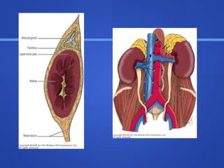
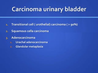
Urological
Malignancies
DR SANTOSH AGRAWAL
Chief Urologist and KidneyTransplant Surgeon
Bansal Hospital , Bhopal, MP
MBBS, MS, MCh (PGI, Chandigarh), FIAGES, FALS
Fellow in Robotic Surgery, Roswell Park Cancer Institute, Buffalo , USA
 Kidney mass/ renal mass
 Carcinoma urinary Bladder
 Prostate cancer
 Penile cancer
 Testicular cancer
Renal mass (neoplasm)
Benign
 Adenoma
 Angiomyolipoma
( AML)
 Oncocytoma
 Simple cyst
 Fibroma
 angioma
Malignent
 Renal cell carcinoma
(RCC)
 Urothelial carcimona
(TCC- transitional cell
carcinoma of renal pelvis)
 Kidney sarcoma
 Squamous cell carcinoma
 wilm’s tumor
Renal cell carcinoma
1. Clear cell carcinoma ( conventional type) – most common
( 70- 80%)
2. Chromophobic carcinoma ( 10-15%)
3. Chromophilic (papillary) 3-5 %
4. Collecting duct carcinoma(1%)
Clinical features
 Asymptomatic
 Hematuria
 Flank mass
 Flank pain
 Dragging sensation in flankm
 Rapidly devoloping left varicocele
 Metastatic symptoms
 Paraneoplastic symptoms/consitutional symtoms
 Fever
 Polycythemia ( increased erythropoetin production)
 Raised ESR
 Hypertension ( increased renin production)
 Hypercalcemia ( increased PTH like substance)
 Stauffer’s syndrome- nonmetastatic hepatic dysfunction (
raised ALP, SGOT/PT, prothombin time)
Investigation
 Ultrasonography
 Contrast enhanced CECT scan of Abdomen± chest
 CXR –PA veiw
 CBC
 Urea/creatinine
 LFT
 Serum calcium
Staging
 Robson’s staging
TNM staging
 T staging-
 T1: tumor < 7 cms , confined to kidney
 T2 : tumor > 7 cms confined to kidney
 T3 : perinephric fat invasion or Renal vein thrombus
 T4: adjacent organ involvement
 N staging
 N0
 N1
 M staging
 Mo
 M1
Treatment
 Surgery - primary therapy
 Radiotherapy and chemotherapy – no role
 Immunotherapy – interlekin 2 and interferon – α
 Newer therapy – sunitinib and sorafenib ( tyrosin kinase
inhibiter)
Surgery
 Localized tumor
 Partial nephrectomy
 Radical nephrectomy ( removal of kidney , perinephric fat,
Gerota’s fascia, upper third of ureter)
 Locally advanced
 Radical nephrectomy with removal of renal vein thrombus or
IVC trombactomy
 Metastatic- palliative nephrectomy
Partial
nephrectomy
Wilm’s tumor
 Most common solid renal tumor of child hood.
 Usually present before 5 years age ( peak age = 3 years)
 Usually unilateral ( 5% bilateral)
 Two forms
1. Familial
2. Non familial
Associated syndomes
 WAGR syndrome
 Beck with Weid man syndrome
 Isolated hemihypertrophy
 Trisomy 18
Gross pathology
 large, multilobulated, and gray or tan in color with focal
areas of hemorrhage and necrosis. A fibrous
pseudocapsule is occasionally seen.
Clinical features
 Abdominal pain/distensioon
 Nausea/vomiting
 Hematuria
 hypertension
Investigation
 Blood investigation –
 CXR
 Contrast enhanced CT scan
treatment
 Nephrectomy
 Chemotherapy followed by nephrectomy
Carcinoma urinary bladder
1. Transitional cell ( urothelial) carcinoma ( > 90%)
2. Squamous cella carcinoma
3. Adenocarcinoma
1. Urachal adenocarcinoma
2. Glandular metaplasia
Etiology
 Smoking
 Anniline dye
 Cyclophosphamide
 bilharziasis
Clinical features
 Painless gross hematuria
 Clot retention of urine
 Flank pain
 Pelvic or perineal pain
 Frequency urgency
 Microscopic hematuria
 Asymptomatic incidentally detected
investigation
 Urine microscopy – RBCs, pus cells
 Urine for malignant Cytology
 Ultrasonography
 IVP ( intravenous pyelography) – not done
 CECT ( contrast enhanced CT scan) – most common and useful
 MRI
 CXR – PA veiw to rule out chest mets
 Blood investigation-
 Bone scan to rule out bony metastasis
Staging of bladder tumor
 Cystoscopy and trans urethral endoscopic resection of
bladder tumor (TURBT)
 Resected tumor and biopsy from tumor base sent for
histopathological examination
 Intravesical Mitomycin- C 40 mg dissolved in 50 ml of NS (
normal saline ) retained for 45 minutes to 1 hr.
1. Superficial disease – tumor not invading detrusor muscle
(Ta-T1 diseas)
1. Low grade
2. High grade
2. Muscle invasive disease (T2 disease)
3. Locally advanced disease (T3-4, N positive)
4. Metastatic disease ( M1 disease)
 Superficial disease -TURBT
 Low grade : intravesical mitomycin C, thiotepa
 Muscle invasive disease
 Radical cystetcomy with formation of ileal conduit
 Or orthotopic neobladder
 Radiotherapy- external beam radiotherapy
 30-50% response rate
 Non surgical candidate
 Chemotherapy
 MVAC( methotrexate, vancomycin, adriamycin,cisplatin)
 GC (gemcitabin, cisplatin)
Prostate carcinoma
 Most comman carcinoma in US after age of 65 years
 TURP for BPH does not give protection
 Arises from peripheral part of prostate.
Clinical features
 Asymptomatic
 Difficulty is passing urine , hematuria, acute retention
 Severe back pain
 Paralysis
 Limb edema
 p/r- hard nodules
Spread of prostate Ca
 Hematogenous
 Mulple osteoblastic bone metastasis
 Lymphatic spread
 Internal iliac to supraclavicular
 Local spread
 urehtra;: retention of urine
 Bladder: hematuria
 Rectum
 Seminal vesicle
Staging prostate Ca(TNM)
 T
 T1a
 T1b
 T2
 T2a
 T2b
 T2c
 T3
 T4
 N
 M
Investigation/ work up
 Serum PSA
 Serene protease enzyme
 Organ specific not cancer specific
 Release from prostate gland into circulation
 Used in screening and assessing treatment response
 Biopsy of prostate
 X ray of bone
 Alkaline phosphatase
 Acid phosphatase
 CT scan and MRI – for local and LN staging
 Bone scan-nuclear medicine scan picks up bone metastasis
Prostate Ca - treatment
 Treatment options
 1. radical prostatectomy
 2. radical radiotherapy
 3. watchful waiting
 4. androgen blockade only
 Treatment depends upon-
 Stage of the disease (TNM staging, gleason score and PSA
level)
 Life expectancy
 Comorbidity
 Patient wishes
 Localized prostate cancer
 Locally advanced
 Metastatic
 Pathological fracure – surgery
 Local radiotherapy
 Hemibody radiation
 Bilateral orchidectomy
 Bicalutamaide
 Honvan ( oral DES – estrogen)
Carcinoma penis
 Etiology
 Phimosis
 Premalignant condition
 Erythroplasia of Queyrat- red velvety lesion
 Bowen’s disease – eczematous plaque
 Buschke Lowenstien tumor- cauliflower like lesion rarely
malignant foci
Clinical features
 Present after the age of 50 years
 Non healing ulcer over penis
 Foul smelling blood stained discharge
 Phimosis
 Inguinal swelling
Spread
1. Direct spread
2. Lymphatic spread
3. Hematogenous spread
Diagnosis
 Wedge biopsy of growth
 FNAC
Treatment
 Primary tumor
 inguinal LN
Primary tumor
 Circumcision
 Partial penectomy
 Total penectomy
Inguinal lymph node
 Superficial lymph node dissection
 Sentinel lymph node biopsy
 Ilio-inguinal lymph node disssection
Testicular tumor

Urological malignancies for MBBS students

  • 1.
    Urological Malignancies DR SANTOSH AGRAWAL ChiefUrologist and KidneyTransplant Surgeon Bansal Hospital , Bhopal, MP MBBS, MS, MCh (PGI, Chandigarh), FIAGES, FALS Fellow in Robotic Surgery, Roswell Park Cancer Institute, Buffalo , USA
  • 2.
     Kidney mass/renal mass  Carcinoma urinary Bladder  Prostate cancer  Penile cancer  Testicular cancer
  • 3.
    Renal mass (neoplasm) Benign Adenoma  Angiomyolipoma ( AML)  Oncocytoma  Simple cyst  Fibroma  angioma Malignent  Renal cell carcinoma (RCC)  Urothelial carcimona (TCC- transitional cell carcinoma of renal pelvis)  Kidney sarcoma  Squamous cell carcinoma  wilm’s tumor
  • 4.
    Renal cell carcinoma 1.Clear cell carcinoma ( conventional type) – most common ( 70- 80%) 2. Chromophobic carcinoma ( 10-15%) 3. Chromophilic (papillary) 3-5 % 4. Collecting duct carcinoma(1%)
  • 5.
    Clinical features  Asymptomatic Hematuria  Flank mass  Flank pain  Dragging sensation in flankm  Rapidly devoloping left varicocele  Metastatic symptoms
  • 6.
     Paraneoplastic symptoms/consitutionalsymtoms  Fever  Polycythemia ( increased erythropoetin production)  Raised ESR  Hypertension ( increased renin production)  Hypercalcemia ( increased PTH like substance)  Stauffer’s syndrome- nonmetastatic hepatic dysfunction ( raised ALP, SGOT/PT, prothombin time)
  • 7.
    Investigation  Ultrasonography  Contrastenhanced CECT scan of Abdomen± chest  CXR –PA veiw  CBC  Urea/creatinine  LFT  Serum calcium
  • 8.
  • 9.
    TNM staging  Tstaging-  T1: tumor < 7 cms , confined to kidney  T2 : tumor > 7 cms confined to kidney  T3 : perinephric fat invasion or Renal vein thrombus  T4: adjacent organ involvement  N staging  N0  N1  M staging  Mo  M1
  • 10.
    Treatment  Surgery -primary therapy  Radiotherapy and chemotherapy – no role  Immunotherapy – interlekin 2 and interferon – α  Newer therapy – sunitinib and sorafenib ( tyrosin kinase inhibiter)
  • 11.
    Surgery  Localized tumor Partial nephrectomy  Radical nephrectomy ( removal of kidney , perinephric fat, Gerota’s fascia, upper third of ureter)  Locally advanced  Radical nephrectomy with removal of renal vein thrombus or IVC trombactomy  Metastatic- palliative nephrectomy
  • 13.
  • 14.
    Wilm’s tumor  Mostcommon solid renal tumor of child hood.  Usually present before 5 years age ( peak age = 3 years)  Usually unilateral ( 5% bilateral)  Two forms 1. Familial 2. Non familial
  • 15.
    Associated syndomes  WAGRsyndrome  Beck with Weid man syndrome  Isolated hemihypertrophy  Trisomy 18
  • 16.
    Gross pathology  large,multilobulated, and gray or tan in color with focal areas of hemorrhage and necrosis. A fibrous pseudocapsule is occasionally seen.
  • 17.
    Clinical features  Abdominalpain/distensioon  Nausea/vomiting  Hematuria  hypertension
  • 18.
    Investigation  Blood investigation–  CXR  Contrast enhanced CT scan
  • 19.
  • 20.
    Carcinoma urinary bladder 1.Transitional cell ( urothelial) carcinoma ( > 90%) 2. Squamous cella carcinoma 3. Adenocarcinoma 1. Urachal adenocarcinoma 2. Glandular metaplasia
  • 21.
    Etiology  Smoking  Annilinedye  Cyclophosphamide  bilharziasis
  • 22.
    Clinical features  Painlessgross hematuria  Clot retention of urine  Flank pain  Pelvic or perineal pain  Frequency urgency  Microscopic hematuria  Asymptomatic incidentally detected
  • 23.
    investigation  Urine microscopy– RBCs, pus cells  Urine for malignant Cytology  Ultrasonography  IVP ( intravenous pyelography) – not done  CECT ( contrast enhanced CT scan) – most common and useful  MRI  CXR – PA veiw to rule out chest mets  Blood investigation-  Bone scan to rule out bony metastasis
  • 24.
    Staging of bladdertumor  Cystoscopy and trans urethral endoscopic resection of bladder tumor (TURBT)  Resected tumor and biopsy from tumor base sent for histopathological examination  Intravesical Mitomycin- C 40 mg dissolved in 50 ml of NS ( normal saline ) retained for 45 minutes to 1 hr.
  • 27.
    1. Superficial disease– tumor not invading detrusor muscle (Ta-T1 diseas) 1. Low grade 2. High grade 2. Muscle invasive disease (T2 disease) 3. Locally advanced disease (T3-4, N positive) 4. Metastatic disease ( M1 disease)
  • 28.
     Superficial disease-TURBT  Low grade : intravesical mitomycin C, thiotepa  Muscle invasive disease  Radical cystetcomy with formation of ileal conduit  Or orthotopic neobladder
  • 29.
     Radiotherapy- externalbeam radiotherapy  30-50% response rate  Non surgical candidate  Chemotherapy  MVAC( methotrexate, vancomycin, adriamycin,cisplatin)  GC (gemcitabin, cisplatin)
  • 30.
    Prostate carcinoma  Mostcomman carcinoma in US after age of 65 years  TURP for BPH does not give protection  Arises from peripheral part of prostate.
  • 31.
    Clinical features  Asymptomatic Difficulty is passing urine , hematuria, acute retention  Severe back pain  Paralysis  Limb edema  p/r- hard nodules
  • 32.
    Spread of prostateCa  Hematogenous  Mulple osteoblastic bone metastasis  Lymphatic spread  Internal iliac to supraclavicular  Local spread  urehtra;: retention of urine  Bladder: hematuria  Rectum  Seminal vesicle
  • 33.
    Staging prostate Ca(TNM) T  T1a  T1b  T2  T2a  T2b  T2c  T3  T4  N  M
  • 34.
    Investigation/ work up Serum PSA  Serene protease enzyme  Organ specific not cancer specific  Release from prostate gland into circulation  Used in screening and assessing treatment response
  • 35.
     Biopsy ofprostate  X ray of bone  Alkaline phosphatase  Acid phosphatase  CT scan and MRI – for local and LN staging  Bone scan-nuclear medicine scan picks up bone metastasis
  • 36.
    Prostate Ca -treatment  Treatment options  1. radical prostatectomy  2. radical radiotherapy  3. watchful waiting  4. androgen blockade only
  • 37.
     Treatment dependsupon-  Stage of the disease (TNM staging, gleason score and PSA level)  Life expectancy  Comorbidity  Patient wishes
  • 38.
     Localized prostatecancer  Locally advanced  Metastatic  Pathological fracure – surgery  Local radiotherapy  Hemibody radiation  Bilateral orchidectomy  Bicalutamaide  Honvan ( oral DES – estrogen)
  • 39.
    Carcinoma penis  Etiology Phimosis  Premalignant condition  Erythroplasia of Queyrat- red velvety lesion  Bowen’s disease – eczematous plaque  Buschke Lowenstien tumor- cauliflower like lesion rarely malignant foci
  • 40.
    Clinical features  Presentafter the age of 50 years  Non healing ulcer over penis  Foul smelling blood stained discharge  Phimosis  Inguinal swelling
  • 41.
    Spread 1. Direct spread 2.Lymphatic spread 3. Hematogenous spread
  • 42.
    Diagnosis  Wedge biopsyof growth  FNAC
  • 43.
  • 44.
    Primary tumor  Circumcision Partial penectomy  Total penectomy
  • 45.
    Inguinal lymph node Superficial lymph node dissection  Sentinel lymph node biopsy  Ilio-inguinal lymph node disssection
  • 46.