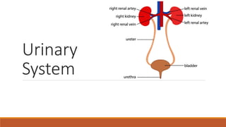
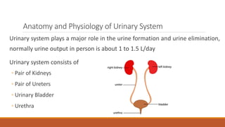
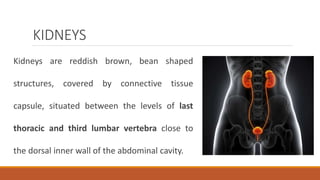
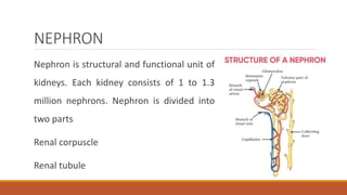
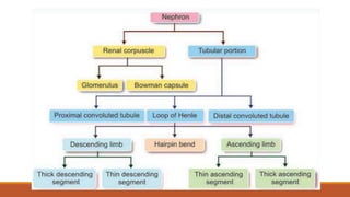
2. Introduction
Excretion is the process through which unwanted/toxic substances and
metabolic wastes are eliminated from the body and maintains our body in
homeostasis stage. Elimination process carry out through digestive system,
lungs, skin, liver and urinary system.
3. Anatomy and Physiology of Urinary System
Urinary system plays a major role in the urine formation and urine elimination,
normally urine output in person is about 1 to 1.5 L/day
Urinary system consists of
◦ Pair of Kidneys
◦ Pair of Ureters
◦ Urinary Bladder
◦ Urethra
4. KIDNEYS
Kidneys are reddish brown, bean shaped
structures, covered by connective tissue
capsule, situated between the levels of last
thoracic and third lumbar vertebra close to
the dorsal inner wall of the abdominal cavity.
5. Dimensions of Kidney
Each kidney of an adult human measures 10-13
cm in length, 5-7.5 cm in width, 2-3 cm in
thickness with an average weight of 120- 170 g.
There is a depression on the medial border of
kidney called hilum, through which renal artery,
renal veins, nerves, and ureters pass
6. Components of Kidney
Renal Capsule - Outer covering of Kidney
Renal cortex - The reddish-brown lining
present in between renal capsule and
renal medulla
Renal medulla - It is innermost lining of
kidney consists of Calyces & Pyramids
7. Functions of Kidneys
Controlling ACID-base balance
Controlling WATER balance
Maintaining ELECTROLYTE balance
Removing TOXINS and waste products from the body
Controlling BLOOD PRESSURE
Producing the hormone ERYTHROPOIETIN
Activating vitamin D
8. Controlling ACID-BASE Balance
Kidney maintains the pH of body. The normal pH of the blood is 7.35
to 7.45. To maintain this healthy range, the kidneys excrete acids (H+)
and bases (HCO3
-) when there’s an excess of them, or retain these
compounds when the body is lacking them.
9. Controlling Water Balance
Kidney maintains the water balance. An increase in osmolarity of the
blood as the result hypothalamus stimulates to release ADH (Anti-
Diuretic Hormone/Vasopressin) which results into reabsorption of
water from the kidney tubules.
10. Maintaining Electrolyte Balance
The kidneys filter some electrolytes from the blood, return part of
them back into circulation, and excrete excess electrolytes into the
urine. The levels of electrolytes like sodium and phosphate are
largely dependent on the health of person’s kidneys
11. Removing toxins and waste products
from the body
The kidneys filter out water-soluble waste products and toxins, flushing them out of the body
with urine.
Ammonia & Urea from Deamination of amino acid
Bilirubin from catabolism of Hemoglobin
Creatinine from the breakdown of creatine phosphate in muscle fibers
Uric Acid from catabolism of nucleic acid
Other – foreign substances from diet i.e. drugs and environmental toxins
12. Controlling Blood Pressure
The kidneys produce an enzyme called renin.
Renin converts angiotensinogen produced in the liver into angiotensin I, which is
later converted into angiotensin II in lungs. Angiotensin II constricts the blood
vessels and increases blood pressure as a result.
On the other hand, when blood pressure is too high, the kidneys produce more
urine to reduce the volume of liquid circulating in the body and somewhat helps
to reduce the high blood pressure.
13. Producing Hormone Erythropoietin
The kidneys produce a hormone called erythropoietin. The main
function of this hormone is to help body create more red blood cells
(erythrocytes), which are essential for the transport of oxygen
throughout all the tissues and organs
14. Activating Vitamin D
The kidneys transform calcifediol into calcitriol, the active form of
vitamin D. Calcitriol circulates in the blood and plays a vital role in
regulating calcium and phosphate balance in the body, which is
essential for healthy bone growth.
16. NEPHRON
Nephron is structural and functional unit of
kidneys. Each kidney consists of 1 to 1.3
million nephrons. Nephron is divided into
two parts
Renal corpuscle
Renal tubule
17.
18. Renal Corpuscle
It is situated near the cortex region of the
kidneys and helps in the filtration of the
blood. It is formed by two portions
Glomerulus — Glomerulus is a bunch of
capillaries enclosed by Bowman’s capsule.
Glomerular capillaries placed between
afferent (in) arteriole on one end and
efferent (out) arteriole on the other end
Bowman’s capsule — It cover the
glomerular capillaries and continue with the
tubular part
19. Renal Tubule
It is continuation part of the bowman’s capsule. On the basis of
structure and function it divided into the three parts.
Proximal convoluted tubule
Loop of Henle (descending limb and ascending limb)
Distal convoluted tubule
Collecting duct
20. Physiology of Urine Formation
When blood passes through glomerular capillaries the plasma is
filter by the filtering membrane into bowman’s capsule. Urine
formation includes three processes.
Glomerular filtration
Tubular reabsorption
Tubular secretion
21. Glomerular Filtration
Filtration takes place through the semi permeable walls of the glomerulus
& glomerular capsule or Bowman’s capsule.
Water and small molecules pass through it.
The afferent renal artery brings blood to the glomerulus and the efferent
artery carries the blood away from it. As the diameter of afferent artery is
more than the efferent artery, a hydrostatic pressure is generated in the
glomerulus (55mm Hg).
22. Glomerular Filtration
This pressure is opposed by osmotic pressure (30 mmHg) and filtrate
hydrostatic pressure in capsule (15mm Hg). The net filtration
pressure is 55-(30+15) = 10 mm of Hg.
It’s also called as Ultrafiltration because minutes particle also filtered
through it except plasma protein and blood cells. The filtered fluid is
called glomerular filtrate.
23. Glomerular Filtration
Glomerular filtration rate (GFR)— GFR
is defined as the total amount of
filtrate form in all the nephrons of both
the kidneys in the given unit of time.
Normal GFR is 125 ml/min or about
180 ml/min
25. Tubular Reabsorption
Needed/Wanted substances are reabsorb by the renal tubules and transported
back to the blood.
Components like glucose, vitamins, amino acids get completely reabsorbed into
the blood. These are called high threshold substances.
Low threshold substances like urea, uric acid are absorbed slightly.
Some substances like creatinine are not reabsorbed at sites of reabsorption
26. Sites of Reabsorption
Proximal Convoluted Tubule— About 85% of the substances reabsorbed by the PCT. Glucose, amino
acids, sodium, potassium, calcium, bicarbonates, chlorides, phosphates, urea, uric acid and water are
the major substances reabsorbed by the PCT
Loop of Henle— Sodium and chloride are main substances reabsorbed from loop of Henle
Distal Convoluted Tubule— Sodium, calcium, bicarbonate, and water are reabsorbed from DCT
Collecting Duct— Condition reabsorption happens in the collecting duct because, water reabsorbed
in the presence of Antidiuretic hormone (ADH), and sodium reabsorbed in the presence of
Aldosterone
28. Tubular Secretion
Substances are transported from blood into renal tubules.
Substances not required & the foreign material which have not got cleared
during filtration due to short time, are secreted into the distal convoluted tubule
& excreted in the urine.
Tubular secretion of Hydrogen ions is important for maintaining pH. H+ ions are
secreted in combination with bicarbonate as carbonic acid, with ammonia as
ammonium chloride & with hydrogen phosphate as dihydrogen phosphate
29. Site of Secretion
Potassium is secreted actively by sodium-potassium pump in
proximal and distal convoluted tubules, and collecting ducts
Ammonia is secreted in the proximal convoluted tubule
Hydrogen ions are secreted in the proximal and distal convoluted
tubules. Maximum hydrogen ion secretion occurs in proximal tubule
Urea is secreted in the loop of Henle
30.
31. Micturition
Micturition is the process by which urine is eliminated from the
urinary bladder. It is a reflex process.
Urinary bladder— It is the triangular hollow organ, in which ureters
open. It is formed by smooth muscles called detrusor muscle and it
open into urethra via internal urethral sphincter.
32.
33. Micturition
Urethra— Urethral structure and function different in male and female. In both,
two urethral sphincters present in urinary tract.
◦ Internal urethral sphincter.
◦ External urethral sphincter.
Urinary bladder and the internal urethral sphincter are supplied by sympathetic
and parasympathetic divisions of autonomic nervous (Pelvic nerve) system. The
external sphincter is supplied by somatic nerves fibers (pudendal nerve).
34. Micturition Reflex
Stimulation of stretch receptors situated on the wall of urinary bladder and
urethra. When about 300 to 400 ml of urine is collected in the urinary bladder.
Then sensory impulses reach to spinal cord through the sensory fibers of pelvic
nerve and motor impulse produce in the bladder and internal urethral sphincter.
Motor impulse cause the contraction in detrusor muscle and relaxation in
internal sphincter so that, urine enters in urethra from the urinary bladder
35. Micturition Reflex
Once urine enters in urethra, then urethral stretch receptors sense the change
and send sensory impulse to spinal cord via pelvic nerve.
Now the impulses generated from spinal centers inhibit pudendal nerve.
So, the external sphincter relaxes and micturition occurs.
During micturition, the flow of urine is facilitated by the increase in the
abdominal pressure due to the voluntary contraction of abdominal muscle.
36. Clearance Test/Renal Function Test.
Properties of urine
◦ Volume— 1-1.5 L/day.
◦ Reaction— Slightly acidic with PH 4.5-6.
◦ Specific gravity—1.010 to 1.025.
◦ Osmolarity— 1200 mOsm/L.
Examination of urine.
◦ Colour- Pale Yellow.
◦ Odour- Mild aromatic and ammonia like on standing.
◦ Total solid content- 30 to 70 g/lit.
◦ Organic Constituents- Urea, creatinine, uric acid, urobilinogen.
37. Physical Examination
Colour: Abnormal colouration of urine than straw colour is due to several
causes such as Jaundice, Haematouria, haemoglobinuria.
Appearance: Turbidity appears in both the physiological and pathological
conditions.
Volume: Increase in urine volume indicates increase in protein catabolism and
renal disorders such as chronic renal failure, diabetes insipidus and glycosuria.
Specific gravity: It is low in diabetes insipidus, and high in diabetes mellitus,
acute renal failure and excess medication.
pH: pH is useful in determining the metabolic or respiratory acidosis or
alkalosis.
38. Microscopic Examination
Red blood cells: presence of red blood cells in urine indicates glomerular
disease such as glomerulonephritis.
White blood cells: Number increase in acute glomerulonephritis, infection of
urinary tract, vagina or cervix.
Crystals: Abnormal crystals such as crystals of cystine and tyrosine appear in
liver diseases.
Bacteria: Bacterial growth appears during the infection’s conditions.
39. Chemical Examination
Glucose: Normal- 180 mg/dL
Protein: Normal- 30 mg/day
Ketone bodies
Urobilinogen: 1 to 3.5 mg/daily
Bile salts, Bilirubin, Blood.
40. Abnormal Constituents of Urine
Glucose: - Presence of glucose in the urine called Glycosuria.
Blood: - Presence of blood in the urine called Haematouria.
Protein: - Presence of protein in the urine called Proteinuria.
Ketone Bodies: - Presence of ketone bodies in the urine called Ketonuria.
Albumin: - Presence of albumin in the urine called Albuminuria.
Bilirubin & Bile Salt: - Presence of bilirubin in the urine called Bilirubinuria.
Pyuria: - Presence of WBCs or pus in urine called Pyuria.
Editor's Notes
homeostasis stage - sensing, processing and responding
Osmolarity - The term osmolarity refers to the number of particles of solute per liter of solution