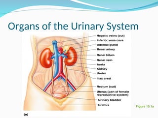
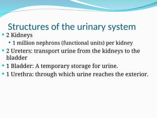
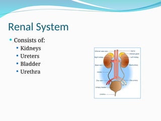
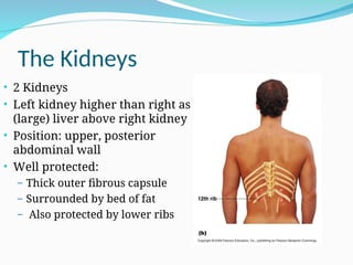
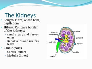
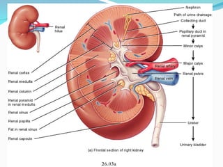
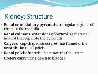
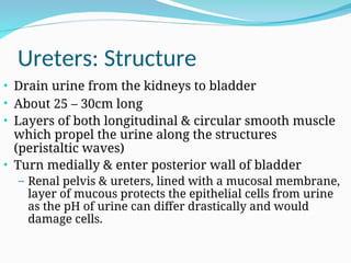
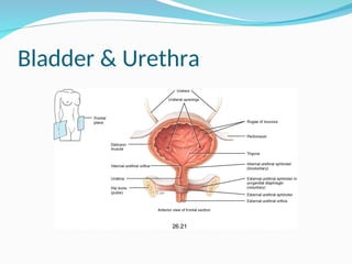
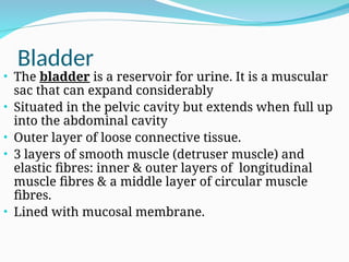
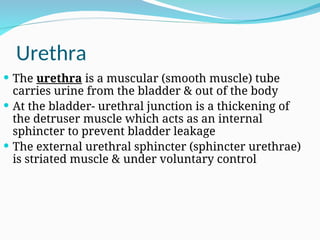
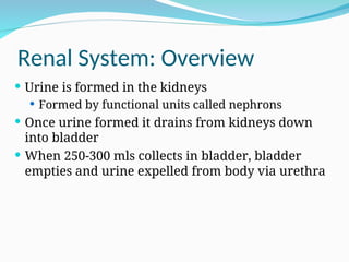
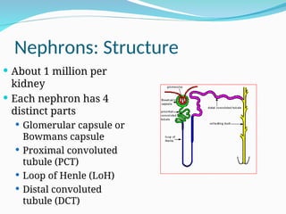
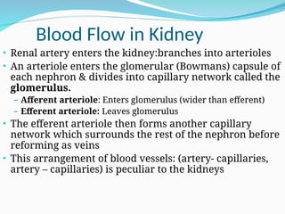
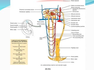
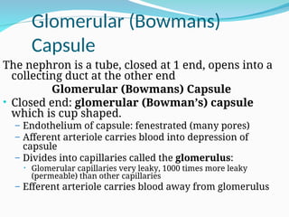
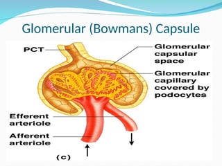
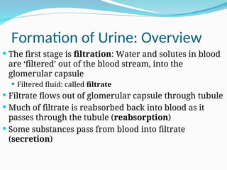
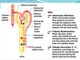
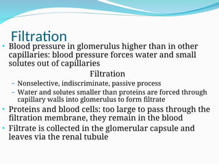
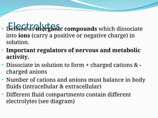
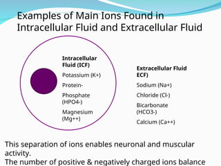
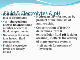
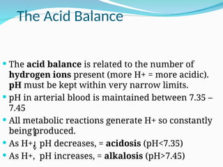
BY. UNIS
Learning Outcomes
• Be able to label the renal system and internal
structure of the kidney
• State the functions of the renal system
• Label the parts of the nephron
• Describe the process of urine formation
– Filtration
– Reabsorption
– Secretion
• State two hormones involved in the regulation
of fluid and electrolyte balance
• Understand the basics of maintaining fluid and
electrolyte balance
Figure 15.1a
Organs of the Urinary System
Structures of the urinary system
 2 Kidneys
 1 million nephrons (functional units) per kidney
 2 Ureters: transport urine from the kidneys to the
bladder
 1 Bladder: A temporary storage for urine.
 1 Urethra: through which urine reaches the exterior.
Functions of the Renal System
 Formation of Urine
 Urine formed in the Kidneys
 Elimination of waste products
 Nitrogenous wastes
 Toxins
 Drugs
Funtions of the Renal System
 Regulates many aspects of homeostasis
 Water (fluid) balance
 Electrolyte balance
 Acid-base balance in the blood
 Involved in blood pressure regulation
 Red blood cell production: Kidneys produce
erythropoetin
 Activation of vitamin D
Renal System
 Consists of:
 Kidneys
 Ureters
 Bladder
 Urethra
The Kidneys
• 2 Kidneys
• Left kidney higher than right as
(large) liver above right kidney
• Position: upper, posterior
abdominal wall
• Well protected:
– Thick outer fibrous capsule
– Surrounded by bed of fat
– Also protected by lower ribs
The Kidneys
• Length 11cm, width 6cm,
depth 3cm
• Hilum: Concave border
of the kidneys:
– renal artery and nerves
enter
– Renal veins and ureters
leave
• 2 main parts
– Cortex (outer)
– Medulla (inner)
Kidney: Structure
• Renal or medullary pyramids: triangular regions of
tissue in the medulla
• Renal columns: extensions of cortex-like material
inward that separate the pyramids
• Calyces: cup-shaped structures that funnel urine
towards the renal pelvis
• Renal pelvis: funnels urine towards the ureter
• Ureters carry urine down to bladder
Ureters: Structure
• Drain urine from the kidneys to bladder
• About 25 – 30cm long
• Layers of both longitudinal & circular smooth muscle
which propel the urine along the structures
(peristaltic waves)
• Turn medially & enter posterior wall of bladder
– Renal pelvis & ureters, lined with a mucosal membrane,
layer of mucous protects the epithelial cells from urine
as the pH of urine can differ drastically and would
damage cells.
Bladder & Urethra
Bladder
• The bladder is a reservoir for urine. It is a muscular
sac that can expand considerably
• Situated in the pelvic cavity but extends when full up
into the abdominal cavity
• Outer layer of loose connective tissue.
• 3 layers of smooth muscle (detruser muscle) and
elastic fibres: inner & outer layers of longitudinal
muscle fibres & a middle layer of circular muscle
fibres.
• Lined with mucosal membrane.
Urethra
 The urethra is a muscular (smooth muscle) tube
carries urine from the bladder & out of the body
 At the bladder- urethral junction is a thickening of
the detruser muscle which acts as an internal
sphincter to prevent bladder leakage
 The external urethral sphincter (sphincter urethrae)
is striated muscle & under voluntary control
Renal System: Overview
 Urine is formed in the kidneys
 Formed by functional units called nephrons
 Once urine formed it drains from kidneys down
into bladder
 When 250-300 mls collects in bladder, bladder
empties and urine expelled from body via urethra
Nephrons: Structure
 About 1 million per
kidney
 Each nephron has 4
distinct parts
 Glomerular capsule or
Bowmans capsule
 Proximal convoluted
tubule (PCT)
 Loop of Henle (LoH)
 Distal convoluted
tubule (DCT)
Blood Flow in Kidney
• Renal artery enters the kidney:branches into arterioles
• An arteriole enters the glomerular (Bowmans) capsule of
each nephron & divides into capillary network called the
glomerulus.
– Afferent arteriole: Enters glomerulus (wider than efferent)
– Efferent arteriole: Leaves glomerulus
• The efferent arteriole then forms another capillary
network which surrounds the rest of the nephron before
reforming as veins
• This arrangement of blood vessels: (artery- capillaries,
artery – capillaries) is peculiar to the kidneys
Glomerular (Bowmans)
Capsule
The nephron is a tube, closed at 1 end, opens into a
collecting duct at the other end
Glomerular (Bowmans) Capsule
• Closed end: glomerular (Bowman’s) capsule
which is cup shaped.
– Endothelium of capsule: fenestrated (many pores)
– Afferent arteriole carries blood into depression of
capsule
– Divides into capillaries called the glomerulus:
• Glomerular capillaries very leaky, 1000 times more leaky
(permeable) than other capillaries
– Efferent arteriole carries blood away from glomerulus
Glomerular (Bowmans) Capsule
Formation of Urine: Overview
 The first stage is filtration: Water and solutes in blood
are ‘filtered’ out of the blood stream, into the
glomerular capsule
 Filtered fluid: called filtrate
 Filtrate flows out of glomerular capsule through tubule
 Much of filtrate is reabsorbed back into blood as it
passes through the tubule (reabsorption)
 Some substances pass from blood into filtrate
(secretion)
Filtration
• Blood pressure in glomerulus higher than in other
capillaries: blood pressure forces water and small
solutes out of capillaries
Filtration
– Nonselective, indiscriminate, passive process
– Water and solutes smaller than proteins are forced through
capillary walls into glomerulus to form filtrate
• Proteins and blood cells: too large to pass through the
filtration membrane, they remain in the blood
• Filtrate is collected in the glomerular capsule and
leaves via the renal tubule
Reabsorption
 As the fluid passes through the tubules much of the
water & solutes that were filtered out of the blood
are reabsorbed back into the blood stream
 Some by passive diffusion & some are actively
transported which is a more selective process and
depends on the bodies needs
Reabsorption
By the time filtrate leaves the PCT all glucose & amino
acids, 80-90% bicarbonate, 65% Na+, K+ & water &
varying amounts of other solutes have been
reabsorbed
In the loop of Henle 15% Water is reabsorbed in the
descending limb. Solutes such as Na+, K+, bicarbonate
& Cl- reabsorbed in the ascending limb
Water & solutes continue to be reabsorbed in the DCT
and collecting ducts
Secretion
 Not all substances are cleared from the blood into the
filtrate in the glomerular capsule, e.g. some drugs, H+,
urea and creatanine are secreted into the filtrate as it
passes along the tubule.
 Secretion of substances is regulated by amounts of
different substances present in the blood e.g. K+
secretion can vary depending on dietary intake, if
high more is secreted.
Hormonal Regulation
• Most water moves back into blood passively with
solutes
• Antidiuretic hormone acts on the collecting ducts, it
increases re-absorption of water from filtrate when
dehydrated so less urine passed
• Aldosterone increases reabsorption of sodium (Na+)
in the DCT and collecting duct, (water moves back
passively (osmosis) with Na+, not a direct effect of
aldosterone)
Filtrate and Urine
• Kidneys are very active 24 hours day, filtration takes place
constantly to maintain health
• The filtration rate of all nephron’s is 125mls/minute, this
is a lot, per hour, 125 X 60 is = 7,500mls/per hour which is
7.5L
• Per day (X 24) this amounts to 180,000 mls or 180L per day
• The average sized adult only passes 1–2L of urine per day,
so most filtrate is reabsorbed.
Urine
 After leaving the collecting duct the processes of re-
absorption & secretion are completed
 The fluid is now classed as ‘urine’
 Urine is what remains after the filtrate has lost most
of its water, nutrients, and necessary ions
 Urine contains nitrogenous waste and substances that
are not needed by the body
Urine
 Yellow colour due to the pigment urochrome (from
the destruction of hemoglobin) and solutes
 Sterile
 Slightly aromatic
 Normal pH of around 6
 Specific gravity of 1.001 to 1.035
Urine Contains
 Urine is mostly water
 Solutes normally found in urine are:
 Sodium and potassium ions
 Urea, uric acid, creatinine
 Ammonia
 Bicarbonate ions
Abnormal Constituents of
Urine
 Solutes NOT normally found in urine
 Glucose: this is filtered but rapidly reabsorbed in the
PCT
 Blood proteins
 Red blood cells
 Haemoglobin
 White blood cells (pus)
 Bile
Fluid Compartments
 Water is the ‘universal solvent’
In health fluid balance is maintained automatically
 Fluid Compartments
2 main compartments
Intracellular (about 66% of total body fluid2/3))
(=40% body weight)
Extracellular (about 33% of total body fluid (1/3))
Interstitial fluid & Blood Plasma
(=80% of ECF) (=20% of ECF)
Fluid Balance
• In health balance is normally well maintained
• When additional fluid is required we become aware
of this (thirst) and drink
• Excess fluid is excreted via the kidneys, urine
becomes more dilute
• To maintain normal levels of electrolytes and rid the
body of toxins and by products of metabolism the
kidneys must constantly remain active (24 hours/day)
Water Input & Output (Average)
Water Input: ml per day Water Output: ml per day
Drink 1500
Food 800
Metabolic Production: 200
Urine: 1400
Evaporation:
Lungs: 500
Skin: 400
(insensible loss, i.e. not
detected by senses)
Faeces: 200
Total 2500 Total 2500
Note: Values will vary with diet and environmental conditions
Electrolytes
• Defined as inorganic compounds which dissociate
into ions (carry a positive or negative charge) in
solution.
• Important regulators of nervous and metabolic
activity.
• Dissociate in solution to form + charged cations & -
charged anions
• Number of cations and anions must balance in body
fluids (intracellular & extracellular)
• Different fluid compartments contain different
electrolytes (see diagram)
Extracellular Fluid
ECF)
Sodium (Na+)
Chloride (Cl-)
Bicarbonate
(HCO3-)
Calcium (Ca++)
Intracellular
Fluid (ICF)
Potassium (K+)
Protein-
Phosphate
(HPO4-)
Magnesium
(Mg++)
Examples of Main Ions Found in
Intracellular Fluid and Extracellular Fluid
This separation of ions enables neuronal and muscular
activity.
The number of positive & negatively charged ions balance
Fluid & Electrolytes & pH
 Body fluids: a complex
mix of electrolytes
 + & - charged ions
always balance (same
number of each) in
fluid compartments.
 Not always the same
ions in each fluid
compartment
 Fluid & electrolyte
levels are closely
linked
 Hydrogen (H+) formed as by
product of metabolism of
amino-acids.
 Concentration of free H+
determines intra &
extracellular fluid pH (acid or
alkaline), it effects the activity
of many enzyme systems
 pH stands for pressure of
hydrogen
The Acid Balance
 The acid balance is related to the number of
hydrogen ions present (more H+ = more acidic).
pH must be kept within very narrow limits.
 pH in arterial blood is maintained between 7.35 –
7.45
 All metabolic reactions generate H+ so constantly
being produced.
 As H+, pH decreases, = acidosis (pH<7.35)
 As H+, pH increases, = alkalosis (pH>7.45)
Fluid and Electrolyte
Imbalance
• Dehydration may increase plasma concentrations of
ions.
• Sodium and Potassium depletion, could be due to:
– ( loss of) Gastric aspirate
– Vomiting, diarrhoea
• Acidosis or Alkalosis (H+)
• In health, the kidneys maintain fluid, electrolyte and
pH within very narrow ranges (balance)
Fluid Balance
• Fluid loss during breathing and from sweat cannot be
measured (800-1000mls/day)
• Fluid balance charts are normally in a ‘positive’ balance
(more in than out) as this fluid loss cannot be measured
• More water lost if temperature is high (pyrexia) resulting
in increased sweating and also increased respiratory rate
so:
• Intake should also be increased
Fluid Balance
 A constant fluid intake is vital to health if blood is to
maintain fluid, electrolyte and acid balance
 Fluid input and output often monitored closely in
hospital
 When unwell fluid intake must be maintained, if oral
intake is limited or restricted intravenous fluids can be
given
 Ensuring adequate intake is very important
Summary of lecture contents
 Structure and functions of the renal system
 Structure and function of the nephron
• Formation of urine
– Filtration
– Reabsorption
– Secretion
 Hormones involved in regulation of fluid and
electrolyte balance
 Basics of fluid and electrolyte balance
THANK YOU

urinary system by Dr Smart.ppt6356372663563

  • 1.
  • 2.
    Learning Outcomes • Beable to label the renal system and internal structure of the kidney • State the functions of the renal system • Label the parts of the nephron • Describe the process of urine formation – Filtration – Reabsorption – Secretion • State two hormones involved in the regulation of fluid and electrolyte balance • Understand the basics of maintaining fluid and electrolyte balance
  • 3.
    Figure 15.1a Organs ofthe Urinary System
  • 4.
    Structures of theurinary system  2 Kidneys  1 million nephrons (functional units) per kidney  2 Ureters: transport urine from the kidneys to the bladder  1 Bladder: A temporary storage for urine.  1 Urethra: through which urine reaches the exterior.
  • 5.
    Functions of theRenal System  Formation of Urine  Urine formed in the Kidneys  Elimination of waste products  Nitrogenous wastes  Toxins  Drugs
  • 6.
    Funtions of theRenal System  Regulates many aspects of homeostasis  Water (fluid) balance  Electrolyte balance  Acid-base balance in the blood  Involved in blood pressure regulation  Red blood cell production: Kidneys produce erythropoetin  Activation of vitamin D
  • 7.
    Renal System  Consistsof:  Kidneys  Ureters  Bladder  Urethra
  • 8.
    The Kidneys • 2Kidneys • Left kidney higher than right as (large) liver above right kidney • Position: upper, posterior abdominal wall • Well protected: – Thick outer fibrous capsule – Surrounded by bed of fat – Also protected by lower ribs
  • 9.
    The Kidneys • Length11cm, width 6cm, depth 3cm • Hilum: Concave border of the kidneys: – renal artery and nerves enter – Renal veins and ureters leave • 2 main parts – Cortex (outer) – Medulla (inner)
  • 11.
    Kidney: Structure • Renalor medullary pyramids: triangular regions of tissue in the medulla • Renal columns: extensions of cortex-like material inward that separate the pyramids • Calyces: cup-shaped structures that funnel urine towards the renal pelvis • Renal pelvis: funnels urine towards the ureter • Ureters carry urine down to bladder
  • 12.
    Ureters: Structure • Drainurine from the kidneys to bladder • About 25 – 30cm long • Layers of both longitudinal & circular smooth muscle which propel the urine along the structures (peristaltic waves) • Turn medially & enter posterior wall of bladder – Renal pelvis & ureters, lined with a mucosal membrane, layer of mucous protects the epithelial cells from urine as the pH of urine can differ drastically and would damage cells.
  • 13.
  • 14.
    Bladder • The bladderis a reservoir for urine. It is a muscular sac that can expand considerably • Situated in the pelvic cavity but extends when full up into the abdominal cavity • Outer layer of loose connective tissue. • 3 layers of smooth muscle (detruser muscle) and elastic fibres: inner & outer layers of longitudinal muscle fibres & a middle layer of circular muscle fibres. • Lined with mucosal membrane.
  • 15.
    Urethra  The urethrais a muscular (smooth muscle) tube carries urine from the bladder & out of the body  At the bladder- urethral junction is a thickening of the detruser muscle which acts as an internal sphincter to prevent bladder leakage  The external urethral sphincter (sphincter urethrae) is striated muscle & under voluntary control
  • 16.
    Renal System: Overview Urine is formed in the kidneys  Formed by functional units called nephrons  Once urine formed it drains from kidneys down into bladder  When 250-300 mls collects in bladder, bladder empties and urine expelled from body via urethra
  • 17.
    Nephrons: Structure  About1 million per kidney  Each nephron has 4 distinct parts  Glomerular capsule or Bowmans capsule  Proximal convoluted tubule (PCT)  Loop of Henle (LoH)  Distal convoluted tubule (DCT)
  • 18.
    Blood Flow inKidney • Renal artery enters the kidney:branches into arterioles • An arteriole enters the glomerular (Bowmans) capsule of each nephron & divides into capillary network called the glomerulus. – Afferent arteriole: Enters glomerulus (wider than efferent) – Efferent arteriole: Leaves glomerulus • The efferent arteriole then forms another capillary network which surrounds the rest of the nephron before reforming as veins • This arrangement of blood vessels: (artery- capillaries, artery – capillaries) is peculiar to the kidneys
  • 20.
    Glomerular (Bowmans) Capsule The nephronis a tube, closed at 1 end, opens into a collecting duct at the other end Glomerular (Bowmans) Capsule • Closed end: glomerular (Bowman’s) capsule which is cup shaped. – Endothelium of capsule: fenestrated (many pores) – Afferent arteriole carries blood into depression of capsule – Divides into capillaries called the glomerulus: • Glomerular capillaries very leaky, 1000 times more leaky (permeable) than other capillaries – Efferent arteriole carries blood away from glomerulus
  • 21.
  • 22.
    Formation of Urine:Overview  The first stage is filtration: Water and solutes in blood are ‘filtered’ out of the blood stream, into the glomerular capsule  Filtered fluid: called filtrate  Filtrate flows out of glomerular capsule through tubule  Much of filtrate is reabsorbed back into blood as it passes through the tubule (reabsorption)  Some substances pass from blood into filtrate (secretion)
  • 24.
    Filtration • Blood pressurein glomerulus higher than in other capillaries: blood pressure forces water and small solutes out of capillaries Filtration – Nonselective, indiscriminate, passive process – Water and solutes smaller than proteins are forced through capillary walls into glomerulus to form filtrate • Proteins and blood cells: too large to pass through the filtration membrane, they remain in the blood • Filtrate is collected in the glomerular capsule and leaves via the renal tubule
  • 25.
    Reabsorption  As thefluid passes through the tubules much of the water & solutes that were filtered out of the blood are reabsorbed back into the blood stream  Some by passive diffusion & some are actively transported which is a more selective process and depends on the bodies needs
  • 26.
    Reabsorption By the timefiltrate leaves the PCT all glucose & amino acids, 80-90% bicarbonate, 65% Na+, K+ & water & varying amounts of other solutes have been reabsorbed In the loop of Henle 15% Water is reabsorbed in the descending limb. Solutes such as Na+, K+, bicarbonate & Cl- reabsorbed in the ascending limb Water & solutes continue to be reabsorbed in the DCT and collecting ducts
  • 27.
    Secretion  Not allsubstances are cleared from the blood into the filtrate in the glomerular capsule, e.g. some drugs, H+, urea and creatanine are secreted into the filtrate as it passes along the tubule.  Secretion of substances is regulated by amounts of different substances present in the blood e.g. K+ secretion can vary depending on dietary intake, if high more is secreted.
  • 28.
    Hormonal Regulation • Mostwater moves back into blood passively with solutes • Antidiuretic hormone acts on the collecting ducts, it increases re-absorption of water from filtrate when dehydrated so less urine passed • Aldosterone increases reabsorption of sodium (Na+) in the DCT and collecting duct, (water moves back passively (osmosis) with Na+, not a direct effect of aldosterone)
  • 30.
    Filtrate and Urine •Kidneys are very active 24 hours day, filtration takes place constantly to maintain health • The filtration rate of all nephron’s is 125mls/minute, this is a lot, per hour, 125 X 60 is = 7,500mls/per hour which is 7.5L • Per day (X 24) this amounts to 180,000 mls or 180L per day • The average sized adult only passes 1–2L of urine per day, so most filtrate is reabsorbed.
  • 31.
    Urine  After leavingthe collecting duct the processes of re- absorption & secretion are completed  The fluid is now classed as ‘urine’  Urine is what remains after the filtrate has lost most of its water, nutrients, and necessary ions  Urine contains nitrogenous waste and substances that are not needed by the body
  • 32.
    Urine  Yellow colourdue to the pigment urochrome (from the destruction of hemoglobin) and solutes  Sterile  Slightly aromatic  Normal pH of around 6  Specific gravity of 1.001 to 1.035
  • 33.
    Urine Contains  Urineis mostly water  Solutes normally found in urine are:  Sodium and potassium ions  Urea, uric acid, creatinine  Ammonia  Bicarbonate ions
  • 34.
    Abnormal Constituents of Urine Solutes NOT normally found in urine  Glucose: this is filtered but rapidly reabsorbed in the PCT  Blood proteins  Red blood cells  Haemoglobin  White blood cells (pus)  Bile
  • 35.
    Fluid Compartments  Wateris the ‘universal solvent’ In health fluid balance is maintained automatically  Fluid Compartments 2 main compartments Intracellular (about 66% of total body fluid2/3)) (=40% body weight) Extracellular (about 33% of total body fluid (1/3)) Interstitial fluid & Blood Plasma (=80% of ECF) (=20% of ECF)
  • 36.
    Fluid Balance • Inhealth balance is normally well maintained • When additional fluid is required we become aware of this (thirst) and drink • Excess fluid is excreted via the kidneys, urine becomes more dilute • To maintain normal levels of electrolytes and rid the body of toxins and by products of metabolism the kidneys must constantly remain active (24 hours/day)
  • 37.
    Water Input &Output (Average) Water Input: ml per day Water Output: ml per day Drink 1500 Food 800 Metabolic Production: 200 Urine: 1400 Evaporation: Lungs: 500 Skin: 400 (insensible loss, i.e. not detected by senses) Faeces: 200 Total 2500 Total 2500 Note: Values will vary with diet and environmental conditions
  • 38.
    Electrolytes • Defined asinorganic compounds which dissociate into ions (carry a positive or negative charge) in solution. • Important regulators of nervous and metabolic activity. • Dissociate in solution to form + charged cations & - charged anions • Number of cations and anions must balance in body fluids (intracellular & extracellular) • Different fluid compartments contain different electrolytes (see diagram)
  • 39.
    Extracellular Fluid ECF) Sodium (Na+) Chloride(Cl-) Bicarbonate (HCO3-) Calcium (Ca++) Intracellular Fluid (ICF) Potassium (K+) Protein- Phosphate (HPO4-) Magnesium (Mg++) Examples of Main Ions Found in Intracellular Fluid and Extracellular Fluid This separation of ions enables neuronal and muscular activity. The number of positive & negatively charged ions balance
  • 40.
    Fluid & Electrolytes& pH  Body fluids: a complex mix of electrolytes  + & - charged ions always balance (same number of each) in fluid compartments.  Not always the same ions in each fluid compartment  Fluid & electrolyte levels are closely linked  Hydrogen (H+) formed as by product of metabolism of amino-acids.  Concentration of free H+ determines intra & extracellular fluid pH (acid or alkaline), it effects the activity of many enzyme systems  pH stands for pressure of hydrogen
  • 41.
    The Acid Balance The acid balance is related to the number of hydrogen ions present (more H+ = more acidic). pH must be kept within very narrow limits.  pH in arterial blood is maintained between 7.35 – 7.45  All metabolic reactions generate H+ so constantly being produced.  As H+, pH decreases, = acidosis (pH<7.35)  As H+, pH increases, = alkalosis (pH>7.45)
  • 42.
    Fluid and Electrolyte Imbalance •Dehydration may increase plasma concentrations of ions. • Sodium and Potassium depletion, could be due to: – ( loss of) Gastric aspirate – Vomiting, diarrhoea • Acidosis or Alkalosis (H+) • In health, the kidneys maintain fluid, electrolyte and pH within very narrow ranges (balance)
  • 43.
    Fluid Balance • Fluidloss during breathing and from sweat cannot be measured (800-1000mls/day) • Fluid balance charts are normally in a ‘positive’ balance (more in than out) as this fluid loss cannot be measured • More water lost if temperature is high (pyrexia) resulting in increased sweating and also increased respiratory rate so: • Intake should also be increased
  • 44.
    Fluid Balance  Aconstant fluid intake is vital to health if blood is to maintain fluid, electrolyte and acid balance  Fluid input and output often monitored closely in hospital  When unwell fluid intake must be maintained, if oral intake is limited or restricted intravenous fluids can be given  Ensuring adequate intake is very important
  • 45.
    Summary of lecturecontents  Structure and functions of the renal system  Structure and function of the nephron • Formation of urine – Filtration – Reabsorption – Secretion  Hormones involved in regulation of fluid and electrolyte balance  Basics of fluid and electrolyte balance
  • 46.