SlideShare a Scribd company logo
Mrs.JEEVA.D
NURSING TUTOR,
GANGA COLLEGE OF NURSING,
COIMBATORE.
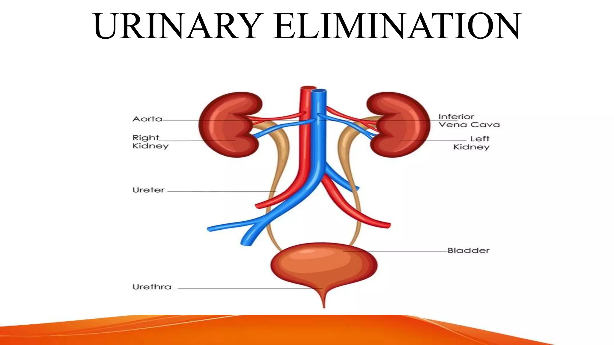
URINARY ELIMINATION
OBJECTIVES
• At end of the session the leaner will be able to
• Understand the factors influencing urination
• discuss the characteristics of normal urine
• elaborate the pathological conditions of urinary elimination
• explain the procedures which facilitates urinary elimination
such as ;
- Providing urinal
- Condom drainage
- Bladder irrigation
- Catheterization with care of Urinary drainage
INTRODUCTION
• Elimination is the expulsion of waste products from the
body through the skin ,lungs, kidneys and rectum
• In specific , Urinary elimination is the removal of waste
products from the body through the urinary system
• The urinary system consists of organs that produce and
excrete urine from the body.
• Urine contains waste: mostly excess water, salts and
nitrogen compounds. Normal adult bladder can store up
to 5 liters.
• Also responsible for regulating blood volume and blood
pressure.
PHYSIOLOGY OF URINE
• The nephrons of the kidneys process blood and create urine through a
process of filtration, reabsorption, and secretion.
• When the urine is collected in the bladder, A desire to void is experienced
due to the stimulation of stretch receptors.
• The stretch receptors transmit the message to the voiding reflex center in
the spinal cord(2nd to 4th sacral vertebrae)
• If the time is appropriate the brain sends message through spinal cord
causing stimulation so that the urine can be released from the bladder.
• If the time and place are appropriate the external sphincter relaxes and the
urination process takes place.
CHARACTERISTICS OF NORMAL URINE
• Volume : An amount of 1000- 2000 ml of urine is excreted in 24 hrs. as well it
depends upon the water intake and change of weather .
• Colour : The normal urine is pale yellowish or
amber in color.
• Appearance : The normal urine is clear with no deposit.
• Odour : The normal urine has an aromatic odour
• Reaction : Reaction of normal urine is slightly acidic
• Specific gravity: It varies from 1.016 - 1.025 with a
normal fluid intake.
Infants: An infant may urinate as often as 20 times a day . It is
colorless and odorless and infants are born without urinary
control.
Preschoolers: Pre-schoolers is able to take responsibility for
independent toileting.
School age children : Their elimination system reaches maturity
and gets proper control over the system.
Older adults: the excretory function of kidneys diminishes with
age due to decreased kidney’s filtering ability & decreased muscle
tone which leads to the poor bladder control.
Factors Influencing Urinary Elimination
PSYCHOLOGICAL
FACTORS
• Anxiety and emotional stress
may cause a sense of urgency
and increased frequency of
urination
• Privacy ,normal positions,
sufficient time can stimulate the
micturition reflex
FLUID AND FOOD INTAKE
• If fluid intake is greatly increased, frequency of
voiding increases. If fluid intake is
decreased,frequency of voiding decreases.
• Ingestion of certain fluids directly affects urine
production and excretion .eg:coffee,tea,cola
drinks
• Increased urine production and elimination
happens if we ingest foods high in fluid
content .eg:fruits,vegetables.
MEDICATIONS
• Cholinergic and diuretics can cause
urinary elimination
• Anticholinergics and opioid analgesics
may cause urinary retention.
• In fact, Some medicine cause change
in color of urine.
To red color —methyldopa
[ Anti hypertensive ]
To orange or pink
– Phenotoin or rifambin
PATHOLOGICAL CONDITIONS
• Diabetes mellitus, Multiple
sclerosis and Parkinsons disease
can cause changes in nerve
functions that can lead to possible
loss of bladder tone.
• Diabetes insipidus: can Increases
urine production and elimination
• Neurogenic bladder: The client
does not perceive bladder fullness
and is unable to control the
urinary sphincter.
DIAGNOSTIC AND SURGICAL PROCEDURES
• Diagnostic procedures:
Eg ; Urethra may swell
following cystoscopy and any
kind of examination involves
urinary system stimulates
micturition.
• Surgical procedures
Eg ; Surgery of lower
abdomen and pelvic structures
sometimes impairs urination
because of local trauma to
surrounding tissue
SOCIO CULTURAL FACTORS
Certain life-style behaviors can affect
urinary elimination such as
 Insufficient time,
 Unavailability of toilet facilities,
 Lack of privacy,
 Inability to assume a normal
position
ALTERATIONS IN URINARY ELIMINATION
• .Polyuria
Production of abnormal large amounts of urine by
the kidney. Polyuria can follow excessive fluid intake - a
condition known as polydipsia.
• Diuresis
It is the another term for production and excretion of
large amounts of urine
• Oliguria
Defined as low urine output usually less than 500 ml a
day or 30 ml an hour.
• .Anuria
Refers to a lack of urine production with no effective urinary
output.
• Nocturia
Voiding of two or more times at night.
• Dysuria
It means voiding that is either painful or difficult.
• Enuresis
Defined as involuntary urination in children.
• Nocturnal enuresis
Involuntary urination during night.
URINARY INCONTINENCE
 UI is the involuntary passage of urine.
 It refers to the inability to control passage of urine.
 It is a symptom not a disease.
 It may be temporary or permanent.
 Leakage may be continuous or intermittent.
Causes:
 Urinary Tract Infection
 Surgery or trauma causing sphincter damage
 Paralysis, Cognitive Impairment and
Unconsciousness
TYPES OF INCONTINENCE
STRESS INCONTINENCE:
Involuntary loss of urine through an intact urethra as a result of
sudden increase in intra abdominal pressure. It is seen mainly in women
who have had vaginal deliveries
URGE INCONTINENCE:
Involuntary loss of urine associated with a strong urge to void that
cannot be suppressed.
REFLEX INCONTINENCE:
Involuntary loss of urine due to hyper reflexia in the absence of
normal sensation .
OVERFLOW INCONTINENCE
Involuntary loss of urine associated with over distention of the bladder
.
MEASURES TO OVERCOME UIC
• Establish a regular voiding schedule for the patient.
• Perineal exercises: To increase the tone of muscles concerned
with the micturition, in particular the perineal and abdominal
muscles.
• Periodic tightening of the perineal muscles, intentionally stopping
and then starting the urine stream etc. Can help in gaining voiding
control.
• Arrange toilet or bedpan within the easy reach of the patient .
• Medical and surgical correction of the causative factors.
• Bladder training program Includes education of the client and
support
• Condom drainage
URINARY RETENTION
The state in which one experiences incomplete emptying of the
bladder.
Causes:
 Obstruction of urine flow due to Prostate gland enlargement,
Pregnancy, Urethral edema, Surgery or Diagnostic
examinations
 Alteration in motor or sensory innervation to the detrusor
muscle and internal sphincter. eg: Spinal cord Injury,
Peripheral nerve trauma, Degeneration of peripheral nerves
 Inability to relax external spinchter due to emotional stress or
anxiety ,muscle tension
PREVENTION & TREATMENT OF URINARY RETENTION
 Assist the patient to his or her normal position for voiding.
 Provide privacy.
 Offer a bedpan or urinal that is warm becoz a bedpan that is cold to touch may cause
contraction of the perineal muscles
 Foster the muscles relaxation by providing necessary physical support to the patient and
by relieving pain.
 Provide any assistance when the patient feels the need to void.
 Running water within the hearing of the patient or flushing the toilet stimulate the
micturition reflex.
 Provide enough time for micturition.
 Reassurance and emotional support are helpful to reflex the patient
 A hot enema if permitted may relieve the retention of the urine. Give fluids freely unless
contra-indicated.
TYPES OF SPECIMEN
 Random specimen
A randomly collected specimen collected at unspecified
times and is often more convenient for the patient
 First morning specimen
 Fasting specimen:
 2-hour postprandial specimen.
 24 hour (timed) specimen.
 Catheterized specimen
Collection of Urine specimen
• Collection and transportation of urine specimens to the
laboratory is important and there are wide variety of
procedures are available for the specimen collection.
Those are
 Urine collection from test tubes
 Urine collection by U-Bag for infants
 Urine collection from catheter
 24 hours urine collection
 Collection of mid stream urine
URINALYSIS or URINE TESTING
PURPOSES :
 To observe urine color and clarity
 To measure urine specific gravity
 To determine the urine acidity and
alkalinity of the urine
 To determine the presence of
glucose and albumin
• ARTICLES
• Container for specimen
• Benedict solution
• Acetic acid
• Test tubes [4-6] & holder.
• Red and blue Litmus paper
• Urinometer
• Kidney tray
• Paper bag
• Spirit lamp with spirit
• Matchbox
• Cotton balls in bowl
Urine analysis is the analysis of urine in order to find out the presence of sugar,
albumin and microorganism.
TESTING FOR GLUCOSE
 Pour 5 ml of benedict’s solution in the test tube.
Boil it to find out the color change, if no change that shows the purity
of benedict’s solution.
 Add 8-10 drops of urine into the solution and reheat it; allow it to
cool.
 Observe the color change which indicates the sugar level.
 Blue : 0% – Green : 1% – Yellow : 2 % – Orange : 3 % – Brick red :
5%
TESTING FOR ALBUMIN
 Fill the test tube 3/4th with urine, check the reaction, if it
is alkaline make it acidic.
 Boil the top one third of the portion.
If there is cloudy appearance it indicates the presence of
albumin or phosphate.
Add 5 drops of acetic acid & reheat.
 If cloudy appearance still presents it indicates albumin
presence and if it disappears it shows phosphate
presence.
PROVIDING URINAL
Meeting urinary elimination need of
bed-ridden male patients using an
urinal.
PURPOSES:
To meet and maintain the elimination
needs of the client
To give perineal care
 To observe and collect specimen for
diagnostic procedure.
• Explain the procedure and provide privacy to patient
• Palpate for distended bladder
• Assess patient knowledge regarding urinal use.
• Wash and done gloves
• Assist patient into appropriate position
• Patient should hold urinal in position
• Once patient has finished voiding remove urinal.
• Collect urine in container if required and empty urine into toilet
and cleanse the urinal.
• Allow patient to wash hand after voiding.
• Remove gloves and wash hands
• Record amount of urine passed if needed.
PROCEDURE TO PROVIDE URINAL
CONDOM DRAINAGE
• It is a soft, pliable rubber sheath applied over the male’s
peni’s.
EQUIPMENTS
A tray containing
• Condom
• Adhesive tape and mission
• Urine collection (drainage) bag with drainage tubing.
• Basin of warm water and soap
• Towel
• Cover sheet
• Protective sheet
• Bed pan
• Kidney tray
GUIDELINES
 When securing the condom, be sure that
a.Adhesive band touches only the condom.
b.The adhesive band is not tight as it will impair the blood supply to the
penis.
 Infection may result from trauma to the urethral meatus or builds up
pressure in the outflow tubing.
 Condom and drainage tubes are changed on alternate days.
 When changing condom, clean the urethral meatus and penis thoroughly
and check for signs of skin infections.
 Check out the tubing frequently for patency.
 Check the twisting of the condom near drainage tubing causing irritation
and obstruction of urine outflow.
PROCEDURE
• Take the supplies to the bedside.
• Explain the procedure to the patient and maintain privacy
• Position the patient in supine
• Cover the upper trunk with cover sheet and total back the top
bed clothes onto the mid of thighs.
• Place protective sheet and cloth across the thighs.
• Clean the genitalia with soap and water thoroughly and dry.
• Tie the urine collecting bag to the bed and bring drainage
tubing from the side or through the side rail.
• Make a hole in the end of the condom.
• Grasp the penis firmly along the shaft with your left hand, hold the condom with your
right hand at the tip of penis and roll the condom on the penial shaft.
• Secure the condom in place with the adhesive strip snugly around the top of the
condom over penial shaft.
• Fix the drainage tube into the hole made in the end of the condom, using an adhesive
strip.
• Place the excess coil tubing on bed and secure to the bottom sheet.
• Remove the cover sheet and cover the patient with top bed clothes. Position the client
comfortable.
• Clean dry and replace the supplies, wash hands.
• Record the date, time and reason of applying condom for drainage in the nurse's
notes.
PROCEDURE
BLADDER IRRIGATION
• Bladder irrigation or wash is defined as washing of the urinary
bladder by directly a stream of solution into the bladder through
the urinary meatus by means of a catheter tubing and funnel.
• It is washing out of the urinary bladder by directing a stream of
solution into the bladder, through the urinary meatus by means
of the catheter.
-Dr.s.Anuradha
BLADDER IRRIGATION
• PURPOSES :
 To cleanse the bladder from
decomposed urine
bacteria,excess mucus and
pus.
 To medicate the lining of the
bladder of antiseptic irrigation.
 To prepare the bladder for
surgery as a preoperative
measure.
 To promote healing.
 To relieve congestion and pain
• SOLUTION USED
1.Normal saline 0.9 percentage
2.Boric acid solution 2 percent
3.Sterile water
4.Acetic acid 1;4000 to treat
pseudomonas infection
5.Sodium nitrate 1;8000 to prevent
clot formation
6.KMnO4 1;5000 – 1;10,000
7.Acriflavin 1;10,000
8.Silver nitrate 1;5000
GENERAL INSTRUCTIONS FOR BLADDER IRRIGATION
 Practice strict aseptic technique. Wash hands before and after the
procedure to prevent cross infection.
 Maintain an accurate records of the amount of fluid used for irrigation and
the total amount of urinary drainage.
 Irrigation are carried out until the return flow is clear.
 The temperature of the solution needed for cleaning purpose body
temperature in enough.
 The temperature of the solution needed for therapeutic purposes ranging
from 100-110*F.
 The maximum amount of solution used for cleaning is 2 pints and also
depends on the patient’s condition.
TYPES OF URINARY CATHETERS USED FOR
BLADDER IRRIGATION
1.Straight Catheters
Metal Catheter
Simple Rubber Catheter
2. Self Retaining/Indwelling/Retention Catheters
Double Lumen Foley’s Catheter
Triple Lumen Foley’s Catheter
Malecot Catheters
Pesser Catheter/Mushroom Tip Catheter
3. Supra-pubic Catheters
4. Ureteric Catheters
EQUIPMENT
A Sterile tray containing;
1.Funnel, tubing 3 feet long which fits the connection screw
clip and glass connection.
2.A small mug or pint measures to pour solution.
3.A sterile pint jug with required solution.
4.Solution thermometer kept in antiseptic solution in a bottle if
available.
5.Medication if ordered.
6.Bucket for emptying the return flow.
7.A mackintosh with towel.
PROCEDURE
• Explain the sequence of the procedure.
• Arrange the articles at the bedside.
• Provide privacy.
• Place the patient in comfortable position (supine position or
semi fowler’s position).
• Place the mackintosh under the buttocks.
• Drape the patient with a bath blanket, expose the perineal
area.
STEPS OF PROCEDURE
• Wash hands thoroughly.
• Wear gloves and empty the bladder keeping outlet of catheter
uncontaminated.
• After urine withdrawal, attach glass, connection tubing and funnel to the
catheter.
• Place bucket or kidney tray conveniently near the meatus.
• Hold the funnel lowered with one hand and with other hand pour 75-100ml of
solution along sides of the funnel.
• Raise the tube and keep the funnel 30 cm above bed level.
• Never allow the funnel to be empty, lower the funnel and slowly invert in over
the bucket.
• At the end of the procedure, clamp tubing disconnect glass connection,
tubing and funnel, gently remove catheter and complete.
CATHETERIZATION
• Urinary catheterization is the insertion of either a single use
catheter or a retention catheter into the bladder through
urethra, using aseptic technique.
• A urinary catheter is a hollow, partially flexible tube that
collects urine from the bladder and leads to a drainage bag.
Urinary catheters come in many sizes and types. They can be
made of rubber, plastic (PVC),silicone material.
TYPES
 INTERMITTENT TECHNIQUE
• Straight single use catheter is introduced for a period long
enough to drain the bladder. When the bladder is empty, the
nurse immediately withdraws the catheter. It can be repeated, as
necessary.
 INDWELLING OR FOLEY’S CATHETER
• It remains in place for an extended period until a client is able to
void completely and voluntarily. It may be necessary to change
indwelling catheters periodically.
• Children's usually require no 8 to 10 French.
• Women require no 14 to 16 French.
• Men no 16 to 18 French.
PURPOSE
To relieve acute or chronic urinary retention.
To prevent bedwetting when the patient is incontinent.
To provide for intermittent or continuous bladder drainage and
irrigation.
To obtain a sterile urine specimen.
To maintain urinary drainage when it is not desirable for the
patient to void e.g after repair of the perineum.
To ensure safety by emptying the bladder before operation of
the pelvic organs.
INDICATIONS
Acute retention of urine in male patients with prostatic
enlargement.
Chronic retention of urine.
Comotosed patients, neurogenic bladder, hypotonic bladder,
spinal cord injury.
Incontinence of urine.
Before surgical intervention of pelvic organs.
Acute retention of urine.
Investigative purpose.
PRINCIPLES INVOLVED IN URINARY CATHETERIZATION
• Microorganisms are present in the environment on the articles and on
the skin.
• Cleaning an area where there is less number of organisms, before
cleaning an area where there are more organisms, minimize the
spread of organisms to the clean area.
• Urinary bladder is a sterile cavity and the urinary meatus acts as a
portal of entry for pathogenic organism.
• Lubrication reduce friction.
• Thorough knowledge of anatomy and physiology of the genitourinary
system facilitates catheterization of the urinary bladder.
• Systemic ways of doing saves time, energy and material.
• Any unfamiliar situations produce anxiety.
PREPARATION OF THE CLIENT
• Explain the procedure to the
patient to gain his cooperation and
confidence.
• Place the patient in a dorsal
recumbent position towards the
edge of the bed near the nurse.
• Arrange the articles at bedside.
• Place a screen or draw curtain
and drape the patient.
EQUIPMENT
ARTICLES PURPOSE
1.A screen, extra sheet/bath blanket. To provide privacy and comfort to the patient.
2.A mackintosh and a towel. To prevent soiling of the bed linen.
3.A bed pan and a perineal care tray, A hand wash
tray.
To wash the vulva thoroughly before catheterization.
4.A kidney tray and a paper bag, a specimen bottle
and form.
To discard the waste.
To send the specimen.
5.A pair of gloves(clean and sterile) To maintain aseptic technique.
6.A drainage bag with tubing,lubricants,plaster. To collect urine if indwelling catheter is inserted.to
lubricate cathetet.plaster for secure it.
7.A sterile catheterization tray containing a sterile
bowl.
To hold normal saline
8.Gauze swabs To clean the perineum.
9.Disecting forceps/sponge holding forceps,
catheters (plain rubber/ foley’s ),a syringe and a
needle.
To hold the swabs to clean the perineum.To
withdraw the urine.to inflate the bulb of foley’s
catheter.
10.A kidney tray To collect urine.
PROCEDURE
1.Explain the procedure to the patient.
2.Close door or draw curtains or screen the patient.
3.Wash hands with soap and water. Wear clean/sterile gloves.
4.Place a mackintosh lined with towel under the patient’s buttocks.
5.Bring the patient towards one side of the bed.
6.Position the patient.
a.Male supine position with thighs slightly abducted.
b.Females supine position with knees flexed.
c. Wash the perineal area with warm soapy swabs and plain water swabs.
d.Rinse and dry the area.
e.Open the sterile catheterization tray using sterile technique.
7.Cleanse the perineal area. Female spread labias with thumb and index finger of one
hand, continue to hold through out the procedure.
8.With the other hand, use forceps to hold cotton balls soaked in antiseptic lotion.
9.Cleanse area from clitoris down to anus, using one swab at a time.
10. MALE with a gloved hand; pick up a cotton ball with forceps and clean the penis
move it in a circular motion from urethral meatus down to the base of glans.
repeat,using one swab once.
11.Sterile catheter is kept covered until ready for use.
12.Pick up the catheter with a gloved hand 3-4inches from the catheter tip.
-Hold one end of the catheter loosely coiled in the palm of the same hand.
-place the distal end in the urine tray receptacle if straight catheterization is being
done.
13.Lubricate the tip of the catheter with a sterile lubricant.
14.Gently insert appropriate size of the catheter 2-4 inches for the female.
For male patient, lift penis from the shaft just below the glans
perpendicular to the client’s body and apply light traction upwards. retract
the foreskin and introduce 7-9inches.
15.Do not use force to insert the catheter
16.If indwelling catheter, inflate ballon with the required amount of sterile
water.
AFTER CARE OF THE PATIENT AND ARTICLES
• Wash hands and dry the perineum to remove any urine spilled on
the skin.
• Remove the drape sheet and replace the bed linen.
• Position the patient in correct body alignment and make him/her
comfortable.
• Measure urine and observe the characteristics of urine and record
it.
• Wash the articles, disinfect and replace in the utility room.
• Send the urine specimen for laboratory investigation.
• Record the procedure on the nurses record (time,purpose,amount
and characteristics of urine drained).
PERINEAL CARE
• Perineal care is the care of perineum which involves
cleaning of the external genitalia and the surrounding
area using surgical aseptic technique.
• Perineal care is bathing the genitalia and surrounding
area . In addition proper assessment and care of the
perineal area will need professional assistance.
INDICATIONS
Clients, who are unable to do self-care to maintain perineal
hygiene.
Clients with genitourinary tract infection to prevent ascending
infection.
Clients with incontinence of urine and stool to prevent
infection and discomfort.
Client with indwelling catheter to prevent ascending infection.
Postpartum clients to maintain hygiene, promote comfort and
prevent infection.
Client with injury, ulcer or surgery on the perineal area or
rectum to prevent infection and promote healing.
EQUIPMENTS
 A sterile tray containing
• Kidney tray
• Bowl
• Cotton swabs
• Gauze pieces
• Perineal pad if needed
• Thumb forceps
• Artery forceps
 A clean tray containing
• Cleaning agent
• Diluted Dettol or savlon(2%) or povidone solution follow hospital policy
• Disposable cloves
• Bath blanket
• Jag with Warm water
• Bed pan
• urinal as required
PROCEDURE
STEPS RATIONAL
1. Assess for client’s risk for developing infection and
client’s ability for self-care.
2.Explain the procedure and its purpose to client Help minimize anxiety which is embarrassing to nurse
and client.
3.Pull curtain around bed. To maintain privacy.
4.Wear disposable gloves Eliminates transmission of organism.
5.If fecal matter is present, use tissue. Clean buttocks
and anus washing from front to back, cleanse rinse and
dry area.
Cleaning reduces transmission of organism from anus to
urethra or genitalia.
6.Change close if soiled.
7.Drape client
8.Keep ready all articles within reach. For easy reach.
STEPS RATIONAL
FOR FEMALE CLIENT
9.Assist client to dorsal recumbent position.
Provide easy access to genitalia.
10.Lower side rails and help client flex knees and spread legs,
not for limitation in client’s positioning.
Provide full exposure to female genitalia.
11.Clean and dry the client’s upper thighs Minimizes transmission of microorganism.
12.Clean the vaginal orifice first proceeding to minora,majora
and then perineum for cleaning retract the folder by use of a
dry gauze.
13.Wipe the area from perineum to rectum. Cleansing method reduces transfer of organism.
14.Each stroke separates swab and clean thoroughly.
15.If client uses bedpan ,pour warm water over perineal area
and dry from cleanest to least clean.
Rinsing removes microorganism and drying removes moisten
which may harbor microorganism.
FOR MALE CLIENT
16.Place the client in supine position wash and dry the upper
things.
17.Gently raise peins. Place towel,grasp shaft of penis and if
client is not circumcised,retract foreskin in.if client has
retraction postpone the procedure.
Build-up of perineal secreation can soil surrounding skin
surface.
STEPS RATIONAL
18.clean tip of the penis at urethral meatus,first using
circular motion cleanse from meatus outside.discard
the washed cloth,repeat with clean cloth until penis is
clean and dry gently and return foreskin to its natural
position.
Direction of cleaning removes contamination to the
area prevents organism from entering urethra.
19.Clean shaft of the penis with gentle but firm
downward strokes and underlying surface of penis
rinse and dry.
Tightening of foreskin around the shaft can cause
local edema and discomfort vigorous massage may
lead to irritation.
20.Gently cleanse scrotum.lift it and clean underlying
skinfold and dry well.
Pressure on scrotum tissue can be painful to client.
21.Provide comfortable position.
22.Replace articles.wash hands.record it in nurses
notes.
REFERENCE
• Potter and Perry, Fundamental of Nursing, 9th edition
2017, page number :1013-1054.
• TNAI, Fundamentals of Nursing A procedure
Manual,10th edition 2016 , page number: 440-465.
• Anuradha, Fundamental of Nursing,1st edition, page no
:10.133 to 10.163.
• Wolters Kluwer, Manual of Nursing Procedures and
Practice,2nd edition, page number 105-110
• www. Google. co.in,,, urinary elimination and
pertaining clinical procedures…
URINARY ELIMINATION.pptx

More Related Content

What's hot

What's hot (20)

Carbohydrates Unit 2nd of Nutrition
Carbohydrates Unit 2nd of Nutrition Carbohydrates Unit 2nd of Nutrition
Carbohydrates Unit 2nd of Nutrition
 
Nursing process,,,Assessment
Nursing process,,,AssessmentNursing process,,,Assessment
Nursing process,,,Assessment
 
NUTRITIONAL NEEDS.pptx
NUTRITIONAL NEEDS.pptxNUTRITIONAL NEEDS.pptx
NUTRITIONAL NEEDS.pptx
 
Health assessment
Health assessmentHealth assessment
Health assessment
 
Urinary elimination
Urinary eliminationUrinary elimination
Urinary elimination
 
nursing process:Implementation and evaluation
nursing process:Implementation and evaluationnursing process:Implementation and evaluation
nursing process:Implementation and evaluation
 
Nutritional need for patient
Nutritional need for patient Nutritional need for patient
Nutritional need for patient
 
Mobility and Immobility
Mobility and ImmobilityMobility and Immobility
Mobility and Immobility
 
cultural diversity & spirituality.pptx
cultural diversity & spirituality.pptxcultural diversity & spirituality.pptx
cultural diversity & spirituality.pptx
 
Principles of Bed making.pptx
Principles of Bed making.pptxPrinciples of Bed making.pptx
Principles of Bed making.pptx
 
Mobility and immobility
Mobility  and  immobilityMobility  and  immobility
Mobility and immobility
 
CARBOHYDRATES AND THEIR METABOLISM for nurses - P.B.Sc.pptx
CARBOHYDRATES AND THEIR METABOLISM for nurses - P.B.Sc.pptxCARBOHYDRATES AND THEIR METABOLISM for nurses - P.B.Sc.pptx
CARBOHYDRATES AND THEIR METABOLISM for nurses - P.B.Sc.pptx
 
Bandages and Binders [Autosaved].pptx
Bandages and Binders [Autosaved].pptxBandages and Binders [Autosaved].pptx
Bandages and Binders [Autosaved].pptx
 
FALL RISK ASSESSMENT.pptx
FALL RISK ASSESSMENT.pptxFALL RISK ASSESSMENT.pptx
FALL RISK ASSESSMENT.pptx
 
Sensory need.pptx
Sensory need.pptxSensory need.pptx
Sensory need.pptx
 
Providing safe and clean environment.pptx
Providing  safe and clean environment.pptxProviding  safe and clean environment.pptx
Providing safe and clean environment.pptx
 
MEETING NUTRITIONAL NEEDS
MEETING NUTRITIONAL NEEDSMEETING NUTRITIONAL NEEDS
MEETING NUTRITIONAL NEEDS
 
Scope of nursing
Scope of nursingScope of nursing
Scope of nursing
 
Sleep and rest
Sleep and restSleep and rest
Sleep and rest
 
unit 7 clinical sociology.pptx
unit 7 clinical sociology.pptxunit 7 clinical sociology.pptx
unit 7 clinical sociology.pptx
 

Similar to URINARY ELIMINATION.pptx

Urinary elimination.pptxmmmmmmkmkkkkkkkii
Urinary elimination.pptxmmmmmmkmkkkkkkkiiUrinary elimination.pptxmmmmmmkmkkkkkkkii
Urinary elimination.pptxmmmmmmkmkkkkkkkii
RawalRafiqLeghari
 
Urinary Elimination
Urinary EliminationUrinary Elimination
Urinary Elimination
Tosca Torres
 
Elimination system (urinary) 26 june 2015
Elimination system (urinary) 26 june 2015Elimination system (urinary) 26 june 2015
Elimination system (urinary) 26 june 2015
Abdullah Hussaini
 
Elimination system (urinary) 26 june 2015
Elimination system (urinary) 26 june 2015Elimination system (urinary) 26 june 2015
Elimination system (urinary) 26 june 2015
sarmadsoomar92
 

Similar to URINARY ELIMINATION.pptx (20)

Concept of Elimination...docx
Concept of Elimination...docxConcept of Elimination...docx
Concept of Elimination...docx
 
Concept of Elimination.pdf
Concept of Elimination.pdfConcept of Elimination.pdf
Concept of Elimination.pdf
 
Urinary elimination.pptxmmmmmmkmkkkkkkkii
Urinary elimination.pptxmmmmmmkmkkkkkkkiiUrinary elimination.pptxmmmmmmkmkkkkkkkii
Urinary elimination.pptxmmmmmmkmkkkkkkkii
 
Concept of Elimination.docx
Concept of Elimination.docxConcept of Elimination.docx
Concept of Elimination.docx
 
Urinary Elimination
Urinary EliminationUrinary Elimination
Urinary Elimination
 
urinary elimination.pptx
urinary elimination.pptxurinary elimination.pptx
urinary elimination.pptx
 
Elimination
EliminationElimination
Elimination
 
Elimination
EliminationElimination
Elimination
 
Retension & Incontinence of Urine.pptx
Retension & Incontinence of Urine.pptxRetension & Incontinence of Urine.pptx
Retension & Incontinence of Urine.pptx
 
urinalysis [Autosaved].pptx how to interpret
urinalysis [Autosaved].pptx how to interpreturinalysis [Autosaved].pptx how to interpret
urinalysis [Autosaved].pptx how to interpret
 
Elimination system (urinary) 26 june 2015
Elimination system (urinary) 26 june 2015Elimination system (urinary) 26 june 2015
Elimination system (urinary) 26 june 2015
 
Elimination system (urinary) 26 june 2015
Elimination system (urinary) 26 june 2015Elimination system (urinary) 26 june 2015
Elimination system (urinary) 26 june 2015
 
Elimination system (urinary) 26 june 2015
Elimination system (urinary) 26 june 2015Elimination system (urinary) 26 june 2015
Elimination system (urinary) 26 june 2015
 
Urine analysis physical
Urine analysis physicalUrine analysis physical
Urine analysis physical
 
Disorders of micturation (sreemayee)
Disorders of micturation (sreemayee)Disorders of micturation (sreemayee)
Disorders of micturation (sreemayee)
 
Presentation on incontinence
Presentation on incontinencePresentation on incontinence
Presentation on incontinence
 
Urinary elimination VI b.pdf
Urinary elimination VI b.pdfUrinary elimination VI b.pdf
Urinary elimination VI b.pdf
 
The nursing care.pptx
The nursing care.pptxThe nursing care.pptx
The nursing care.pptx
 
Analysis of Normal Urine.pptx
Analysis of Normal Urine.pptxAnalysis of Normal Urine.pptx
Analysis of Normal Urine.pptx
 
elimination
eliminationelimination
elimination
 

Recently uploaded

THORACOTOMY . SURGICAL PERSPECTIVES VOL 1
THORACOTOMY . SURGICAL PERSPECTIVES VOL 1THORACOTOMY . SURGICAL PERSPECTIVES VOL 1
THORACOTOMY . SURGICAL PERSPECTIVES VOL 1
DR SETH JOTHAM
 
Alcohol_Dr. Jeenal Mistry MD Pharmacology.pdf
Alcohol_Dr. Jeenal Mistry MD Pharmacology.pdfAlcohol_Dr. Jeenal Mistry MD Pharmacology.pdf
Alcohol_Dr. Jeenal Mistry MD Pharmacology.pdf
Dr Jeenal Mistry
 

Recently uploaded (20)

Non-Invasive assessment of arterial stiffness in advanced heart failure patie...
Non-Invasive assessment of arterial stiffness in advanced heart failure patie...Non-Invasive assessment of arterial stiffness in advanced heart failure patie...
Non-Invasive assessment of arterial stiffness in advanced heart failure patie...
 
THORACOTOMY . SURGICAL PERSPECTIVES VOL 1
THORACOTOMY . SURGICAL PERSPECTIVES VOL 1THORACOTOMY . SURGICAL PERSPECTIVES VOL 1
THORACOTOMY . SURGICAL PERSPECTIVES VOL 1
 
The hemodynamic and autonomic determinants of elevated blood pressure in obes...
The hemodynamic and autonomic determinants of elevated blood pressure in obes...The hemodynamic and autonomic determinants of elevated blood pressure in obes...
The hemodynamic and autonomic determinants of elevated blood pressure in obes...
 
Aptopadesha Pramana / Pariksha: The Verbal Testimony
Aptopadesha Pramana / Pariksha: The Verbal TestimonyAptopadesha Pramana / Pariksha: The Verbal Testimony
Aptopadesha Pramana / Pariksha: The Verbal Testimony
 
Multiple sclerosis diet.230524.ppt3.pptx
Multiple sclerosis diet.230524.ppt3.pptxMultiple sclerosis diet.230524.ppt3.pptx
Multiple sclerosis diet.230524.ppt3.pptx
 
US E-cigarette Summit: Taming the nicotine industrial complex
US E-cigarette Summit: Taming the nicotine industrial complexUS E-cigarette Summit: Taming the nicotine industrial complex
US E-cigarette Summit: Taming the nicotine industrial complex
 
Arterial health throughout cancer treatment and exercise rehabilitation in wo...
Arterial health throughout cancer treatment and exercise rehabilitation in wo...Arterial health throughout cancer treatment and exercise rehabilitation in wo...
Arterial health throughout cancer treatment and exercise rehabilitation in wo...
 
ANATOMY OF THE LOWER URINARY TRACT AND MALE [Autosaved] [Autosaved].pptx
ANATOMY OF THE LOWER URINARY TRACT AND MALE [Autosaved] [Autosaved].pptxANATOMY OF THE LOWER URINARY TRACT AND MALE [Autosaved] [Autosaved].pptx
ANATOMY OF THE LOWER URINARY TRACT AND MALE [Autosaved] [Autosaved].pptx
 
5cl adbb 5cladba cheap and fine Telegram: +85297504341
5cl adbb 5cladba cheap and fine Telegram: +852975043415cl adbb 5cladba cheap and fine Telegram: +85297504341
5cl adbb 5cladba cheap and fine Telegram: +85297504341
 
Young at heart: Cardiovascular health stations to empower healthy lifestyle b...
Young at heart: Cardiovascular health stations to empower healthy lifestyle b...Young at heart: Cardiovascular health stations to empower healthy lifestyle b...
Young at heart: Cardiovascular health stations to empower healthy lifestyle b...
 
Alcohol_Dr. Jeenal Mistry MD Pharmacology.pdf
Alcohol_Dr. Jeenal Mistry MD Pharmacology.pdfAlcohol_Dr. Jeenal Mistry MD Pharmacology.pdf
Alcohol_Dr. Jeenal Mistry MD Pharmacology.pdf
 
Gauri Gawande(9) Constipation Final.pptx
Gauri Gawande(9) Constipation Final.pptxGauri Gawande(9) Constipation Final.pptx
Gauri Gawande(9) Constipation Final.pptx
 
DECIPHERING COMMON ECG FINDINGS IN ED.pptx
DECIPHERING COMMON ECG FINDINGS IN ED.pptxDECIPHERING COMMON ECG FINDINGS IN ED.pptx
DECIPHERING COMMON ECG FINDINGS IN ED.pptx
 
Why invest into infodemic management in health emergencies
Why invest into infodemic management in health emergenciesWhy invest into infodemic management in health emergencies
Why invest into infodemic management in health emergencies
 
Retinal consideration in cataract surgery
Retinal consideration in cataract surgeryRetinal consideration in cataract surgery
Retinal consideration in cataract surgery
 
Is preeclampsia and spontaneous preterm delivery associate with vascular and ...
Is preeclampsia and spontaneous preterm delivery associate with vascular and ...Is preeclampsia and spontaneous preterm delivery associate with vascular and ...
Is preeclampsia and spontaneous preterm delivery associate with vascular and ...
 
Antiulcer drugs Advance Pharmacology .pptx
Antiulcer drugs Advance Pharmacology .pptxAntiulcer drugs Advance Pharmacology .pptx
Antiulcer drugs Advance Pharmacology .pptx
 
Compare home pulse pressure components collected directly from home
Compare home pulse pressure components collected directly from homeCompare home pulse pressure components collected directly from home
Compare home pulse pressure components collected directly from home
 
Presentació "Advancing Emergency Medicine Education through Virtual Reality"
Presentació "Advancing Emergency Medicine Education through Virtual Reality"Presentació "Advancing Emergency Medicine Education through Virtual Reality"
Presentació "Advancing Emergency Medicine Education through Virtual Reality"
 
TEST BANK For Wong’s Essentials of Pediatric Nursing, 11th Edition by Marilyn...
TEST BANK For Wong’s Essentials of Pediatric Nursing, 11th Edition by Marilyn...TEST BANK For Wong’s Essentials of Pediatric Nursing, 11th Edition by Marilyn...
TEST BANK For Wong’s Essentials of Pediatric Nursing, 11th Edition by Marilyn...
 

URINARY ELIMINATION.pptx

  • 3. OBJECTIVES • At end of the session the leaner will be able to • Understand the factors influencing urination • discuss the characteristics of normal urine • elaborate the pathological conditions of urinary elimination • explain the procedures which facilitates urinary elimination such as ; - Providing urinal - Condom drainage - Bladder irrigation - Catheterization with care of Urinary drainage
  • 4. INTRODUCTION • Elimination is the expulsion of waste products from the body through the skin ,lungs, kidneys and rectum • In specific , Urinary elimination is the removal of waste products from the body through the urinary system • The urinary system consists of organs that produce and excrete urine from the body. • Urine contains waste: mostly excess water, salts and nitrogen compounds. Normal adult bladder can store up to 5 liters. • Also responsible for regulating blood volume and blood pressure.
  • 5. PHYSIOLOGY OF URINE • The nephrons of the kidneys process blood and create urine through a process of filtration, reabsorption, and secretion. • When the urine is collected in the bladder, A desire to void is experienced due to the stimulation of stretch receptors. • The stretch receptors transmit the message to the voiding reflex center in the spinal cord(2nd to 4th sacral vertebrae) • If the time is appropriate the brain sends message through spinal cord causing stimulation so that the urine can be released from the bladder. • If the time and place are appropriate the external sphincter relaxes and the urination process takes place.
  • 6. CHARACTERISTICS OF NORMAL URINE • Volume : An amount of 1000- 2000 ml of urine is excreted in 24 hrs. as well it depends upon the water intake and change of weather . • Colour : The normal urine is pale yellowish or amber in color. • Appearance : The normal urine is clear with no deposit. • Odour : The normal urine has an aromatic odour • Reaction : Reaction of normal urine is slightly acidic • Specific gravity: It varies from 1.016 - 1.025 with a normal fluid intake.
  • 7. Infants: An infant may urinate as often as 20 times a day . It is colorless and odorless and infants are born without urinary control. Preschoolers: Pre-schoolers is able to take responsibility for independent toileting. School age children : Their elimination system reaches maturity and gets proper control over the system. Older adults: the excretory function of kidneys diminishes with age due to decreased kidney’s filtering ability & decreased muscle tone which leads to the poor bladder control. Factors Influencing Urinary Elimination
  • 8. PSYCHOLOGICAL FACTORS • Anxiety and emotional stress may cause a sense of urgency and increased frequency of urination • Privacy ,normal positions, sufficient time can stimulate the micturition reflex
  • 9. FLUID AND FOOD INTAKE • If fluid intake is greatly increased, frequency of voiding increases. If fluid intake is decreased,frequency of voiding decreases. • Ingestion of certain fluids directly affects urine production and excretion .eg:coffee,tea,cola drinks • Increased urine production and elimination happens if we ingest foods high in fluid content .eg:fruits,vegetables.
  • 10. MEDICATIONS • Cholinergic and diuretics can cause urinary elimination • Anticholinergics and opioid analgesics may cause urinary retention. • In fact, Some medicine cause change in color of urine. To red color —methyldopa [ Anti hypertensive ] To orange or pink – Phenotoin or rifambin
  • 11. PATHOLOGICAL CONDITIONS • Diabetes mellitus, Multiple sclerosis and Parkinsons disease can cause changes in nerve functions that can lead to possible loss of bladder tone. • Diabetes insipidus: can Increases urine production and elimination • Neurogenic bladder: The client does not perceive bladder fullness and is unable to control the urinary sphincter.
  • 12. DIAGNOSTIC AND SURGICAL PROCEDURES • Diagnostic procedures: Eg ; Urethra may swell following cystoscopy and any kind of examination involves urinary system stimulates micturition. • Surgical procedures Eg ; Surgery of lower abdomen and pelvic structures sometimes impairs urination because of local trauma to surrounding tissue
  • 13. SOCIO CULTURAL FACTORS Certain life-style behaviors can affect urinary elimination such as  Insufficient time,  Unavailability of toilet facilities,  Lack of privacy,  Inability to assume a normal position
  • 14. ALTERATIONS IN URINARY ELIMINATION • .Polyuria Production of abnormal large amounts of urine by the kidney. Polyuria can follow excessive fluid intake - a condition known as polydipsia. • Diuresis It is the another term for production and excretion of large amounts of urine • Oliguria Defined as low urine output usually less than 500 ml a day or 30 ml an hour.
  • 15. • .Anuria Refers to a lack of urine production with no effective urinary output. • Nocturia Voiding of two or more times at night. • Dysuria It means voiding that is either painful or difficult. • Enuresis Defined as involuntary urination in children. • Nocturnal enuresis Involuntary urination during night.
  • 16. URINARY INCONTINENCE  UI is the involuntary passage of urine.  It refers to the inability to control passage of urine.  It is a symptom not a disease.  It may be temporary or permanent.  Leakage may be continuous or intermittent. Causes:  Urinary Tract Infection  Surgery or trauma causing sphincter damage  Paralysis, Cognitive Impairment and Unconsciousness
  • 17. TYPES OF INCONTINENCE STRESS INCONTINENCE: Involuntary loss of urine through an intact urethra as a result of sudden increase in intra abdominal pressure. It is seen mainly in women who have had vaginal deliveries URGE INCONTINENCE: Involuntary loss of urine associated with a strong urge to void that cannot be suppressed. REFLEX INCONTINENCE: Involuntary loss of urine due to hyper reflexia in the absence of normal sensation . OVERFLOW INCONTINENCE Involuntary loss of urine associated with over distention of the bladder .
  • 18. MEASURES TO OVERCOME UIC • Establish a regular voiding schedule for the patient. • Perineal exercises: To increase the tone of muscles concerned with the micturition, in particular the perineal and abdominal muscles. • Periodic tightening of the perineal muscles, intentionally stopping and then starting the urine stream etc. Can help in gaining voiding control. • Arrange toilet or bedpan within the easy reach of the patient . • Medical and surgical correction of the causative factors. • Bladder training program Includes education of the client and support • Condom drainage
  • 19. URINARY RETENTION The state in which one experiences incomplete emptying of the bladder. Causes:  Obstruction of urine flow due to Prostate gland enlargement, Pregnancy, Urethral edema, Surgery or Diagnostic examinations  Alteration in motor or sensory innervation to the detrusor muscle and internal sphincter. eg: Spinal cord Injury, Peripheral nerve trauma, Degeneration of peripheral nerves  Inability to relax external spinchter due to emotional stress or anxiety ,muscle tension
  • 20. PREVENTION & TREATMENT OF URINARY RETENTION  Assist the patient to his or her normal position for voiding.  Provide privacy.  Offer a bedpan or urinal that is warm becoz a bedpan that is cold to touch may cause contraction of the perineal muscles  Foster the muscles relaxation by providing necessary physical support to the patient and by relieving pain.  Provide any assistance when the patient feels the need to void.  Running water within the hearing of the patient or flushing the toilet stimulate the micturition reflex.  Provide enough time for micturition.  Reassurance and emotional support are helpful to reflex the patient  A hot enema if permitted may relieve the retention of the urine. Give fluids freely unless contra-indicated.
  • 21. TYPES OF SPECIMEN  Random specimen A randomly collected specimen collected at unspecified times and is often more convenient for the patient  First morning specimen  Fasting specimen:  2-hour postprandial specimen.  24 hour (timed) specimen.  Catheterized specimen
  • 22. Collection of Urine specimen • Collection and transportation of urine specimens to the laboratory is important and there are wide variety of procedures are available for the specimen collection. Those are  Urine collection from test tubes  Urine collection by U-Bag for infants  Urine collection from catheter  24 hours urine collection  Collection of mid stream urine
  • 23. URINALYSIS or URINE TESTING PURPOSES :  To observe urine color and clarity  To measure urine specific gravity  To determine the urine acidity and alkalinity of the urine  To determine the presence of glucose and albumin • ARTICLES • Container for specimen • Benedict solution • Acetic acid • Test tubes [4-6] & holder. • Red and blue Litmus paper • Urinometer • Kidney tray • Paper bag • Spirit lamp with spirit • Matchbox • Cotton balls in bowl Urine analysis is the analysis of urine in order to find out the presence of sugar, albumin and microorganism.
  • 24. TESTING FOR GLUCOSE  Pour 5 ml of benedict’s solution in the test tube. Boil it to find out the color change, if no change that shows the purity of benedict’s solution.  Add 8-10 drops of urine into the solution and reheat it; allow it to cool.  Observe the color change which indicates the sugar level.  Blue : 0% – Green : 1% – Yellow : 2 % – Orange : 3 % – Brick red : 5%
  • 25. TESTING FOR ALBUMIN  Fill the test tube 3/4th with urine, check the reaction, if it is alkaline make it acidic.  Boil the top one third of the portion. If there is cloudy appearance it indicates the presence of albumin or phosphate. Add 5 drops of acetic acid & reheat.  If cloudy appearance still presents it indicates albumin presence and if it disappears it shows phosphate presence.
  • 26. PROVIDING URINAL Meeting urinary elimination need of bed-ridden male patients using an urinal. PURPOSES: To meet and maintain the elimination needs of the client To give perineal care  To observe and collect specimen for diagnostic procedure.
  • 27. • Explain the procedure and provide privacy to patient • Palpate for distended bladder • Assess patient knowledge regarding urinal use. • Wash and done gloves • Assist patient into appropriate position • Patient should hold urinal in position • Once patient has finished voiding remove urinal. • Collect urine in container if required and empty urine into toilet and cleanse the urinal. • Allow patient to wash hand after voiding. • Remove gloves and wash hands • Record amount of urine passed if needed. PROCEDURE TO PROVIDE URINAL
  • 28. CONDOM DRAINAGE • It is a soft, pliable rubber sheath applied over the male’s peni’s.
  • 29. EQUIPMENTS A tray containing • Condom • Adhesive tape and mission • Urine collection (drainage) bag with drainage tubing. • Basin of warm water and soap • Towel • Cover sheet • Protective sheet • Bed pan • Kidney tray
  • 30. GUIDELINES  When securing the condom, be sure that a.Adhesive band touches only the condom. b.The adhesive band is not tight as it will impair the blood supply to the penis.  Infection may result from trauma to the urethral meatus or builds up pressure in the outflow tubing.  Condom and drainage tubes are changed on alternate days.  When changing condom, clean the urethral meatus and penis thoroughly and check for signs of skin infections.  Check out the tubing frequently for patency.  Check the twisting of the condom near drainage tubing causing irritation and obstruction of urine outflow.
  • 31. PROCEDURE • Take the supplies to the bedside. • Explain the procedure to the patient and maintain privacy • Position the patient in supine • Cover the upper trunk with cover sheet and total back the top bed clothes onto the mid of thighs. • Place protective sheet and cloth across the thighs. • Clean the genitalia with soap and water thoroughly and dry. • Tie the urine collecting bag to the bed and bring drainage tubing from the side or through the side rail. • Make a hole in the end of the condom.
  • 32. • Grasp the penis firmly along the shaft with your left hand, hold the condom with your right hand at the tip of penis and roll the condom on the penial shaft. • Secure the condom in place with the adhesive strip snugly around the top of the condom over penial shaft. • Fix the drainage tube into the hole made in the end of the condom, using an adhesive strip. • Place the excess coil tubing on bed and secure to the bottom sheet. • Remove the cover sheet and cover the patient with top bed clothes. Position the client comfortable. • Clean dry and replace the supplies, wash hands. • Record the date, time and reason of applying condom for drainage in the nurse's notes. PROCEDURE
  • 33. BLADDER IRRIGATION • Bladder irrigation or wash is defined as washing of the urinary bladder by directly a stream of solution into the bladder through the urinary meatus by means of a catheter tubing and funnel. • It is washing out of the urinary bladder by directing a stream of solution into the bladder, through the urinary meatus by means of the catheter. -Dr.s.Anuradha
  • 34. BLADDER IRRIGATION • PURPOSES :  To cleanse the bladder from decomposed urine bacteria,excess mucus and pus.  To medicate the lining of the bladder of antiseptic irrigation.  To prepare the bladder for surgery as a preoperative measure.  To promote healing.  To relieve congestion and pain • SOLUTION USED 1.Normal saline 0.9 percentage 2.Boric acid solution 2 percent 3.Sterile water 4.Acetic acid 1;4000 to treat pseudomonas infection 5.Sodium nitrate 1;8000 to prevent clot formation 6.KMnO4 1;5000 – 1;10,000 7.Acriflavin 1;10,000 8.Silver nitrate 1;5000
  • 35. GENERAL INSTRUCTIONS FOR BLADDER IRRIGATION  Practice strict aseptic technique. Wash hands before and after the procedure to prevent cross infection.  Maintain an accurate records of the amount of fluid used for irrigation and the total amount of urinary drainage.  Irrigation are carried out until the return flow is clear.  The temperature of the solution needed for cleaning purpose body temperature in enough.  The temperature of the solution needed for therapeutic purposes ranging from 100-110*F.  The maximum amount of solution used for cleaning is 2 pints and also depends on the patient’s condition.
  • 36. TYPES OF URINARY CATHETERS USED FOR BLADDER IRRIGATION 1.Straight Catheters Metal Catheter Simple Rubber Catheter 2. Self Retaining/Indwelling/Retention Catheters Double Lumen Foley’s Catheter Triple Lumen Foley’s Catheter Malecot Catheters Pesser Catheter/Mushroom Tip Catheter 3. Supra-pubic Catheters 4. Ureteric Catheters
  • 37. EQUIPMENT A Sterile tray containing; 1.Funnel, tubing 3 feet long which fits the connection screw clip and glass connection. 2.A small mug or pint measures to pour solution. 3.A sterile pint jug with required solution. 4.Solution thermometer kept in antiseptic solution in a bottle if available. 5.Medication if ordered. 6.Bucket for emptying the return flow. 7.A mackintosh with towel.
  • 38. PROCEDURE • Explain the sequence of the procedure. • Arrange the articles at the bedside. • Provide privacy. • Place the patient in comfortable position (supine position or semi fowler’s position). • Place the mackintosh under the buttocks. • Drape the patient with a bath blanket, expose the perineal area.
  • 39. STEPS OF PROCEDURE • Wash hands thoroughly. • Wear gloves and empty the bladder keeping outlet of catheter uncontaminated. • After urine withdrawal, attach glass, connection tubing and funnel to the catheter. • Place bucket or kidney tray conveniently near the meatus. • Hold the funnel lowered with one hand and with other hand pour 75-100ml of solution along sides of the funnel. • Raise the tube and keep the funnel 30 cm above bed level. • Never allow the funnel to be empty, lower the funnel and slowly invert in over the bucket. • At the end of the procedure, clamp tubing disconnect glass connection, tubing and funnel, gently remove catheter and complete.
  • 40. CATHETERIZATION • Urinary catheterization is the insertion of either a single use catheter or a retention catheter into the bladder through urethra, using aseptic technique. • A urinary catheter is a hollow, partially flexible tube that collects urine from the bladder and leads to a drainage bag. Urinary catheters come in many sizes and types. They can be made of rubber, plastic (PVC),silicone material.
  • 41. TYPES  INTERMITTENT TECHNIQUE • Straight single use catheter is introduced for a period long enough to drain the bladder. When the bladder is empty, the nurse immediately withdraws the catheter. It can be repeated, as necessary.  INDWELLING OR FOLEY’S CATHETER • It remains in place for an extended period until a client is able to void completely and voluntarily. It may be necessary to change indwelling catheters periodically. • Children's usually require no 8 to 10 French. • Women require no 14 to 16 French. • Men no 16 to 18 French.
  • 42. PURPOSE To relieve acute or chronic urinary retention. To prevent bedwetting when the patient is incontinent. To provide for intermittent or continuous bladder drainage and irrigation. To obtain a sterile urine specimen. To maintain urinary drainage when it is not desirable for the patient to void e.g after repair of the perineum. To ensure safety by emptying the bladder before operation of the pelvic organs.
  • 43. INDICATIONS Acute retention of urine in male patients with prostatic enlargement. Chronic retention of urine. Comotosed patients, neurogenic bladder, hypotonic bladder, spinal cord injury. Incontinence of urine. Before surgical intervention of pelvic organs. Acute retention of urine. Investigative purpose.
  • 44. PRINCIPLES INVOLVED IN URINARY CATHETERIZATION • Microorganisms are present in the environment on the articles and on the skin. • Cleaning an area where there is less number of organisms, before cleaning an area where there are more organisms, minimize the spread of organisms to the clean area. • Urinary bladder is a sterile cavity and the urinary meatus acts as a portal of entry for pathogenic organism. • Lubrication reduce friction. • Thorough knowledge of anatomy and physiology of the genitourinary system facilitates catheterization of the urinary bladder. • Systemic ways of doing saves time, energy and material. • Any unfamiliar situations produce anxiety.
  • 45. PREPARATION OF THE CLIENT • Explain the procedure to the patient to gain his cooperation and confidence. • Place the patient in a dorsal recumbent position towards the edge of the bed near the nurse. • Arrange the articles at bedside. • Place a screen or draw curtain and drape the patient.
  • 46. EQUIPMENT ARTICLES PURPOSE 1.A screen, extra sheet/bath blanket. To provide privacy and comfort to the patient. 2.A mackintosh and a towel. To prevent soiling of the bed linen. 3.A bed pan and a perineal care tray, A hand wash tray. To wash the vulva thoroughly before catheterization. 4.A kidney tray and a paper bag, a specimen bottle and form. To discard the waste. To send the specimen. 5.A pair of gloves(clean and sterile) To maintain aseptic technique. 6.A drainage bag with tubing,lubricants,plaster. To collect urine if indwelling catheter is inserted.to lubricate cathetet.plaster for secure it. 7.A sterile catheterization tray containing a sterile bowl. To hold normal saline 8.Gauze swabs To clean the perineum. 9.Disecting forceps/sponge holding forceps, catheters (plain rubber/ foley’s ),a syringe and a needle. To hold the swabs to clean the perineum.To withdraw the urine.to inflate the bulb of foley’s catheter. 10.A kidney tray To collect urine.
  • 47. PROCEDURE 1.Explain the procedure to the patient. 2.Close door or draw curtains or screen the patient. 3.Wash hands with soap and water. Wear clean/sterile gloves. 4.Place a mackintosh lined with towel under the patient’s buttocks. 5.Bring the patient towards one side of the bed. 6.Position the patient. a.Male supine position with thighs slightly abducted. b.Females supine position with knees flexed. c. Wash the perineal area with warm soapy swabs and plain water swabs. d.Rinse and dry the area. e.Open the sterile catheterization tray using sterile technique.
  • 48. 7.Cleanse the perineal area. Female spread labias with thumb and index finger of one hand, continue to hold through out the procedure. 8.With the other hand, use forceps to hold cotton balls soaked in antiseptic lotion. 9.Cleanse area from clitoris down to anus, using one swab at a time. 10. MALE with a gloved hand; pick up a cotton ball with forceps and clean the penis move it in a circular motion from urethral meatus down to the base of glans. repeat,using one swab once. 11.Sterile catheter is kept covered until ready for use. 12.Pick up the catheter with a gloved hand 3-4inches from the catheter tip. -Hold one end of the catheter loosely coiled in the palm of the same hand. -place the distal end in the urine tray receptacle if straight catheterization is being done.
  • 49. 13.Lubricate the tip of the catheter with a sterile lubricant. 14.Gently insert appropriate size of the catheter 2-4 inches for the female. For male patient, lift penis from the shaft just below the glans perpendicular to the client’s body and apply light traction upwards. retract the foreskin and introduce 7-9inches. 15.Do not use force to insert the catheter 16.If indwelling catheter, inflate ballon with the required amount of sterile water.
  • 50. AFTER CARE OF THE PATIENT AND ARTICLES • Wash hands and dry the perineum to remove any urine spilled on the skin. • Remove the drape sheet and replace the bed linen. • Position the patient in correct body alignment and make him/her comfortable. • Measure urine and observe the characteristics of urine and record it. • Wash the articles, disinfect and replace in the utility room. • Send the urine specimen for laboratory investigation. • Record the procedure on the nurses record (time,purpose,amount and characteristics of urine drained).
  • 51. PERINEAL CARE • Perineal care is the care of perineum which involves cleaning of the external genitalia and the surrounding area using surgical aseptic technique. • Perineal care is bathing the genitalia and surrounding area . In addition proper assessment and care of the perineal area will need professional assistance.
  • 52. INDICATIONS Clients, who are unable to do self-care to maintain perineal hygiene. Clients with genitourinary tract infection to prevent ascending infection. Clients with incontinence of urine and stool to prevent infection and discomfort. Client with indwelling catheter to prevent ascending infection. Postpartum clients to maintain hygiene, promote comfort and prevent infection. Client with injury, ulcer or surgery on the perineal area or rectum to prevent infection and promote healing.
  • 53. EQUIPMENTS  A sterile tray containing • Kidney tray • Bowl • Cotton swabs • Gauze pieces • Perineal pad if needed • Thumb forceps • Artery forceps  A clean tray containing • Cleaning agent • Diluted Dettol or savlon(2%) or povidone solution follow hospital policy • Disposable cloves • Bath blanket • Jag with Warm water • Bed pan • urinal as required
  • 54. PROCEDURE STEPS RATIONAL 1. Assess for client’s risk for developing infection and client’s ability for self-care. 2.Explain the procedure and its purpose to client Help minimize anxiety which is embarrassing to nurse and client. 3.Pull curtain around bed. To maintain privacy. 4.Wear disposable gloves Eliminates transmission of organism. 5.If fecal matter is present, use tissue. Clean buttocks and anus washing from front to back, cleanse rinse and dry area. Cleaning reduces transmission of organism from anus to urethra or genitalia. 6.Change close if soiled. 7.Drape client 8.Keep ready all articles within reach. For easy reach.
  • 55. STEPS RATIONAL FOR FEMALE CLIENT 9.Assist client to dorsal recumbent position. Provide easy access to genitalia. 10.Lower side rails and help client flex knees and spread legs, not for limitation in client’s positioning. Provide full exposure to female genitalia. 11.Clean and dry the client’s upper thighs Minimizes transmission of microorganism. 12.Clean the vaginal orifice first proceeding to minora,majora and then perineum for cleaning retract the folder by use of a dry gauze. 13.Wipe the area from perineum to rectum. Cleansing method reduces transfer of organism. 14.Each stroke separates swab and clean thoroughly. 15.If client uses bedpan ,pour warm water over perineal area and dry from cleanest to least clean. Rinsing removes microorganism and drying removes moisten which may harbor microorganism. FOR MALE CLIENT 16.Place the client in supine position wash and dry the upper things. 17.Gently raise peins. Place towel,grasp shaft of penis and if client is not circumcised,retract foreskin in.if client has retraction postpone the procedure. Build-up of perineal secreation can soil surrounding skin surface.
  • 56. STEPS RATIONAL 18.clean tip of the penis at urethral meatus,first using circular motion cleanse from meatus outside.discard the washed cloth,repeat with clean cloth until penis is clean and dry gently and return foreskin to its natural position. Direction of cleaning removes contamination to the area prevents organism from entering urethra. 19.Clean shaft of the penis with gentle but firm downward strokes and underlying surface of penis rinse and dry. Tightening of foreskin around the shaft can cause local edema and discomfort vigorous massage may lead to irritation. 20.Gently cleanse scrotum.lift it and clean underlying skinfold and dry well. Pressure on scrotum tissue can be painful to client. 21.Provide comfortable position. 22.Replace articles.wash hands.record it in nurses notes.
  • 57. REFERENCE • Potter and Perry, Fundamental of Nursing, 9th edition 2017, page number :1013-1054. • TNAI, Fundamentals of Nursing A procedure Manual,10th edition 2016 , page number: 440-465. • Anuradha, Fundamental of Nursing,1st edition, page no :10.133 to 10.163. • Wolters Kluwer, Manual of Nursing Procedures and Practice,2nd edition, page number 105-110 • www. Google. co.in,,, urinary elimination and pertaining clinical procedures…