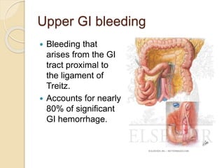
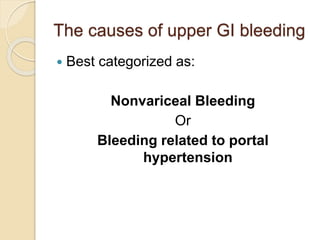
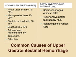
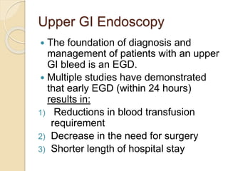
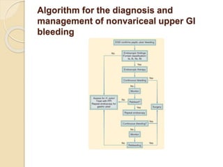
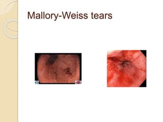
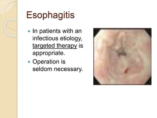
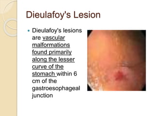
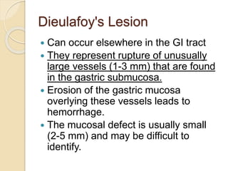
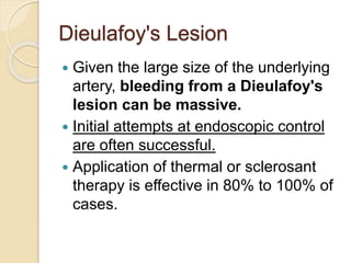
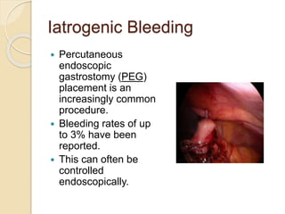
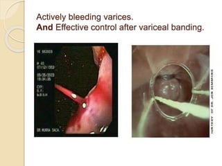
3. Upper GI bleeding
Bleeding that
arises from the GI
tract proximal to
the ligament of
Treitz.
Accounts for nearly
80% of significant
GI hemorrhage.
4. The causes of upper GI bleeding
Best categorized as:
Nonvariceal Bleeding
Or
Bleeding related to portal
hypertension
5. The nonvariceal causes account for about
80% of such bleeding
PUD is the most common cause.
In the remaining 20% of patients, most of
them have cirrhosis and portal
hypertension
which can lead to the development of:
- Gastroesophageal varices,
-Isolated gastric varices,
-Hypertensive portal gastropathy,
All can be the source of an acute upper GI
bleed.
6. The patients with cirrhosis are at high
risk for developing variceal bleeding,
Even in these patients, nonvariceal
sources account for most of the
episodes of GI hemorrhage.[
Because of the greater morbidity and
mortality of variceal bleeding,
Patients with cirrhosis is generally
assumed to have variceal bleeding
and appropriate therapy initiated
8. Upper GI Endoscopy
The foundation of diagnosis and
management of patients with an upper
GI bleed is an EGD.
Multiple studies have demonstrated
that early EGD (within 24 hours)
results in:
1) Reductions in blood transfusion
requirement
2) Decrease in the need for surgery
3) Shorter length of hospital stay
9. Upper GI Endoscopy
Endoscopic identification of the source of
bleeding also permits:
1) Estimate the risk for subsequent or
persistent hemorrhage
2) Facilitating operative planning
20% to 35% of patients undergoing
EGD will require a therapeutic
endoscopic intervention.
5% to 10% will eventually require
surgery.
10. Although the best tool for localization
of the bleeding source is an EGD
1% to 2% of patients the source
cannot be identified
Excessive blood impair visualization
of the mucosal surface.
Aggressive lavage of the stomach with
room temperature normal saline
solution before the procedure can be
helpful.
11. Recent evidence has suggested that a
single bolus injection of intravenous
erythromycin, stimulates gastric
emptying, and can significantly
improve visualization.
If identification of the source is still not
possible:
Angiography may be appropriate in
the reasonably stable patient
12. Although operative intervention must
be seriously considered if the blood
loss is extreme or the patient
hemodynamically unstable.
Tagged RBC scan is seldom
necessary with a confirmed upper GI
bleed.
Contrast studies are usually
contraindicated because they will
interfere with subsequent maneuvers.
13. Specific Causes of Upper
Gastrointestinal Hemorrhage
Nonvariceal Bleeding
14. Peptic Ulcer Disease
PUD is the most frequent cause of
upper GI hemorrhage.
Accounting for about 40% of all cases.
About 10% to 15% of patients with
PUD develop bleeding at some point
in the course of their disease.
Bleeding develops as a consequence
of acid-peptic erosion of the mucosal
surface.
15. Peptic Ulcer Disease
Although chronic blood loss is
common with any ulcer,
Significant bleeding results when there
is involvement of an artery:
1) Either of the submucosa
2) Or, penetration of the ulcer a larger
vessel.
16. Peptic Ulcer Disease
Duodenal ulcers are more common
But gastric ulcers bleed more
commonly
The most significant hemorrhage
occurs when:
duodenal or gastric ulcers penetrate
into branches of the gastroduodenal
artery or left gastric artery.
17. Management
Management Strategies depend on
the appearance of the lesion at
endoscopy.
Endoscopic therapy is instituted if:
1) Bleeding is active
2) Bleeding has already stopped but
there is a significant risk for
rebleeding.
18. The ability to predict the risk for
rebleeding permits:
1. Prophylactic therapy
2. Closer monitoring
3. Earlier detection of hemorrhage in
high-risk patients.
The Forrest classification assess
the risk based on endoscopic
findings, and stratify the patients into
low-, intermediate-, and high-risk
groups
19. Forrest Classification of Endoscopic Findings
and Rebleeding Risks in Peptic Ulcer Disease
CLASSIFICATION REBLEEDING RISK
Grade Ia: Active, pulsatile bleeding
Grade Ib: Active, nonpulsatile
bleeding
Grade IIa: Nonbleeding visible
vessel
Grade IIb: Adherent clot
Grade IIc: Ulcer with black spot
Grade III: Clean, nonbleeding ulcer
bed
High
High
High
Intermediate
Low
Low
20. Algorithm for the diagnosis and
management of nonvariceal upper GI
bleeding
21. Endoscopic therapy
Recommended in:
1)active bleeding
2)visible vessel (Forrest I to IIa).
In cases of an adherent clot
(Forrest IIb): the clot is removed and
the underlying lesion evaluated.
22. Endoscopic therapy
Ulcers with a clean base or a black
spot, secondary to hematin
deposition, are generally not treated
endoscopically.
23. Medical Management In cases
of an acute peptic ulcer bleed
PPIs:
reduce the risk for rebleeding
the need for surgical intervention
patients with a suspected or confirmed
bleeding ulcer are started on a PPI.
24. Association between H. pylori
infection and bleeding
Unlike perforated ulcers, which are
commonly associated with H. pylori
infection,
the association between H. pylori
infection and bleeding is less strong.
Only 60% to 70% of patients with a
bleeding ulcer test positive for H.
pylori.
25. Association between H. pylori
infection and bleeding
This has generated some controversy
as to the importance of H. pylori
treatment in patients with a bleeding
peptic ulcer. However,
several studies and a large meta-
analysis have shown that H. pylori
treatment and eradication, in
patients who test positive for the
infection, results in decreased
rebleeding.
26. Importantly
After the H. pylori has been
eradicated:
No need for long-term acid
suppression.
No increased risk for further bleeding.
27. Patients taking ulcerogenic
medications
Such as NSAIDs or SSRIs,
Who present with a bleeding GI
lesion
These medications are stopped, and
The patient is started on a
nonulcerogenic alternative.
29. Epinephrine injection
Epinephrine injection (1:10,000) to all
four quadrants of the lesion is very
successful in controlling the
hemorrhage.
It has been shown that large-volume
injection (>13 mL) is associated with
better hemostasis,
This suggesting that the endoscopic
injection works in part by compressing
the bleeding vessel and inducing
tamponade.
30. Combination therapy
Epinephrine injection alone is
associated with a high rebleeding
rate
The standard practice is to provide
combination therapy.
This usually means the addition of
thermal therapy to the injection.
31. The thermal energy
A combination of injection with thermal
therapy achieves hemostasis in 90%
of bleeding PUD cases.
The sources of thermal energy can be
heater probes, monopolar or bipolar
electrocoagulation, laser, or argon
plasma coagulator.
32. The thermal energy
The most commonly used therapies
are :-
Electrocoagulation for bleeding ulcers
Argon plasma coagulator for
superficial lesions.
33. Hemoclips
The role of hemoclips is less clear
Several studies have reported mixed
results.
Hemoclips, which can be difficult to
apply, may be particularly effective
when dealing with a spurting vessel
because they provide immediate
control of hemorrhage.
34. Rebleeding of an ulcer
Rebleeding of an ulcer is associated
with a significant increase in mortality
Careful observation of patients at high
risk for rebleeding is important.
35. Second Endoscopy
In those that rebleed, the role of a second
attempt at endoscopic control has been
controversial but recently validated.
Recent study demonstrated that a second
attempt at endoscopic hemostasis is
successful in 75% of patients.Although this
will fail in 25% of patients who will then
require emergent surgery
There is no increase in morbidity or mortality.
Therefore, most clinicians would now
encourage a second attempt at endoscopic
control before surgical intervention.
36. Surgical Management
Despite significant advances in
endoscopic therapy, about 10% of
patients with bleeding ulcers still
require surgical intervention for
effective hemostasis
several clinical and endoscopic
parameters have been employed to
identify patients at high risk for failed
endoscopic therapy
37. Indications for Surgery in
Gastrointestinal Hemorrhage
Hemodynamic instability despite vigorous resuscitation
(>6 units transfusion)
Failure of endoscopic techniques to arrest hemorrhage
Recurrent hemorrhage after initial stabilization (with up
to two attempts at obtaining endoscopic hemostasis)
Shock associated with recurrent hemorrhage
Continued slow bleeding with a transfusion requirement
exceeding 3 units/day
38. Surgical Management
The first priority at operation is
control of the haemorrhage.
Then
A decision must be made regarding
the need for a definitive acid-reducing
procedure.
39. Surgical Management
(Duodenal Ulcer)
The first step in the operation for
duodenal ulcer is exposure of the
bleeding site.
Because most of these lesions are in
the duodenal bulb, longitudinal
duodenotomy or duodenal
pyloromyotomy is performed.
Hemorrhage can typically be controlled
initially with pressure and then direct
suture ligation with nonabsorbable
suture.
40. Surgical Management
(Duodenal Ulcer)
When ulcers are positioned anteriorly,
typically four-quadrant suture ligation
suffices.
A posterior ulcer eroding into the
pancreaticoduodenal or
gastroduodenal artery may require
suture ligature of the vessel proximal
and distal to the ulcer.
41. Surgical Management
(Duodenal Ulcer)
After the bleeding has been
addressed, a definitive acid-reducing
operation is considered.
With the identification of the role of H.
pylori infection in the pathogenesis of
duodenal ulcers
There is an argument that simple
closure and subsequent treatment for
H. pylori is sufficient to prevent
recurrence.
42. Surgical Management
(Duodenal Ulcer)
At the present time an acid-reducing
procedure still appears appropriate in
most patients.
The choice between various
operative procedures has been
based on the hemodynamic condition
of the patient and whether there is a
long-standing history of refractory
ulcer disease.
43. Surgical Management
(Duodenal Ulcer)
As the pylorus has often been opened
in a longitudinal fashion to control the
bleeding,
Closure as a pyloroplasty combined
with truncal vagotomy is the most
frequently used operation for bleeding
duodenal ulcer.
45. Surgical Management
(Duodenal Ulcer)
There is some evidence to suggest
that parietal cell vagotomy may
represent a better therapy for a
bleeding duodenal ulcer in the stable
patient
Surgeon experience with this
procedure may be the determining
factor.
46. Surgical Management
(Duodenal Ulcer)
In a patient who has a known history
of refractory duodenal ulcer disease or
who has failed more conservative
surgery, antrectomy with truncal
vagotomy may be more appropriate.
This procedure is more complex and
is rarely undertaken in a
hemodynamically unstable patient.
47. Surgical Management
(Bleeding gastric ulcers)
Similar to bleeding duodenal ulcers,
control of bleeding is the immediate
priority
This may require gastrotomy and suture
ligation, which, if no other procedure is
performed, is associated with about a
30% risk for rebleeding.
In addition, because of the approximate
10% incidence of malignancy, gastric
ulcer resection is generally indicated.
48. Surgical Management
(Bleeding gastric ulcers)
Simple excision alone is associated
with rebleeding in as many as 20% of
patients,
so that distal gastrectomy is generally
preferred,
although excision combined with
vagotomy and pyloroplasty may be
considered in the high-risk patient.
49. Surgical Management
(Bleeding gastric ulcers)
Bleeding ulcers of the proximal
stomach near the gastroesophageal
junction are more difficult to manage.
Proximal or near-total gastrectomy is
associated with a particularly high
morbidity in the setting of acute
hemorrhage.
50. Surgical Management
(Bleeding gastric ulcers)
Options include:
Distal gastrectomy combined with
resection of a tongue of proximal
stomach to include the ulcer
Or
Vagotomy and pyloroplasty combined
with either wedge resection or simple
oversewing of the ulcer.
51. Mallory-Weiss tears
Mucosal and submucosal tears that
occur near the gastroesophageal
junction.
Classically, these lesions develop in
alcoholic patients after a period of
intense retching and vomiting after
binge drinking,
Can occur in any patient who has a
history of repeated emesis.
52. Mallory-Weiss tears
The mechanism,
proposed by Mallory
and Weiss in 1929, is
forceful contraction of
the abdominal wall
against an unrelaxed
cardia, resulting in
mucosal laceration of
the proximal cardia as
a result of the increase
in intragastric
pressure.
Account for 5% to
10% of cases of upper
GI bleeding
53. Mallory-Weiss tears
Usually diagnosed based on history.
Endoscopy is frequently employed to
confirm the diagnosis. To avoid
missing the lesion.
Important to perform a retroflexion
maneuver and view the area just
below the gastroesophageal junction.
Most tears occur along the lesser
curvature.
55. Mallory-Weiss tears
Supportive therapy is often all that is
necessary because 90% of bleeding
episodes are self-limited, and the
mucosa often heals within 72 hours.
In rare cases of severe ongoing
bleeding, local endoscopic therapy
with injection or electrocoagulation
may be effective.
56. Mallory-Weiss tears
Angiographic embolization, with
absorbable material such as gelatin
sponge, has been successfully employed
If these maneuvers fail, high
gastrotomy and suturing of the
mucosal tear is indicated.
It is important to rule out the diagnosis of
variceal bleeding.
Recurrent bleeding from a Mallory-Weiss
tear is uncommon.
57. Stress- gastritis
Stress-related gastritis is characterized
by the appearance of multiple superficial
erosions of the entire stomach, most
commonly in the body.
It is thought to result from the
combination of acid and pepsin injury in
the context of ischemia from
hypoperfusion states, although NSAIDs
produce a very similar appearance.
58. Stress- gastritis
In the 1960s and 1970s, it was a
commonly encountered lesion in
critically ill patients, with significant
morbidity and mortality from bleeding.
Today, with improvements in the
management of shock and sepsis, as
well as widespread use of acid-
suppressive therapy, significant
bleeding from such lesions is rarely
encountered
59. Esophagitis
The esophagus is infrequently the
source of significant hemorrhage. When
it does occur, it is most commonly the
result of esophagitis.
Esophageal inflammation secondary to
repeated exposure of the esophageal
mucosa to the acidic gastric secretions in
gastroesophageal reflux disease (GERD)
leads to an inflammatory response,
which can result in chronic blood loss.
60. Esophagitis
Ulceration may accompany but the
superficial mucosal ulcerations
generally do not bleed acutely, but
rather present as anemia or guaiac-
positive stools.
A variety of infectious agents may also
cause esophagitis, particularly in the
immunocompromised host .
With infection, hemorrhage can
occasionally be massive.
61. Esophagitis
Other causes of esophageal bleeding
include medications, Crohn's disease,
and radiation.
Treatment typically includes acid-
suppressive therapy.
Endoscopic control of the
hemorrhage, usually with
electrocoagulation or heater probe, is
often successful.
62. Esophagitis
In patients with an
infectious etiology,
targeted therapy is
appropriate.
Operation is
seldom necessary.
63. Dieulafoy's Lesion
Dieulafoy's lesions
are vascular
malformations
found primarily
along the lesser
curve of the
stomach within 6
cm of the
gastroesophageal
junction
64. Dieulafoy's Lesion
Can occur elsewhere in the GI tract
They represent rupture of unusually
large vessels (1-3 mm) that are found
in the gastric submucosa.
Erosion of the gastric mucosa
overlying these vessels leads to
hemorrhage.
The mucosal defect is usually small
(2-5 mm) and may be difficult to
identify.
65. Dieulafoy's Lesion
Given the large size of the underlying
artery, bleeding from a Dieulafoy's
lesion can be massive.
Initial attempts at endoscopic control
are often successful.
Application of thermal or sclerosant
therapy is effective in 80% to 100% of
cases.
66. Dieulafoy's Lesion
In cases that fail endoscopic therapy,
angiographic coil embolization can be
successful.
If these approaches fail, surgical
intervention may be necessary.
Because of the difficulties in
visualization and palpation of these
lesions, prior endoscopic tattooing can
facilitate the procedure.
67. Dieulafoy's Lesion
A gastrotomy is performed, and
attempts are made at identifying the
bleeding source.
In cases in which the bleeding point is
not identified, a partial gastrectomy
may be necessary.
68. Gastric Antral Vascular Ectasia
Also known as
“watermelon stomach,”
Gastric antral vascular
ectasia (GAVE) is
characterized by a
collection of dilated
venules appearing as
linear red streaks
converging on the
antrum in longitudinal
fashion, giving it the
appearance of a
watermelon.
69. Gastric Antral Vascular Ectasia
Acute severe
hemorrhage is rare
in GAVE
Most patients
present with
persistent, iron
deficiency anemia
from continued
occult blood loss.
70. Gastric Antral Vascular Ectasia
Endoscopic therapy is indicated for
persistent, transfusion-dependent
bleeding and has been reportedly
successful in up to 90% of patients.
The preferred endoscopic therapy is
argon plasma coagulation.
Those failing endoscopic therapy are
considered for antrectomy.
71. Malignancy
Malignancies of the upper GI tract are
usually associated with chronic
anemia or hemoccult-positive stool
rather than episodes of significant
hemorrhage.
On occasion, malignancies present as
ulcerative lesions that bleed
persistently.
This is perhaps most characteristic of
the GI stromal tumor (GIST)
72. Malignancy
Bleeding may occur with a variety of
other lesions, including leiomyomas
and lymphomas.
Although endoscopic therapy is often
successful in controlling hemorrhage,
the rebleeding rate is high.
Therefore, when a malignancy is
diagnosed, surgical resection is
indicated.
73. Malignancy
The extent of resection is dependent
on the specific lesion and on whether
the resection is curative or palliative.
Palliative resections for control of
bleeding usually entail wedge
resections.
Standard cancer operations are
indicated when possible, although this
may depend on the hemodynamic
stability of the patient.
74. Aortoenteric Fistula
Primary aortoduodenal fistulas are
rare lesions developing in up to 1% of
aortic graft cases.
They typically develop in the setting of
a previous abdominal aortic aneurysm
repair.
May occur as a result of an
inflammatory or infectious aortitis.
75. Aortoenteric Fistula
The interval between surgery and
hemorrhage can be days to years.
The median interval is about 3 years.
The sequence is thought to involve
development of a pseudoaneurysm at
the proximal anastomotic suture line in
the setting of an infection,
With subsequent fistulization into the
overlying duodenum.
76. Aortoenteric Fistula
Aortoenteric fistula is considered in
all patients with GI hemorrhage
with a known abdominal aortic
aneurysm or a previous prosthetic
aneurysm repair.
Hemorrhage is often massive and fatal
unless immediate surgical intervention
is undertaken.
77. Aortoenteric Fistula
Typically, patients with bleeding from
an aortoenteric fistula will present first
with a “sentinel bleed.”
This is a self-limited episode that
heralds the subsequent massive, and
often fatal, hemorrhage.
This prompts urgent EGD because
diagnosis at this stage can be
lifesaving.
78. Aortoenteric Fistula
Any evidence of bleeding in the distal
duodenum (3rd or 4th portion) is
considered diagnostic.
CT scan with IV contrast will
demonstrate air around the graft
(suggestive of an infection),
Possible pseudoaneurysm,
Rarely the presence of IV contrast in
the duodenal lumen.
80. Aortoenteric Fistula
Therapy includes :
Ligation of the aorta proximal to the
graft.
Removal of the infected prosthesis.
Extra-anatomic bypass.
The defect in the duodenum is often
small and can be repaired primarily.
o This is a complex and often morbid
procedure.
81. Hemobilia
Difficult diagnosis to make.
Typically associated with trauma,
recent instrumentation of the biliary
tree, or hepatic neoplasms.
Unusual cause of GI bleeding.
Suspected in anyone who presents
with hemorrhage, right upper quadrant
pain, and jaundice.
Unfortunately, this triad is seen in less
than half of patients
82. Hemobilia
Endoscopy can be helpful by
demonstrating blood at the ampulla.
Angiography is the diagnostic
procedure of choice.
If diagnosis is confirmed:
Angiographic embolization is the
preferred treatment.
83. Hemosuccus Pancreaticus
Rare cause of upper GI bleeding.
Bleeding from the pancreatic duct, or
hemosuccus pancreaticus.
Typically caused by erosion of a
pancreatic pseudocyst into the splenic
artery.
Presents with abdominal pain and
hematochezia.
84. Hemosuccus Pancreaticus
Difficult diagnosis to make and
requires a high index of suspicion.
In patients with abdominal pain, blood
loss, and a past history of pancreatitis.
Angiography is diagnostic and permits
embolization, which is often
therapeutic.
In cases that are amenable to a distal
pancreatectomy, this procedure often
results in cure.
85. Iatrogenic Bleeding
Upper GI bleeding may follow
therapeutic or diagnostic procedures.
Hemobilia may be iatrogenic in nature,
particularly after percutaneous
transhepatic procedures.
Another common cause of iatrogenic
bleeding is endoscopic
sphincterotomy. This can occur in up
to 2% of cases.
87. Iatrogenic Bleeding
Upper GI bleeding can also be seen in
patients who have recently undergone
upper GI surgery.
In cases in which a resection and
anastomosis have been performed,
the source of the bleeding may be the
suture or the staple line.
It is safe to perform a diagnostic or
even therapeutic EGD
89. Bleeding Related to Portal
Hypertension
Upper GI bleeding is a serious
complication of portal hypertension.
Most often in the setting of cirrhosis.
Hemorrhage related to portal
hypertension is most commonly the
result of bleeding from varices.
90. The Varices
These dilated submucosal veins
develop in response to the portal
hypertension.
Providing a collateral pathway for
decompression of the portal system
into the systemic venous circulation.
Most common in the distal esophagus.
May develop in the stomach and the
hemorrhoidal plexus of the rectum.
91. The Varices
They can reach sizes of 1 to 2 cm.
As they enlarge, the overlying mucosa
becomes increasingly tenuous and
bleeding occurring with minimal
trauma.
92. Portal hypertensive gastropathy
Diffuse dilation of the mucosal and
submucosal venous plexus of the
stomach associated with overlying
gastritis.
Incompletely understood entity.
Endoscopically, the stomach acquires
a snakeskin-like appearance with
cherry-red spots.
This entity uncommonly produces
major hemorrhage.
93. Gastroesophageal varices
Develop in about 30% of patients with
cirrhosis and portal hypertension.
30% in this group develop variceal
bleeding.
Compared with nonvariceal bleeding,
variceal hemorrhage is associated with :
Increased risk for rebleeding
Increased need for transfusions
increased hospital stay, and mortality
94. Gastroesophageal varices
Hemorrhage is frequently massive,
accompanied by hematemesis and
hemodynamic instability.
The hepatic functional reserve,
estimated by Child's criteria.
Despite improvements in the medical
management of these patients, the 6-
week mortality rate after the first
hemorrhage is about 20%.
96. Algorithm for diagnosis and management of GI hemorrhage related to portal
hypertension. EGD, esophagogastroduodenoscopy; TIPS, transjugular
intrahepatic portosystemic shunt.
97. Medical Management
For Bleeding GE varices
In patients with cirrhosis:
pharmacologic therapy to reduce portal
hypertension even while preparing for
emergent EGD.
Vasopressin produces splanchnic
vasoconstriction but also cardiac
vasoconstruction and can lead to MI.
Somatostatin or its synthetic analogue,
Octreotide, is now the vasoactive agent of
choice without cardiac side effects.
Studies have demonstrated its efficacy in
controlling variceal bleeding.
98. Endoscopic Management
For Bleeding GE varices
Early EGD is critical to evaluate the
source of bleeding.
If bleeding esophageal varices are
identified:
Both sclerotherapy and variceal
banding have been shown to control
hemorrhage effectively.
Banding appears to have a lower
complication rate and, when expertise
is available, is the therapy of choice
99. Endoscopic Management
For Bleeding GE varices
Endoscopic approaches, sometimes
with as many as three treatments over
24 hours, control the hemorrhage in
up to 90% of patients with esophageal
varices.
Unfortunately, gastric varices are not
effectively managed by endoscopic
techniques.
101. Other Management
For Bleeding GE varices
In cases in which pharmacologic or
endoscopic therapies fail to control the
hemorrhage:
Tamponade can be successful in
temporizing the hemorrhage.
The Sengstaken-Blakemore tube
consists of a gastric tube with
esophageal and gastric balloons. The
Sengstaken-Blakemore tube consists of
a gastric tube with esophageal and
gastric balloons.
102. Other Management
For Bleeding GE varices
The gastric balloon is inflated, and
tension is applied to the
gastroesophageal junction.
If this does not control the hemorrhage,
the esophageal balloon is inflated as
well. compressing the venous plexus
between them.
These tubes are associated with a high
rate of complications with both aspiration
and inappropriate placement with
esophageal perforation.
103. Other Management
For Bleeding GE varices
Hemorrhage recurs on deflation in up
to 50% of patients.
Currently, balloon tamponade is
reserved for patients with massive
hemorrhage to permit more definitive
therapies.
In cases of severe variceal bleeding
that cannot be controlled
endoscopically, emergent portal
decompression is indicated.
104. percutaneous transjugular
intrahepatic portosystemic shunt
(TIPS)
Required in about 10% of patients with
variceal bleeding.
TIPS procedure can be lifesaving in
patients who are hemodynamically
unstable from refractory variceal
bleeding.
Associated with significantly less
morbidity and mortality than surgical
decompression.
105. percutaneous transjugular
intrahepatic portosystemic shunt
(TIPS)
Studies have shown that TIPS can
control bleeding in 95% of cases.
Rebleeding occurs in up to 20% within
the first month, usually related to
occlusion.
In cases in which TIPS is not available
or fails, emergent surgical intervention
is indicated.
106. Isolated gastric varices
Managed in much the same way as
esophageal varices, although
endoscopic therapy tends to be less
successful.
Pharmacotherapy is primarily
indicated, but when this fails, portal
decompression by means of TIPS or a
surgical shunt is recommended.
107. Isolated gastric varices
Rarely, isolated gastric varices occur after
splenic vein thrombosis.
Most commonly seen in the setting of
pancreatitis.
portal pressures are normal, but left-sided
hypertension, decompressed from the spleen
through the short gastric vessels, produces
the varices.
splenectomy was routinely recommended,
recent data suggest that the incidence of
variceal bleeding is low (4%)
splenectomy is not routinely undertaken now.
108. Portal hypertensive gastropathy
Unlike variceal hemorrhage, bleeding
from portal hypertensive gastropathy is
not amenable to endoscopic treatment.
Because of the diffuse nature of the
mucosal abnormalities. The underlying
pathology involves elevated portal
venous pressures;
Therefore, pharmacologic therapies
aimed at reducing portal venous
pressure are indicated.
If pharmacologic therapy fails to control
acute bleeding, TIPS is considered.