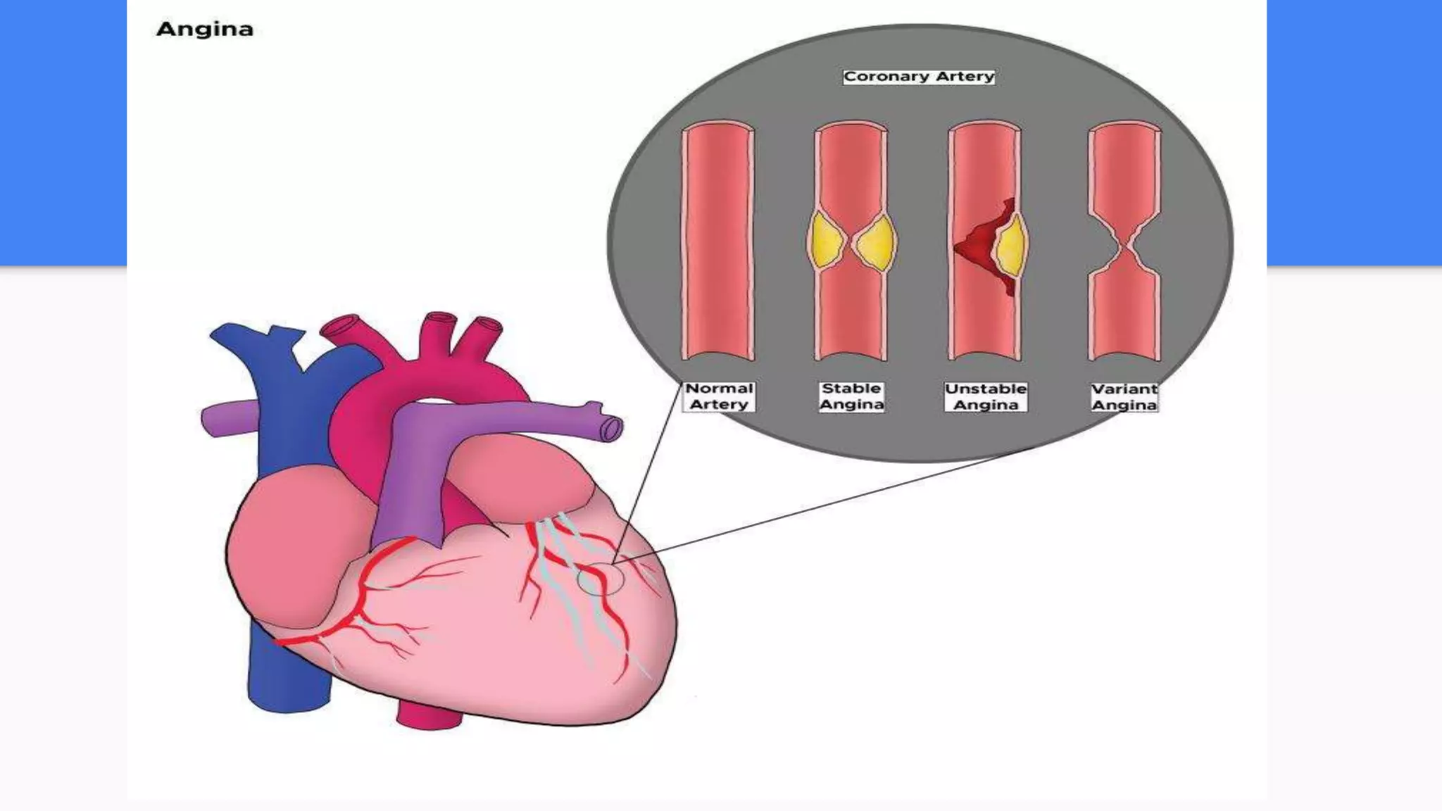
Unstable angina is chest pain caused by reduced blood flow to the heart, often due to a partially blocked artery. It can occur at rest and is considered a medical emergency. The main causes are atherosclerotic plaques that rupture or spasm of the coronary arteries. Patients experience chest pressure or pain that may radiate to the arms or jaw. Evaluation involves ECG, cardiac enzymes, and cardiac stress testing to determine the risk of a heart attack. Treatment focuses on improving blood flow through aspirin, nitroglycerin, and sometimes angioplasty or stenting of blocked arteries.
2. Unstable Angina
Unstable angina is chest discomfort or pain caused by an insufficient flow of
blood and oxygen to the heart. It is part of the acute coronary syndromes and
may lead up to a heart attack
3. Etiology
The most common cause of unstable angina is due to coronary artery
narrowing due to a thrombus that develops on a disrupted atherosclerotic
plaque and is nonocclusive.
A less common cause is vasospasm of a coronary artery (variant Prinzmetal
angina).
6. History and physical
Patients will often present with chest pain, shortness of breath.
1. The chest pain will often be described as pressure-like, tightness, burning, sharp type
of pain
2. The pain will often radiate to the jaw or arms, both left and right sides can be affected.
3. Constitutional symptoms such as nausea, vomiting, diaphoresis, dizziness, and
palpitations may also be present.
4. Exertion may worsen pain and rest can ease the pain.
5. Nitroglycerin and aspirin administration may also improve the pain.
7. Physical Examination
The exam will likely be normal, although the patient may be clutching at their chest, sweating, have
labored breathing, their heart sounds may be tachycardic, and rales may be heard due to pulmonary
edema.
Findings suggestive of a high-risk situation include:
Dyskinetic apex
Elevated JVP
Presence of S3 or S4
New apical systolic murmur
Presence of rales and crackles
Hypotension
8. Evaluation
The patient should have an ECG to evaluate for ischemic signs or possible STEMI.
The ECG in unstable angina may show hyperacute T-wave, flattening of the T-waves,
inverted T-waves, and ST depression.
Any number of arrhythmias may be present in acute coronary syndrome including
junctional rhythms, sinus tachycardia, ventricular tachycardia, ventricular fibrillation, left
bundle branch block, and others.
9. The patient should also have lab work that includes:
1. complete blood count evaluating for anemia,
2. platelet count, and
3. basic metabolic profile evaluating for electrolyte abnormalities.
10. A troponin test should be performed to determine if any of the myocardium has
infarcted.
A pro-brain natriuretic peptide (Pro-BNP) can also be checked, as an elevated
level is associated with higher mortality.
Coagulation studies may be appropriate if the patient will be anticoagulated or
anticoagulation is anticipated.
Often, a chest x-ray will show the heart size and the size of the mediastinum so
the physician may screen for dissection and other explanations of chest pain.
11. The patient should be kept on a cardiac monitor to evaluate for any rhythm
changes.
Further testing may include any number of cardiac stress tests (walking
treadmill stress test, stress echocardiogram, myocardial perfusion imaging,
cardiac CT/MRI, or the gold standard, cardiac catheterization).
12. Treatment
The mainstay of treatment focuses on improving perfusion of the
coronary arteries. This is done in several ways:
1. Patients are often treated with aspirin for its antiplatelet therapies, 162 to
325 mg orally, or 300 mg rectally if the patient is unable to swallow.
The aspirin should be administered with 30 minutes.
13. 2. Nitroglycerin improves perfusion by vasodilation of the coronaries allowing
improved flow and improved blood pressure.
This will decrease the amount of work the heart has to perform, which
decreases the energy demand of the heart.
14. Clopidogrel is an option for patients not able to tolerate aspirin.
Prasugrel is more effective than clopidogrel but is associated with a higher risk
of bleeding.
15. Supplemental oxygen should be given as well via nasal cannula to maintain
appropriate oxygen saturation.
Anticoagulation with low or high molecular weight heparin.
Beta-blockers also can decrease the energy demand by decreasing blood
pressure and heart rate.
16. Cardiac angiography is indicated in unstable angina if the patient has:
● Cardiogenic shock
● Depressed ejection fraction
● Angina refractory to pharmacological therapy
● New MR
● Unstable arrhythmias
● Early PCI in NSTEMI (within 6 hours) has been shown to have lower mortality
than those who undergo delayed PCI.