
Recommended
More Related Content
What's hot
What's hot (18)
Similar to Coronary Artery Disease Diagnosis and Types
Similar to Coronary Artery Disease Diagnosis and Types (20)
Recently uploaded
Recently uploaded (20)
Coronary Artery Disease Diagnosis and Types
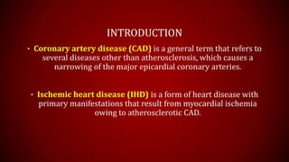
- 1. INTRODUCTION • Coronary artery disease (CAD) is a general term that refers to several diseases other than atherosclerosis, which causes a narrowing of the major epicardial coronary arteries. • Ischemic heart disease (IHD) is a form of heart disease with primary manifestations that result from myocardial ischemia owing to atherosclerotic CAD.
- 4. ETIOLOGY 1. Decreased Blood Flow a) Atherosclerosis b) Coronary artery Spasm c) Traumatic injury d) Embolic events 2. Increased oxygen demand in the presence of fixed restricted oxygen supply a) Diastole b) Systole • Contractile (inotropic) state of the heart • Increased systolic wall tension • Lengthening of ejection time • Changes in heart rate 3. Reduced blood oxygenation
- 7. ANGINA PECTORIS • The term angina pectoris is applied to varying forms of transient chest discomfort that are attributable to insufficient myocardial oxygen. 1. Angina is a clinical syndrome characterized by discomfort in the chest, jaw, shoulder, back, and arms, which is usually aggravated by exertion or stress and relieved by nitroglycerin. 2. Angina can occur in patients with valvular heart disease, uncontrolled hypertension, as well as in noncardiac organ systems such as the chest wall, esophagus, or lungs.
- 8. COMMON CAUSES • Major Cause: – Atherosclerotic lesions • Others: – Tachycardia – Anemia – Hyperthyroidism – Hypotension – Arterial hypoxemia
- 9. TYPES 1. Stable (Classic) Angina 2. Unstable Angina 3. Angina Decubitus 4. Prinzmetal Angina (Vasospastic or Variant Angina)
- 10. STABLE (CLASSIC) ANGINA • In this most common form, has a more predictable pattern, which is brought on by exertion, Emotional stress, Or a heavy meal which is usually relieved By rest, nitroglycerin, Or both • Five components are usually considered: 1. quality, 2. location, and 3. duration of pain; 4. factors provoking Pain and 5. factors that Relieve pain. • Pain has been referred to as “squeezing”, ”grip-like,” “pressure-like”, “suffocating”, and “heavy” and is usually referred to as a discomfort rather than “pain”. • The anginal episode typically lasts for “minutes” and is usually substernal but has a tendency to radiate to the neck, jaw, epigastrium, or arms. • Characteristically, the discomfort builds to a peak, radiating to the jaw, neck, shoulder, and arms, and then subsides without residual sensation. Angina is normally related to physical exertion, and the discomfort usually subsides quickly (i.e., in 3 to 5 mins) with rest; if precipitated by emotional stress, the episode tends to last longer (i.e., about 10 mins). • Stable angina is characteristically the result of a fixed obstruction in a coronary artery.
- 11. UNSTABLE ANGINA a) In many patients who experience unstable angina, symptoms will be caused by significant coronary artery disease. Angina is considered unstable and requires further evaluation if patients experience 1) Rest angina, which usually is prolonged 20 mins occurring within a week of presentation 2) Severe new-onset angina refers to angina of at least Canadian Cardiovascular Society Classification (CCSC) to class III severity, with onset within 2 months of initial presentation 3) Increasing angina refers to previously diagnosed angina that is distinctly more frequent, longer in duration, or lower in threshold 4) Decreased response to rest or nitroglycerin b) Unstable angina predicts a higher short-term risk, represents a progressive clinical entity, may signal incipient MI, is referred to as an acute coronary syndrome, and should be reported promptly to a physician.
- 12. ANGINA DECUBITUS (NOCTURNAL ANGINA) • This angina occurs in the recumbent position and is not specifically related to either rest or exertion. • Gravitational forces shift fluids within the body with a resultant increase in ventricular volume, which increases oxygen needs and produces angina decubitus, and which may indicate cardiac decompensation. • Diuretics alone or in combination effectively reduce left ventricular volume and may aid the patient. • Nitrates such as nitroglycerin may relieve the paroxysmal nocturnal dyspnea (PND) associated with angina decubitus by reducing preload, owing to venous pooling, and improving left ventricular dysfunction. • PND refers to a condition where fluid accumulation in the lungs, normally due to gravitational forces when one is in the recumbent position, makes it very difficult for a patient to breath. Some have referred to this as “cardiac asthma, as the patient is unable to breath and it gets progressively worse. Sleeping with several additional pillows might allow gravity to work on the added fluid in the lungs, with a resultant decrease in symptoms. However, the underlying cause needs to be corrected or the PND will remain.
- 13. PRINZMETAL ANGINA (VASOSPASTIC ANGINA) • Coronary artery spasm that reduces blood flow precipitates this angina. Th e spasm may be superimposed on a coronary artery that already has a fixed obstruction owing to thrombi or plaque formation. • It usually occurs at rest (i.e., pain may disrupt sleep) rather than with exertion or emotional stress, and usually resolves without progression to an acute MI. However, if the attack is prolonged, MI, life- threatening ventricular arrhythmias, and sudden cardiac death can occur. • Characteristically, an electrocardiogram (EKG/ECG) taken during an attack reveals a transient ST- segment elevation, which returns toward normal after the acute attack. The gold standard for identifying Prinzmetal angina is the use of coronary angiography with the administration of agents capable of inducing vasospasm, such as ergot alkaloids. • Calcium-channel blockers, rather than -blockers, are most effective for this form of angina. Nitroglycerin may not provide relief, depending on the cause of vasospasm
- 14. ECG Changes during STEMI
- 15. DIAGNOSTIC TEST RESULTS 1. EKG/ECG: The EKG/ECG is normal in 50% or more of patients with stable angina pectoris, and a normal resting EKG/ECG does not exclude severe IHD. However, an EKG/ECG with evidence of left ventricular hypertrophy or ST-T-wave changes consistent with myocardial ischemia favors the diagnosis of angina pectoris. Th e presence of Q waves from a previous MI makes the diagnosis of IHD very likely. An EKG/ECG obtained during chest pain is abnormal in 50% of patients with angina who have a normal resting EKG/ECG. Th e ST segment can be either elevated or depressed. 2. STRESS TESTING (EXERCISE EKG/ECG) It is a well-established procedure, which aids the diagnosis in patients who have normal resting EKGs/ECGs. The most commonly used definition for a positive test is a 1-mm ST- segment depression or elevation for 60 to 80 msec either during or after exercise. Exercise stress testing is preferable to other variations of the stress test (pharmacological) in patients who are able to exercise.
- 16. DIAGNOSTIC TEST RESULTS 3. PHARMACOLOGICAL STRESS TESTING: • performed in suspected IHD patients when they are not able to perform more than moderate exercise due to various reasons (i.e., severe arthritis, prior injury, reduced exercise tolerance as a result of debilitating illnesses, etc.), • or in patients who are unable to increase the heart rate. a) Intravenous DIPYRIDAMOLE (Persantine; coronary vasodilation), phosphodiesterase inhibition and TXA2 inhibition. b) ADENOSINE (Adenocard; direct coronary vasodilation) by inhibiting cellular uptake and degradation of adenosine increase coronary blood flow, and c) high-dose DOBUTAMINE (Dobutrexョ; 20 to 40 mcg/kg/min) increase oxygen demand through increased heart rate, systolic blood pressure, and myocardial contractility 4. STRESS PERFUSION IMAGING: [with thallium-201 or more recently, technetium-99m (Tc) ] – can diagnose multivessel disease, localized ischemia, and may be able to determine myocardial viability. – Coronary arteriography and cardiac catheterization are very specific and sensitive but are also invasive,
- 17. DIAGNOSTIC TEST RESULTS 5. Various drugs can have an effect on the EKG/ECG and should be considered before, during, and after an exercise test is carried out. Examples include a) Digoxin produces abnormal exercise-induced ST depression in 25% to 40% of apparently healthy, normal subjects without ischemia. b) Beta-Adrenergic blockers may delay the development of an abnormal EKG/ECG if patients receive them before or during a stress test. If possible, therapy should be slowly withheld from the patient at least four to five half-lives before the exercise testing. c) Antihypertensives such as vasodilators can alter the stress test by altering the normal hemodynamic response of blood pressure. In addition, short-term use of nitrates can attenuate angina and ST-segment changes associated with myocardial ischemia.
- 20. ACUTE CORONARY SYNDROME (ACS) • DEFINITION: – ACS is a relatively new term that has been introduced into the medical literature to describe any pattern of clinical symptoms that reflects the development of acute MI (Figure below). This category includes the symptoms related to STEMI, NSTEMI, and unstable angina. Adapted from Vanscoy G. Integrating new fibrinolytic findings into AMI reperfusion and combination therapy: 2002 and beyond. Paper presented at the meeting of the Delaware Valley Chapter of the Pennsylvania Society of Health-Systems Pharmacists; March 7, 2002.
- 21. ACUTE CORONARY SYNDROME (ACS) • The appropriate determination of prognosis as well as clinical interventions depends upon correct classification of patients presenting with presumed ACS. • If a portion of the cardiac muscle suffers a severe and prolonged restriction of oxygenated coronary blood, It leads to ACS such as STEMI and NSTEMI. • In most patients, the cause is an occlusive or near-occlusive thrombus overlying or adjacent to a ruptured atherosclerotic plaque. • This results in cellular ischemia, tissue injury, and tissue necrosis. About 1.5 million people suffer an acute myocardial infarction (AMI) each year. UA is believed to indicate an imminent AMI, and the goal of treatment is to prevent the development of the AMI.
- 22. ACUTE CORONARY SYNDROME (ACS) STEMI • Requires immediate reperfusion therapy – Thrombolysis – Percutaneous Coronary Intervention (PCI) • To remove the offending thrombus UA and NSTEMI • No benefit from the reperfusion therapy • Patients may develop Q-wave MI or non- Q-wave MI. • 25% of patients with both NSTEMI and elevated cardiac enzymes eventually develop Q-wave MI.
- 23. ACUTE CORONARY SYNDROME (ACS) (DIAGNOSIS) • ECG/EKG is at the center of the decision pathway • Confirmed with serial cardiac markers in >90% of patients presenting with ST-elevation. • Patients without ST-elevation UA or NSTEMI – The final diagnosis is made after the presence or absence of serial cardiac markers. • Diagnostic test results confirm the presence of ACS, locale and extent of myocardial damage based on – Patient’s complaints – 12-lead EKG/ECG – Cardiac Enzymes – Cardiac Imaging
- 24. ACUTE CORONARY SYNDROME (ACS) (DIAGNOSIS) • Serial 12-lead EKG: – Abnormal ECG may be absent or inconclusive during first few hours after ACS in 15% of the cases. – If present findings show progressive changes a) First, ST-elevation (injury current) appears injured area. a) Peaked upright or inverted T waves myocardial injury (transmural Q-wave MI) b) Persistant ST-depression non-Q-wave MI. b) Q-waves developing indicate Necrosis but can be seen in other conditions as well. c) Unequivocal diagnosis is only made in all three abnormalities. a) E.g. in non-Q-wave MI only ST-depression may appear. d) The most serious arrhythmic complication of an AMI is Ventricular fibrillation. e) Ventricular premature beats (VPBs) commonly occurring arrhythmia & require treatment.
- 25. ACUTE CORONARY SYNDROME (ACS) (DIAGNOSIS) • Cardiac Enzymes: – Creatinine Kinase –Heart muscle (CK-MB) • 3-12 hours (after pain) elevated • 24 hours Peak level • Returns to baseline 48 – 72 hours (2-3 days) • Typical rise and fall only seen in MI. – Cardiac Troponin I (cTnI) and Cardiac toponin T (cTnT) • More sensitive (after 6 hours of onset) and specific test. • If negative repeat after 12-16 hours • 3 – 12 hours (after pain) elevated • 24 – 48 hours peak level • Returns to baseline 5 – 14 days. – Lactate dehydrogenase (LDH) • Ratio of LDH1 : LDH2 is helpful in diagnosis. • Replaced by cTnT assays. • Cardiac Imaging: – 99mTc-pyrophosphate scintigraphy, – Myocardial perfusion imaging, – 2-D echocardiography – Coronary angiography
- 26. OVERALL TREATMENT GOALS 1. To relieve chest pain and anxiety 2. To reduce cardiac workload and stabilize cardiac rhythm 3. To prevent/reduce myocardial damage by limiting the area affected and preserving pump function 4. To prevent or arrest complications, such as lethal arrhythmias, AMI, HF, or sudden death 5. To reopen (or reperfuse) closed coronary vessels with thrombolytic drugs and/or PCI
- 27. EVALUATION OF ACUTE CORONARY SYNDROME PATIENT.