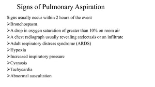
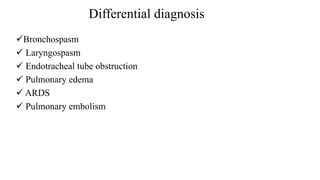
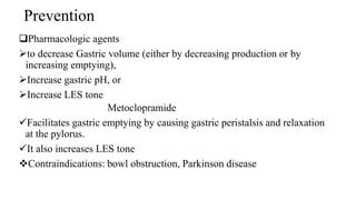
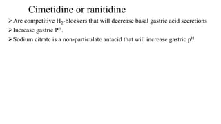
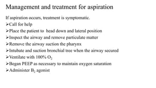
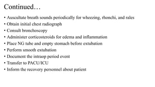
Aspiration Management
Prepared by: Wasihun Aragie
NOV 2019
Definition
ASPIRATION is defined as the inhalation of oropharyngeal or gastric
contents into the larynx and lower respiratory tract.
The nature of the aspirated material, volume of the aspirated material, and
state of the host defenses are three important determinants of the extent and
severity of aspiration pneumonia.
Aspiration can occur at any time during the perioperative period.
Classification of aspiration
Aspiration pneumonitis is defined as acute lung injury after the inhalation
of regurgitated gastric contents.
 Aspiration pneumonitis (Mendelson’s syndrome) is a chemical injury
caused by the inhalation of sterile gastric contents.
This condition involves lung tissue damage as a result of aspiration of non-
infective but very acidic gastric fluid .
Continued…
The pH value of less than 2.5 as a threshold for chemical pneumonitis and
the critical volume for severe pneumonitis is estimated at 0.8 mL/kg
Most common causes are drug overdose, seizures, a massive
cerebrovascular accident, or the use of anesthesia.
Continued…
Aspiration pneumonia develops after the inhalation of colonized
oropharyngeal material.
Any condition that increases the volume or bacterial burden of
oropharyngeal secretions in a person with impaired defense mechanisms
may lead to aspiration pneumonia.
Exogenous lipoid pneumonia (ELP) is a rare form of pneumonia
caused by inhalation or aspiration of a fatty substance. ELP has been
reported with inhalation or ingestion of petroleum jelly, mineral oils, “nasal
drops,” and even intravenous injection of olive oil.
Mechanisms for Protection of Reflux and Aspiration in the
Awake Patient
Lower esophageal sphincter tone (LES): is the primary barrier to gastro-esophageal reflux
Gastro-esophageal angle
Upper esophageal sphincter
Air way reflexes like:
Sneezing
Apnea
Swallowing
 Laryngeal closure
 Coughing
Risk factors for regurgitation and pulmonary
aspiration under general anesthesia
 Obesity
 Depressed level of consciousness
 History of gastritis/ulcer
 Bowel obstruction
 Pregnancy – greater than 12 weeks gestation
 Pain/stress
 Emergency surgery
 ASA IV-V
Continued…
 Ileus
 Trauma
 Concurrent opioid administration
 Symptomatic hiatal hernia
 Esophageal disorders/previous esophageal surgery
 Recent meal
 Diabetes mellitus
Signs of Pulmonary Aspiration
Signs usually occur within 2 hours of the event
Bronchospasm
A drop in oxygen saturation of greater than 10% on room air
A chest radiograph usually revealing atelectasis or an infiltrate
Adult respiratory distress syndrome (ARDS)
Hypoxia
Increased inspiratory pressure
Cyanosis
Tachycardia
Abnormal auscultation
Differential diagnosis
Bronchospasm
 Laryngospasm
 Endotracheal tube obstruction
 Pulmonary edema
 ARDS
 Pulmonary embolism
Prevention
Pharmacologic agents
to decrease Gastric volume (either by decreasing production or by
increasing emptying),
Increase gastric pH, or
Increase LES tone
Metoclopramide
Facilitates gastric emptying by causing gastric peristalsis and relaxation
at the pylorus.
It also increases LES tone
Contraindications: bowl obstruction, Parkinson disease
Cimetidine or ranitidine
Are competitive H2-blockers that will decrease basal gastric acid secretions
Increase gastric PH.
Sodium citrate is a non-particulate antacid that will increase gastric pH.
Continued…
Omeprazole,Rabrazole,Lansoprazole are proton pump inhibitors that block
H+-K+-adenosine triphosphates' activity at the secretory surface of the
parietal cells in the stomach. These drugs decrease the volume and increase
the pH of gastric secretions
Glycopyrrolate, an anticholinergic, will increase gastric pH by inhibiting
vagal mediated gastric acid production. Atropine, however, is ineffective.
Continued…
Nasogastric tube placement
Preoperative fasting
Applying cricoid pressure during rapid sequence induction
Case
• A 23 years old male patient comes to DRH with compliant of two episode of
vomiting, severe abdominal pain of 6 hours duration and diagnosis as acute
appendicitis and scheduled for appendectomy. The anesthetist perform rapid
assessment and found the following.
History no previous anesthesia and surgery exposure. He has history of burn 5
years later at the neck area chest as well recently eat food.
PE slight limitation of neck movement 2 fingerS admit and mallampati 3 the
patient was anesthetized and intubated successfully with the second attempt
using stylate and applying cricoid pressure in the middle of procedure saturation
decrease from 95 to 80%. The anesthetist notice gastric content in the mouth
area and bilateral crepitation on auscultation and suspect aspiration of gastric
content into the lung.
Manage this patient following appropriate steps of aspiration
management
Required resource
Emergency drug(adrenalin)
Monitors (BP apparatus, stethoscope and pulse oxymetry)
GA equipment's(oxygen source, laryngoscope, ETT, IV anesthetic drugs,
suction machine, suction tube, stylate, airway mask)
Management and treatment for aspiration
If aspiration occurs, treatment is symptomatic.
Call for help
Place the patient to head down and lateral position
Inspect the airway and remove particulate matter
Remove the airway suction the pharynx
Intubate and suction bronchial tree when the airway secured
Ventilate with 100% O2
Began PEEP as necessary to maintain oxygen saturation
Administer B2 agonist
Continued…
• Auscultate breath sounds periodically for wheezing, rhonchi, and rales
• Obtain initial chest radiograph
• Consult bronchoscopy
• Administer corticosteroids for edema and inflammation
• Place NG tube and empty stomach before extubation
• Perform smooth extubation
• Document the intraop period event
• Transfer to PACU/ICU
• Inform the recovery personnel about patient
Thank you

Aspiration management

  • 1.
    Aspiration Management Prepared by:Wasihun Aragie NOV 2019
  • 2.
    Definition ASPIRATION is definedas the inhalation of oropharyngeal or gastric contents into the larynx and lower respiratory tract. The nature of the aspirated material, volume of the aspirated material, and state of the host defenses are three important determinants of the extent and severity of aspiration pneumonia. Aspiration can occur at any time during the perioperative period.
  • 3.
    Classification of aspiration Aspirationpneumonitis is defined as acute lung injury after the inhalation of regurgitated gastric contents.  Aspiration pneumonitis (Mendelson’s syndrome) is a chemical injury caused by the inhalation of sterile gastric contents. This condition involves lung tissue damage as a result of aspiration of non- infective but very acidic gastric fluid .
  • 4.
    Continued… The pH valueof less than 2.5 as a threshold for chemical pneumonitis and the critical volume for severe pneumonitis is estimated at 0.8 mL/kg Most common causes are drug overdose, seizures, a massive cerebrovascular accident, or the use of anesthesia.
  • 5.
    Continued… Aspiration pneumonia developsafter the inhalation of colonized oropharyngeal material. Any condition that increases the volume or bacterial burden of oropharyngeal secretions in a person with impaired defense mechanisms may lead to aspiration pneumonia. Exogenous lipoid pneumonia (ELP) is a rare form of pneumonia caused by inhalation or aspiration of a fatty substance. ELP has been reported with inhalation or ingestion of petroleum jelly, mineral oils, “nasal drops,” and even intravenous injection of olive oil.
  • 6.
    Mechanisms for Protectionof Reflux and Aspiration in the Awake Patient Lower esophageal sphincter tone (LES): is the primary barrier to gastro-esophageal reflux Gastro-esophageal angle Upper esophageal sphincter Air way reflexes like: Sneezing Apnea Swallowing  Laryngeal closure  Coughing
  • 7.
    Risk factors forregurgitation and pulmonary aspiration under general anesthesia  Obesity  Depressed level of consciousness  History of gastritis/ulcer  Bowel obstruction  Pregnancy – greater than 12 weeks gestation  Pain/stress  Emergency surgery  ASA IV-V
  • 8.
    Continued…  Ileus  Trauma Concurrent opioid administration  Symptomatic hiatal hernia  Esophageal disorders/previous esophageal surgery  Recent meal  Diabetes mellitus
  • 9.
    Signs of PulmonaryAspiration Signs usually occur within 2 hours of the event Bronchospasm A drop in oxygen saturation of greater than 10% on room air A chest radiograph usually revealing atelectasis or an infiltrate Adult respiratory distress syndrome (ARDS) Hypoxia Increased inspiratory pressure Cyanosis Tachycardia Abnormal auscultation
  • 10.
    Differential diagnosis Bronchospasm  Laryngospasm Endotracheal tube obstruction  Pulmonary edema  ARDS  Pulmonary embolism
  • 11.
    Prevention Pharmacologic agents to decreaseGastric volume (either by decreasing production or by increasing emptying), Increase gastric pH, or Increase LES tone Metoclopramide Facilitates gastric emptying by causing gastric peristalsis and relaxation at the pylorus. It also increases LES tone Contraindications: bowl obstruction, Parkinson disease
  • 12.
    Cimetidine or ranitidine Arecompetitive H2-blockers that will decrease basal gastric acid secretions Increase gastric PH. Sodium citrate is a non-particulate antacid that will increase gastric pH.
  • 13.
    Continued… Omeprazole,Rabrazole,Lansoprazole are protonpump inhibitors that block H+-K+-adenosine triphosphates' activity at the secretory surface of the parietal cells in the stomach. These drugs decrease the volume and increase the pH of gastric secretions Glycopyrrolate, an anticholinergic, will increase gastric pH by inhibiting vagal mediated gastric acid production. Atropine, however, is ineffective.
  • 14.
    Continued… Nasogastric tube placement Preoperativefasting Applying cricoid pressure during rapid sequence induction
  • 15.
    Case • A 23years old male patient comes to DRH with compliant of two episode of vomiting, severe abdominal pain of 6 hours duration and diagnosis as acute appendicitis and scheduled for appendectomy. The anesthetist perform rapid assessment and found the following. History no previous anesthesia and surgery exposure. He has history of burn 5 years later at the neck area chest as well recently eat food. PE slight limitation of neck movement 2 fingerS admit and mallampati 3 the patient was anesthetized and intubated successfully with the second attempt using stylate and applying cricoid pressure in the middle of procedure saturation decrease from 95 to 80%. The anesthetist notice gastric content in the mouth area and bilateral crepitation on auscultation and suspect aspiration of gastric content into the lung.
  • 16.
    Manage this patientfollowing appropriate steps of aspiration management Required resource Emergency drug(adrenalin) Monitors (BP apparatus, stethoscope and pulse oxymetry) GA equipment's(oxygen source, laryngoscope, ETT, IV anesthetic drugs, suction machine, suction tube, stylate, airway mask)
  • 17.
    Management and treatmentfor aspiration If aspiration occurs, treatment is symptomatic. Call for help Place the patient to head down and lateral position Inspect the airway and remove particulate matter Remove the airway suction the pharynx Intubate and suction bronchial tree when the airway secured Ventilate with 100% O2 Began PEEP as necessary to maintain oxygen saturation Administer B2 agonist
  • 18.
    Continued… • Auscultate breathsounds periodically for wheezing, rhonchi, and rales • Obtain initial chest radiograph • Consult bronchoscopy • Administer corticosteroids for edema and inflammation • Place NG tube and empty stomach before extubation • Perform smooth extubation • Document the intraop period event • Transfer to PACU/ICU • Inform the recovery personnel about patient
  • 19.

Editor's Notes

  • #18 Salbutamol : oral 2-4 mg/ 0.25 – 0.5 mg i.m /s.c 100-200 μg inhalation Terbutaline : oral 5 mg/ 0.25 mg s.c./ 250 μg inhalation
  • #19 A wheeze is a continuous, coarse, whistling sound produced in the respiratory airways during breathing.