This document provides an introduction to anatomy and physiology. It defines anatomy and physiology, discusses the relationship between the two, and outlines several key topics including the levels of biological organization, anatomical position and directional terms, body cavities, and systems. It also describes methods for dividing the abdominal-pelvic region into nine regions and four quadrants to identify the location of internal organs. The overall purpose is to introduce foundational concepts of anatomy and physiology.


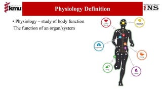


![TAU Anatomical Terminologies [Autosaved]77.pptx](https://cdn.slidesharecdn.com/ss_thumbnails/tauanatomicalterminologiesautosaved77-230127105554-2f0ad404-thumbnail.jpg?width=640&height=640&fit=bounds)


