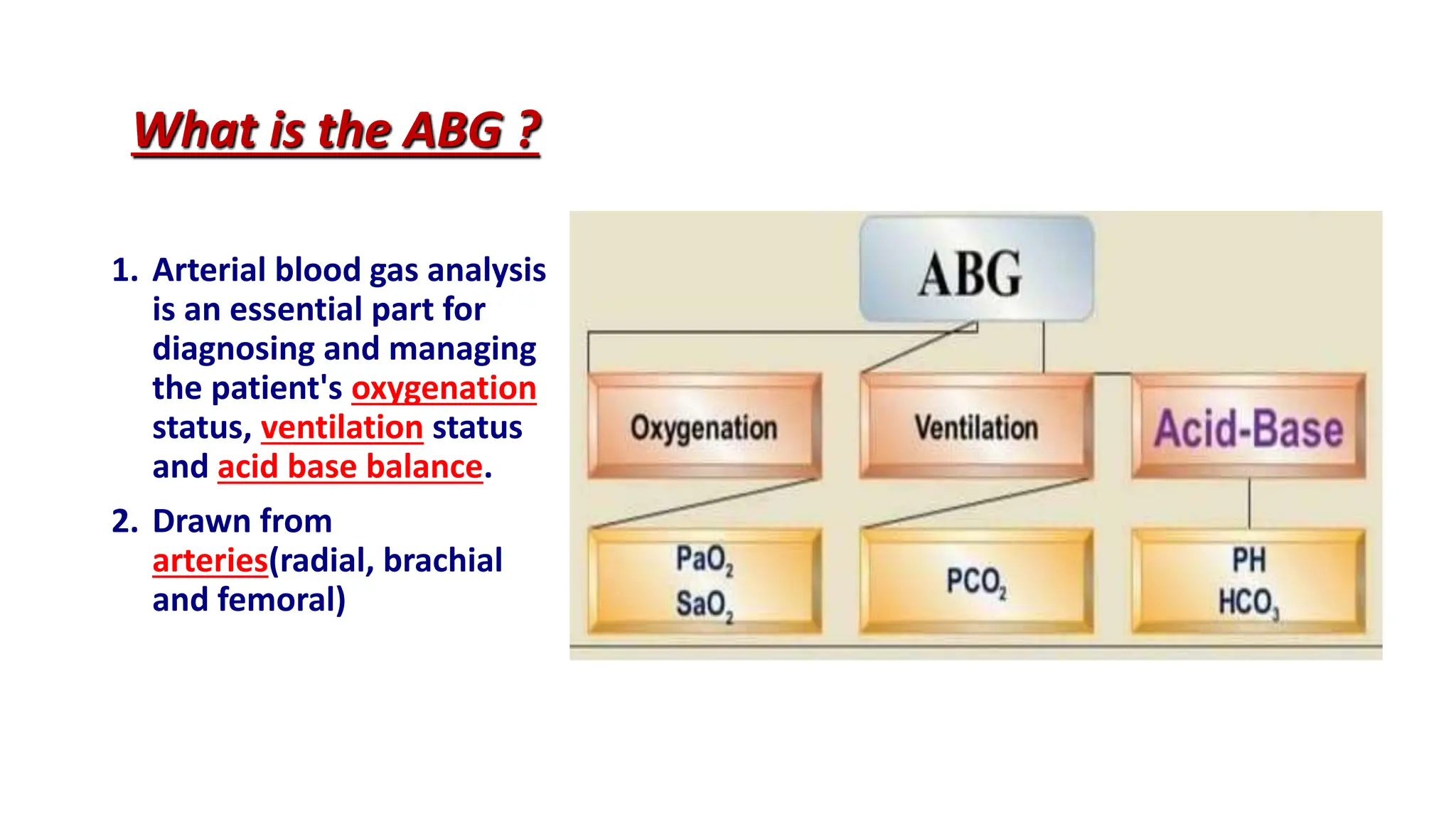
This presentation provides a comprehensive overview of Arterial Blood Gas (ABG) analysis, a vital tool in clinical practice for assessing acid-base balance and respiratory function. Beginning with an introduction to ABG analysis, the presentation delves into the interpretation of key components such as pH, PaO2, PaCO2, HCO3-, and SaO2, elucidating their normal ranges and physiological significance. Through case studies and examples, attendees will gain practical insights into interpreting ABG results in various clinical scenarios, including respiratory and metabolic disturbances. By the end, participants will have a solid understanding of ABG analysis and its crucial role in clinical decision-making.
Contact: jayraut1213@gmail.com
2. INTRODUCTION
• Arterial blood gases are an important routine
investigation to monitor the acid-base imbalance of
the patient. They may help to make diagnosis,
indicate the severity of condition and help to assess
treatment. Blood for ABG analysis can be obtained
by arterial puncture usually from radial and femoral
artery. Bicarbonate helps in maintaining blood pH
value. Kidney and lungs work in our body as
compensatory mechanism.
3. What is the ABG ?
1. Arterial blood gas analysis
is an essential part for
diagnosing and managing
the patient's oxygenation
status, ventilation status
and acid base balance.
2. Drawn from
arteries(radial, brachial
and femoral)
4. PURPOSE
1. To determine the presence and type of acid -
base balance.
2. To check for severe breathing problem and lungs
diseases.
3. Assessment of the response to the therapeutic
intervention such as mechanical ventilator.
7. ACID - BASE BALANCE
1. The primary aim of keeping this delicate balance is to
preserve the Homeostasis i.e. the highly complex
interactions that maintain all body systems to functioning
within a normal range.
2. Any extreme change in this balance ( PH < 6.8 or > 7.8)
may result in disastrous changes e.g. denaturation of
proteins & shut down of all enzymatic and metabolic
processes. Such disturbed environment would be
incompatible with life.
8. Types of Acid - Base Imbalance
1.Respiratory Acidosis
2.Respiratory Alkalosis
3.Metabolic Acidosis
4.Metabolic Alkalosis
9. (I) Respiratory Acidosis
1. It is defined as a pH less than 7.35 with a PaCO2 greater
than 45 mmHg.
2. Acidosis is the accumulation of co₂ which combines with
water in the body to produce carbonic acid, thus lowering
the pH of the blood.
ABG pH PaCO2 HCO3
Respiratory
Acidosis
Normal
12. (II) Respiratory Alkalosis
1. It is defined as a pH greater than 7.45 with a PaCO2
lesser than 35 mmHg.
2. Alkalosis is due to excessive wash of co₂ 2
(hyperventilation), thus increasing the pH of the
blood.
ABG pH PaCO2 HCO3
Respiratory
Alkalosis
Normal
15. (III) Metabolic Acidosis
• It is defined as a pH less than 7.35 with a HCO3 less than 22 mEq/L.
• Toxic Causes: Any disorder that will lead to tissue hypoperfusion
whatever the cause will lead eventually to increase in lactic acid
production resulting in Metabolic Acidosis.
ABG pH PaCO2 HCO3
Metabolic
Acidosis
Normal
1. Late salicylate
2. Methanol
3. Ethylene glycol
4. Iron
16. Causes Metabolic Acidosis
• Alcohol
• Cancer
• Diarrhea
• Liver failure
• Poisoning by aspirin and methanol
• Sever dehydration
• Seizure
18. (IV) Metabolic Alkalosis
• It is defined as a pH greater than 7.45 with HCO3 greater
than 28 mEq/L
• Causes
• It is due to excessive acid loss (repeated vomiting and
nasogastric suction) OR bicarbonate retention e.g. overuse
of sodium bicarbonate.
ABG pH PaCO2 HCO3
Metabolic
Alkalosis
Normal
22. Equipment Required
A clean tray containing:
• Gloves
• Antiseptic solution
• Local anesthesia
• 0.5 ml sodium heparin (1:1000)
• 2 - 3 ml syringe with 20, 23 and 25 gauze needle.
• Paper bag
23.
24. Preparation of client
■ To record the temperature of the client.
■ Record inspired oxygen concentration of the
client.
■ Heparnised the syringe to prevent blood
clotting.
■ To reduce anxiety of the client.
■ Allen's test
25.
26. Allen's Test
1. Patient clenches fist
2. Apply firm pressure to
radial & ulnar arteries
3. Patient relaxes hand
4. Release pressure on the
ulnar artery
5. Palm should flush within
5-15 secs
Compress Radial
Artery
Compress Ulnar
Artery
27. Sample Collection
• Wash hands and wear gloves.
• Place pillow under the patient's wrist.
• Palpate the artery i.e radial, brachial or femoral to be
punctured.
• Obliterate both radial and ulnar arteries at wrist by pressing
them with both thumbs.
• Ask the patient to clench and unclench the fist until
blanching of skin occurs.
28. • Release the pressure on the ulnar artery by removing
the thumb on it.
• Watch for return of circulation to skin with in 15 sec.
• Palpate the radial artery for pulsation.
• Puncture the artery at 45 - 60° deg angle.
• The arterial blood rushes into the syringe with a
great force.
• We should withdraw 2-3 ml of blood for sample.
29. • Once the sample has been taken, withdraw the
needle and apply firm pressure over the site of
puncture with dry sponge.
• Remove the air bubble from syringe and needle.
• Capping the needle.
• Place the capped syringe into an ice container.
• Maintain firm pressure on puncture site for 5 min.
• If patient is on anticoagulants use the high pressure
dressing.
30. Follow-up
•Send the collected sample to the
laboratory immediately.
•Assess for cold hands numbness. or
•Change the ventilator settings.
31. Complication of ABG
• Bleeding
• Infection at puncture site
• Blood accumulating under skin
• Local pain
• Thrombus in artery
• Feeling faint
• Numbness of hand