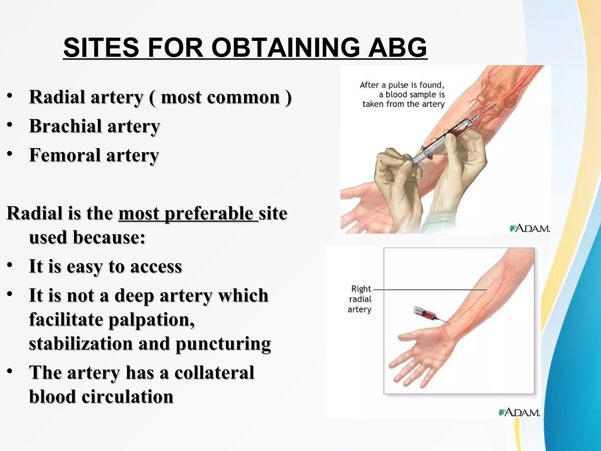
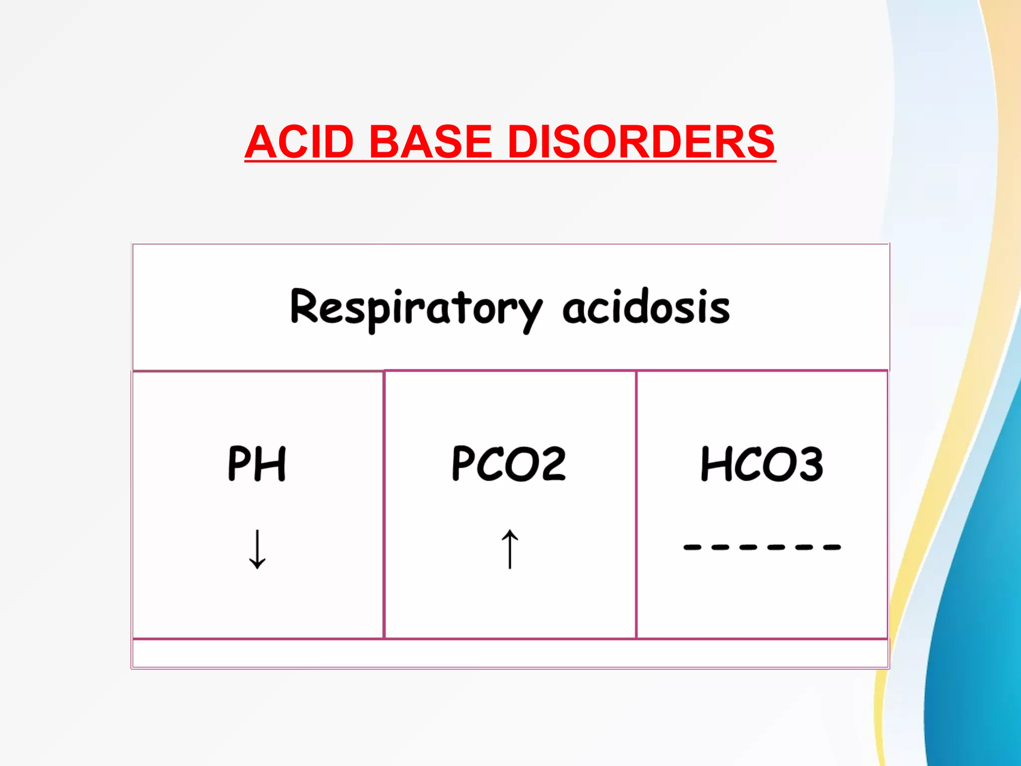
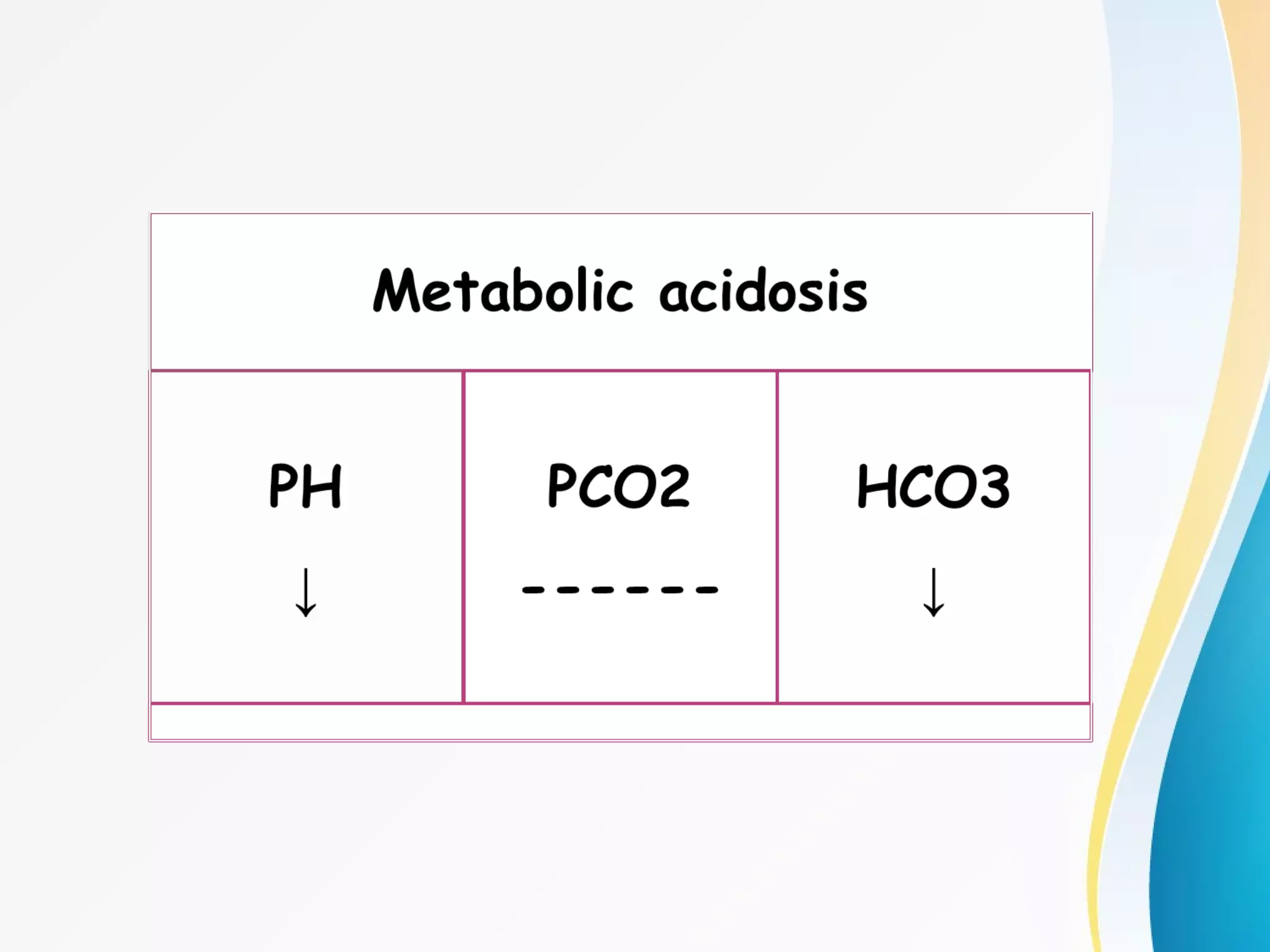
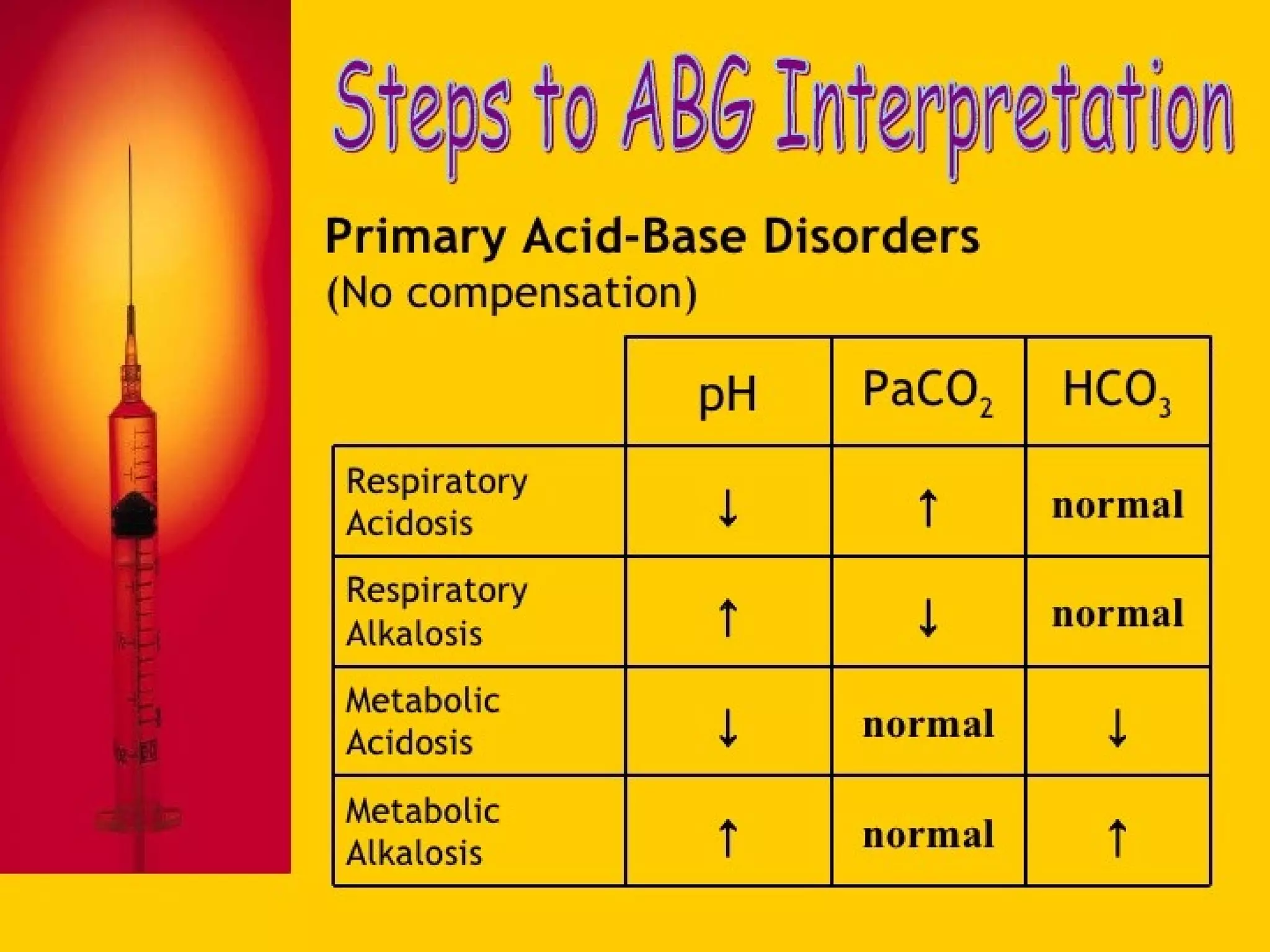
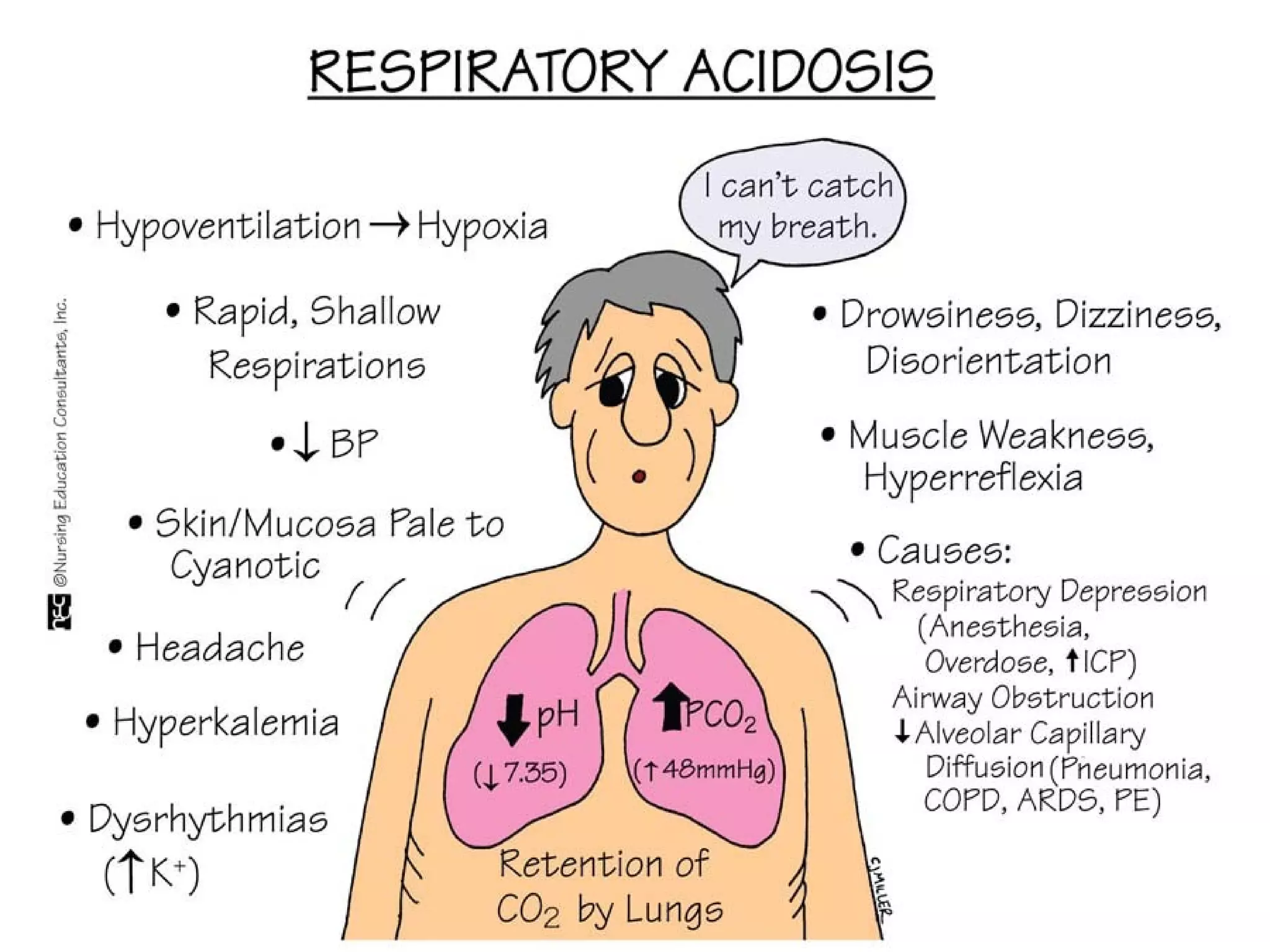
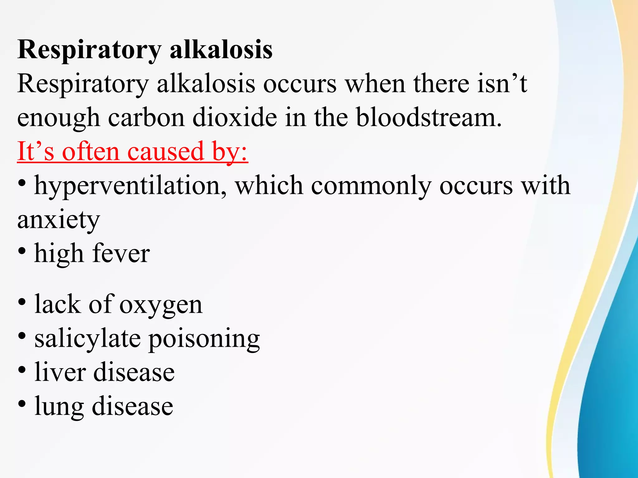
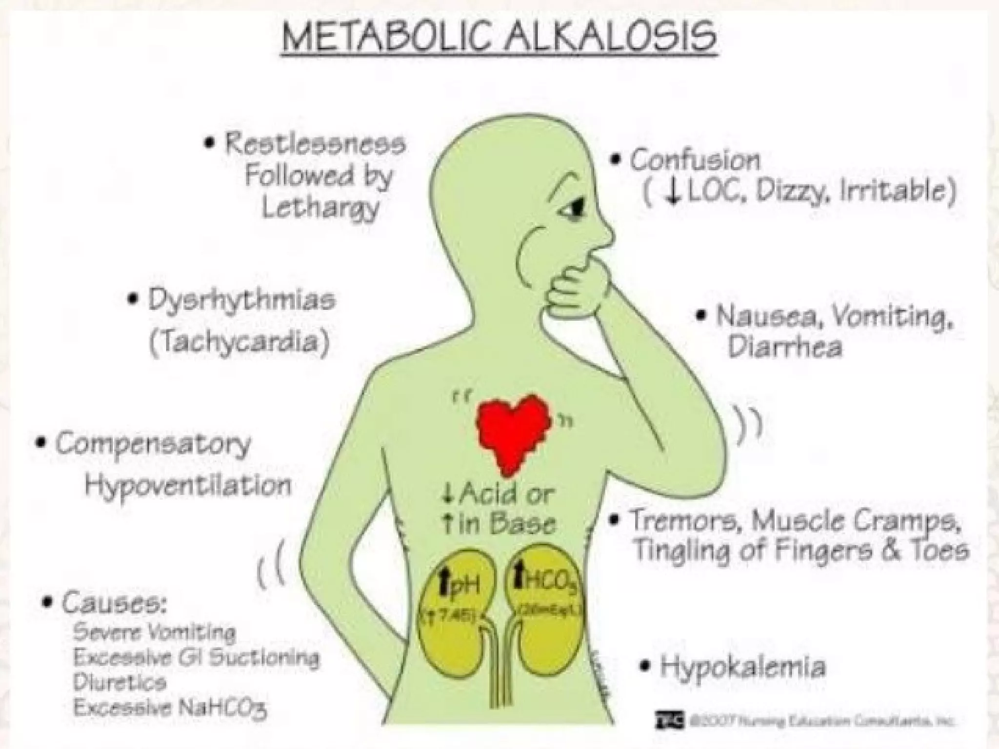
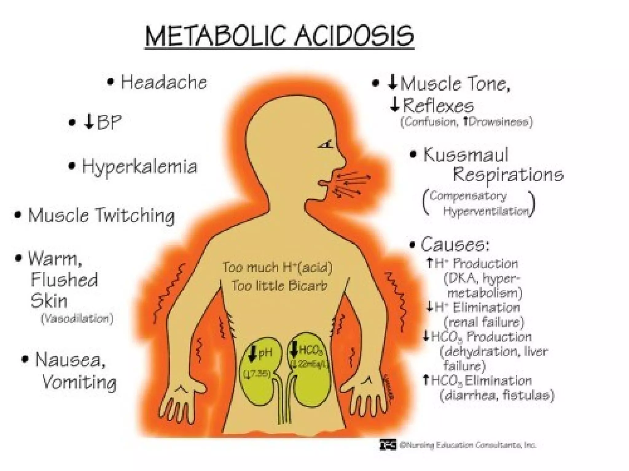
ABG
Shemil
Clinical Instructor
DM WIMS
ArterialBloodGasSampling(ABG)-OSCEGuideNEW.avi
DEFINITION
It is a diagnostic procedure in which a blood is obtained
from an artery directly by an arterial puncture or accessed
by a way of indwelling arterial catheter
INDICATION
• To obtain information about patient ventilation (PCO2) ,
oxygenation (PO2) and acid base balance
• Monitor gas exchange and acid base abnormalities for patient
on mechanical ventilator or not
• To evaluate response to clinical intervention and diagnostic
evaluation ( oxygen therapy )
• An ABG test may be most useful when a person's breathing rate
is increased or decreased or when the person has very high
blood sugar levels, a severe infection, or heart failure
ABG COMPONENT
• PH:
Measures hydrogen ion concentration in the blood, it shows
blood’s acidity or alkalinity.
• PCO2 :
It is the partial pressure of CO2 that is carried by the blood
for excretion by the lungs, known as respiratory parameter.
• PO2:
It is the partial pressure of O2 that is dissolved in the blood ,
it reflects the body ability to pick up oxygen from the lungs.
• HCO3 :
Known as the metabolic parameter, it reflects the kidney’s
ability to retain and excrete bicarbonate. Helps pH of blood
from becoming too acidic or too basic
NORMAL VALUES:
• PH = 7.35 – 7.45
• PCO2 = 35 – 45 mmhg
• PO2 = 80 – 100 mmhg
• HCO3 = 22 – 28 meq/L
EQUIPMENT
Blood gas kit OR
• 1ml syringe
• 23-26 gauge needle
• Stopper or cap
• Alcohol swab
• Disposable gloves
• Plastic bag & crushed ice
• Lidocaine (optional)
• Vial of heparin (1:1000)
• Par code or label
PREPARATORY PHASE:
• Record patient inspired oxygen concentration
• Explain the procedure to the patient
• Provide privacy for client
• If not using heparinized syringe , heparinize the needle
• Perform Allen's test
• Wait at least 20 minutes before drawing blood for ABG
after initiating, changing, or discontinuing oxygen
therapy, or settings of mechanical ventilation, after
suctioning the patient or after Extubation.
ALLEN’S TEST
It is a test done to determine that collateral
circulation is present from the ulnar artery in case
thrombosis occur in the radial artery.
SITES FOR OBTAINING ABG
• Radial artery ( most common )Radial artery ( most common )
• Brachial arteryBrachial artery
• Femoral arteryFemoral artery
Radial is theRadial is the most preferablemost preferable sitesite
used because:used because:
• It is easy to accessIt is easy to access
• It is not a deep artery whichIt is not a deep artery which
facilitate palpation,facilitate palpation,
stabilization and puncturingstabilization and puncturing
• The artery has a collateralThe artery has a collateral
blood circulationblood circulation
PERFORMANCE PHASE:
• Wash hands
• Put on gloves
• Palpate the artery for maximum pulsation
• If radial, perform Allen's test
• Place a small towel roll under the patient wrist
• Instruct the patient to breath normally during the test and warn him
that he may feel brief cramping or throbbing pain at the puncture
site
• Clean with alcohol swab in circular motion
• Skin and subcutaneous tissue may be infiltrated with local
anesthetic agent if needed
• Insert needle at 45 radial ,60 brachial and 90 femoral
• Withdraw the needle and apply digital pressure
• Check bubbles in syringe
• Place the capped syringe in the container of ice
immediately (consumption of oxygen by the leukocytes
and platelets in the sample)
• Maintain firm pressure on the puncture site for 5
minutes, if patient has coagulation abnormalities apply
pressure for 10 – 15 minutes
FOLLOW UP PHASE:
• Send labeled, iced specimen to the lab immediately
• Palpate the pulse distal to the puncture site
• Assess for cold hands, numbness, tingling or discoloration
• Documentation include: results of Allen's test, time the sample was
drawn, temperature, puncture site, time pressure was applied and if
O2 therapy is there
• Make sure it’s noted on the slip whether the patient is breathing room
air or oxygen. If oxygen, document the number of liters . If the
patient is receiving mechanical ventilation, FIO2 should be
documented
COMPLICATION
• Arteriospasm
• Hematoma
• Hemorrhage
• Distal ischemia
• Infection
• Numbness
A LOOK AT ACIDS AND BASES
• The body constantly works to maintain a balance
(homeostasis) between acids and bases. Without
that balance, cells can’t function properly. As
cells use nutrient to produce the energy, two by-
products are formed H+ & CO2. acid-base
balance depends on the regulation of the free
hydrogen ions
• Even slight imbalance can affect metabolism and
essential body functions. Several conditions as
infection or trauma and medications can affect
acid-base balance
ACID BASE DISORDERS
20
A.Y.T
BASE excess
• Base excess and base deficit refer to an
excess or deficit, respectively, in the amount
of base present in the blood.
• A typical reference range for base excess
is −2 to +2 mEq/L
• Positive numbers indicating an excess of
base and negative a deficit
• Base excess beyond the reference range
indicates
• metabolic alkalosis if too high (more than
+2 mEq/L)
• metabolic acidosis if too low (less than −2
mEq/L)
Respiratory acidosis
Respiratory acidosis occurs when too much CO2
builds up in the body. Normally, the lungs remove
CO2 while exhaling.
This may happen due to:
•Chronic airway conditions, like asthma
•Injury to the chest
•Obesity, which can make breathing difficult
•Sedative misuse
•Muscle weakness in the chest
•Problems with the nervous system
•Deformed chest structure
Respiratory alkalosis
Respiratory alkalosis occurs when there isn’t
enough carbon dioxide in the bloodstream.
It’s often caused by:
• hyperventilation, which commonly occurs with
anxiety
• high fever
• lack of oxygen
• salicylate poisoning
• liver disease
• lung disease
Metabolic alkalosis
Metabolic alkalosis develops when body loses too
much acid or gains too much base. This can be
attributed to:
• Excess vomiting, which causes electrolyte loss
• Overuse of diuretics
• A large loss of potassium or sodium in a short
amount of time
• Antacids
• Accidental ingestion of bicarbonate, which can be
found in baking soda
• Laxatives
• Alcohol abuse
Metabolic acidosis :starts in the kidneys instead of the lungs.
It occurs when they can’t eliminate enough acid or when they
get rid of too much base.
Diabetic acidosis occurs in people with diabetes that’s
poorly controlled. When body lacks
enough insulin, ketone build up in the body and acidify
blood.
•Hyperchloremic acidosis results from a loss of sodium
bicarbonate. This base helps to keep the blood neutral.
Both diarrhea and vomiting can cause this type of acidosis.
•Lactic acidosis occurs when there’s too much lactic acid in
body. Causes can include chronic alcohol
use, cancer, seizures, liver failure, prolonged lack of oxygen,
and low blood sugar.
•Renal tubular acidosis occurs when the kidneys are unable
to excrete acids into the urine. This causes the blood to
THANK YOU….
A.Y.T
32
QUESTONS ?

ABG

  • 1.
  • 2.
  • 3.
    DEFINITION It is adiagnostic procedure in which a blood is obtained from an artery directly by an arterial puncture or accessed by a way of indwelling arterial catheter
  • 4.
    INDICATION • To obtaininformation about patient ventilation (PCO2) , oxygenation (PO2) and acid base balance • Monitor gas exchange and acid base abnormalities for patient on mechanical ventilator or not • To evaluate response to clinical intervention and diagnostic evaluation ( oxygen therapy ) • An ABG test may be most useful when a person's breathing rate is increased or decreased or when the person has very high blood sugar levels, a severe infection, or heart failure
  • 5.
    ABG COMPONENT • PH: Measureshydrogen ion concentration in the blood, it shows blood’s acidity or alkalinity. • PCO2 : It is the partial pressure of CO2 that is carried by the blood for excretion by the lungs, known as respiratory parameter. • PO2: It is the partial pressure of O2 that is dissolved in the blood , it reflects the body ability to pick up oxygen from the lungs. • HCO3 : Known as the metabolic parameter, it reflects the kidney’s ability to retain and excrete bicarbonate. Helps pH of blood from becoming too acidic or too basic
  • 6.
    NORMAL VALUES: • PH= 7.35 – 7.45 • PCO2 = 35 – 45 mmhg • PO2 = 80 – 100 mmhg • HCO3 = 22 – 28 meq/L
  • 7.
    EQUIPMENT Blood gas kitOR • 1ml syringe • 23-26 gauge needle • Stopper or cap • Alcohol swab • Disposable gloves • Plastic bag & crushed ice • Lidocaine (optional) • Vial of heparin (1:1000) • Par code or label
  • 8.
    PREPARATORY PHASE: • Recordpatient inspired oxygen concentration • Explain the procedure to the patient • Provide privacy for client • If not using heparinized syringe , heparinize the needle • Perform Allen's test • Wait at least 20 minutes before drawing blood for ABG after initiating, changing, or discontinuing oxygen therapy, or settings of mechanical ventilation, after suctioning the patient or after Extubation.
  • 9.
    ALLEN’S TEST It isa test done to determine that collateral circulation is present from the ulnar artery in case thrombosis occur in the radial artery.
  • 10.
    SITES FOR OBTAININGABG • Radial artery ( most common )Radial artery ( most common ) • Brachial arteryBrachial artery • Femoral arteryFemoral artery Radial is theRadial is the most preferablemost preferable sitesite used because:used because: • It is easy to accessIt is easy to access • It is not a deep artery whichIt is not a deep artery which facilitate palpation,facilitate palpation, stabilization and puncturingstabilization and puncturing • The artery has a collateralThe artery has a collateral blood circulationblood circulation
  • 12.
    PERFORMANCE PHASE: • Washhands • Put on gloves • Palpate the artery for maximum pulsation • If radial, perform Allen's test • Place a small towel roll under the patient wrist • Instruct the patient to breath normally during the test and warn him that he may feel brief cramping or throbbing pain at the puncture site • Clean with alcohol swab in circular motion • Skin and subcutaneous tissue may be infiltrated with local anesthetic agent if needed
  • 13.
    • Insert needleat 45 radial ,60 brachial and 90 femoral • Withdraw the needle and apply digital pressure • Check bubbles in syringe • Place the capped syringe in the container of ice immediately (consumption of oxygen by the leukocytes and platelets in the sample) • Maintain firm pressure on the puncture site for 5 minutes, if patient has coagulation abnormalities apply pressure for 10 – 15 minutes
  • 14.
    FOLLOW UP PHASE: •Send labeled, iced specimen to the lab immediately • Palpate the pulse distal to the puncture site • Assess for cold hands, numbness, tingling or discoloration • Documentation include: results of Allen's test, time the sample was drawn, temperature, puncture site, time pressure was applied and if O2 therapy is there • Make sure it’s noted on the slip whether the patient is breathing room air or oxygen. If oxygen, document the number of liters . If the patient is receiving mechanical ventilation, FIO2 should be documented
  • 15.
    COMPLICATION • Arteriospasm • Hematoma •Hemorrhage • Distal ischemia • Infection • Numbness
  • 16.
    A LOOK ATACIDS AND BASES • The body constantly works to maintain a balance (homeostasis) between acids and bases. Without that balance, cells can’t function properly. As cells use nutrient to produce the energy, two by- products are formed H+ & CO2. acid-base balance depends on the regulation of the free hydrogen ions • Even slight imbalance can affect metabolism and essential body functions. Several conditions as infection or trauma and medications can affect acid-base balance
  • 17.
  • 20.
  • 21.
    BASE excess • Baseexcess and base deficit refer to an excess or deficit, respectively, in the amount of base present in the blood. • A typical reference range for base excess is −2 to +2 mEq/L • Positive numbers indicating an excess of base and negative a deficit
  • 22.
    • Base excessbeyond the reference range indicates • metabolic alkalosis if too high (more than +2 mEq/L) • metabolic acidosis if too low (less than −2 mEq/L)
  • 24.
    Respiratory acidosis Respiratory acidosisoccurs when too much CO2 builds up in the body. Normally, the lungs remove CO2 while exhaling. This may happen due to: •Chronic airway conditions, like asthma •Injury to the chest •Obesity, which can make breathing difficult •Sedative misuse •Muscle weakness in the chest •Problems with the nervous system •Deformed chest structure
  • 26.
    Respiratory alkalosis Respiratory alkalosisoccurs when there isn’t enough carbon dioxide in the bloodstream. It’s often caused by: • hyperventilation, which commonly occurs with anxiety • high fever • lack of oxygen • salicylate poisoning • liver disease • lung disease
  • 28.
    Metabolic alkalosis Metabolic alkalosisdevelops when body loses too much acid or gains too much base. This can be attributed to: • Excess vomiting, which causes electrolyte loss • Overuse of diuretics • A large loss of potassium or sodium in a short amount of time • Antacids • Accidental ingestion of bicarbonate, which can be found in baking soda • Laxatives • Alcohol abuse
  • 30.
    Metabolic acidosis :startsin the kidneys instead of the lungs. It occurs when they can’t eliminate enough acid or when they get rid of too much base. Diabetic acidosis occurs in people with diabetes that’s poorly controlled. When body lacks enough insulin, ketone build up in the body and acidify blood. •Hyperchloremic acidosis results from a loss of sodium bicarbonate. This base helps to keep the blood neutral. Both diarrhea and vomiting can cause this type of acidosis. •Lactic acidosis occurs when there’s too much lactic acid in body. Causes can include chronic alcohol use, cancer, seizures, liver failure, prolonged lack of oxygen, and low blood sugar. •Renal tubular acidosis occurs when the kidneys are unable to excrete acids into the urine. This causes the blood to
  • 32.